
Figure 1.
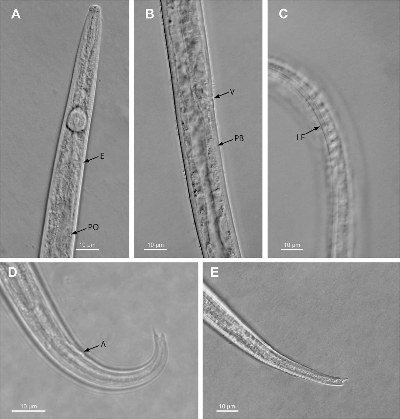
Figure 2.
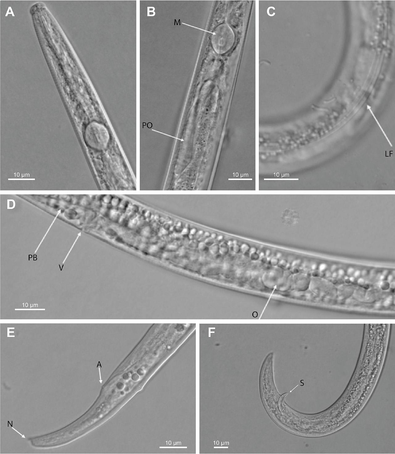
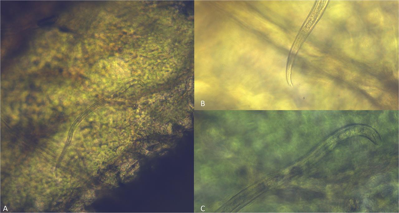
Figure 3.
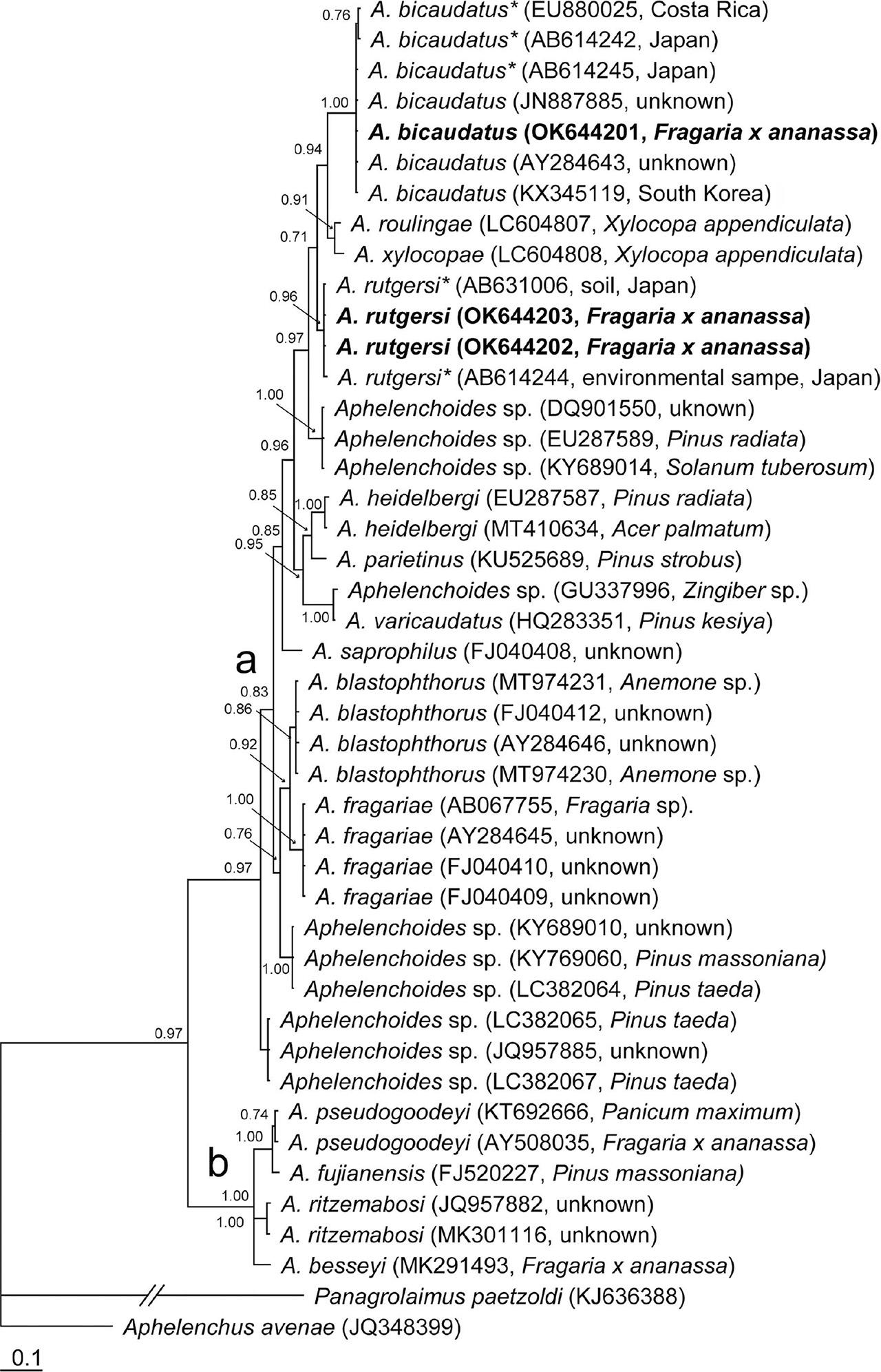
Figure 4.
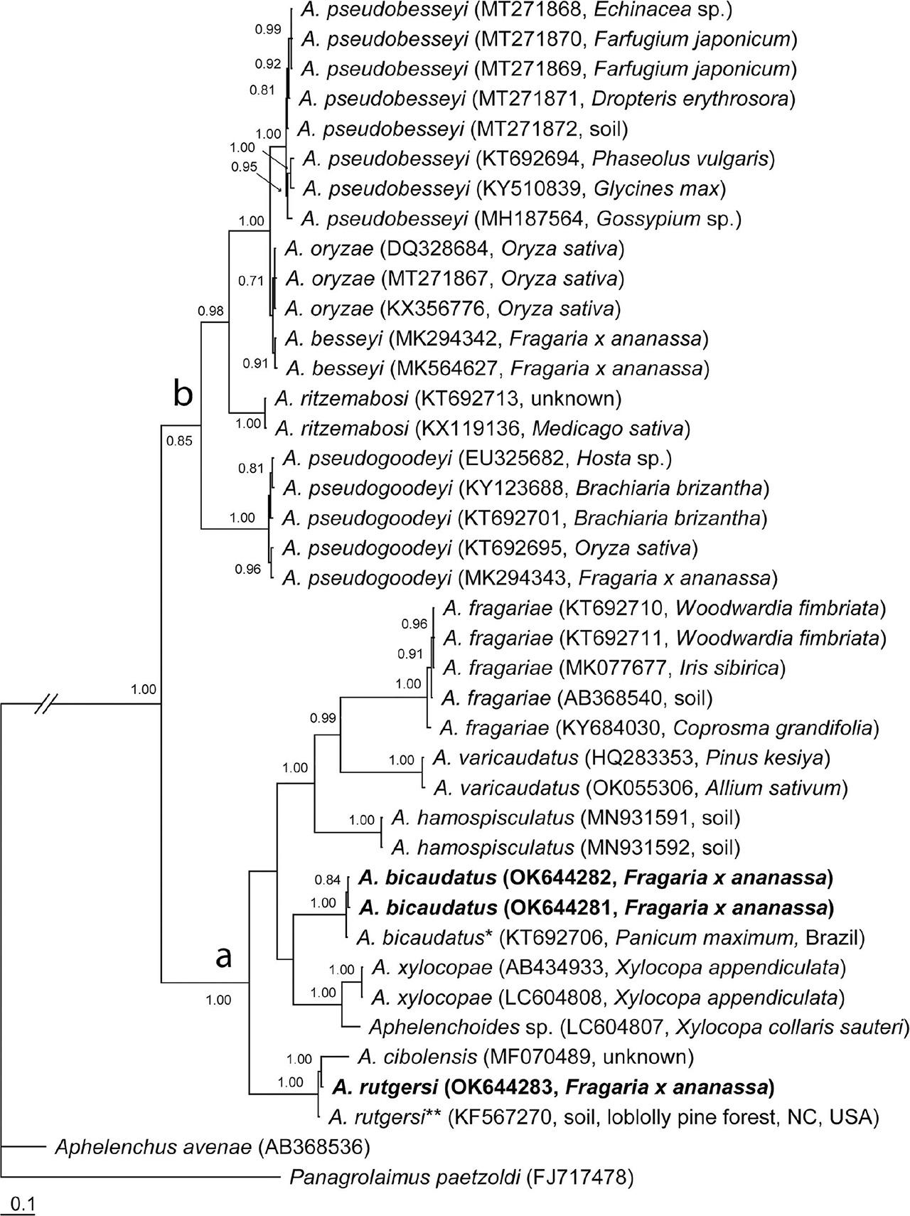
Figure 5.
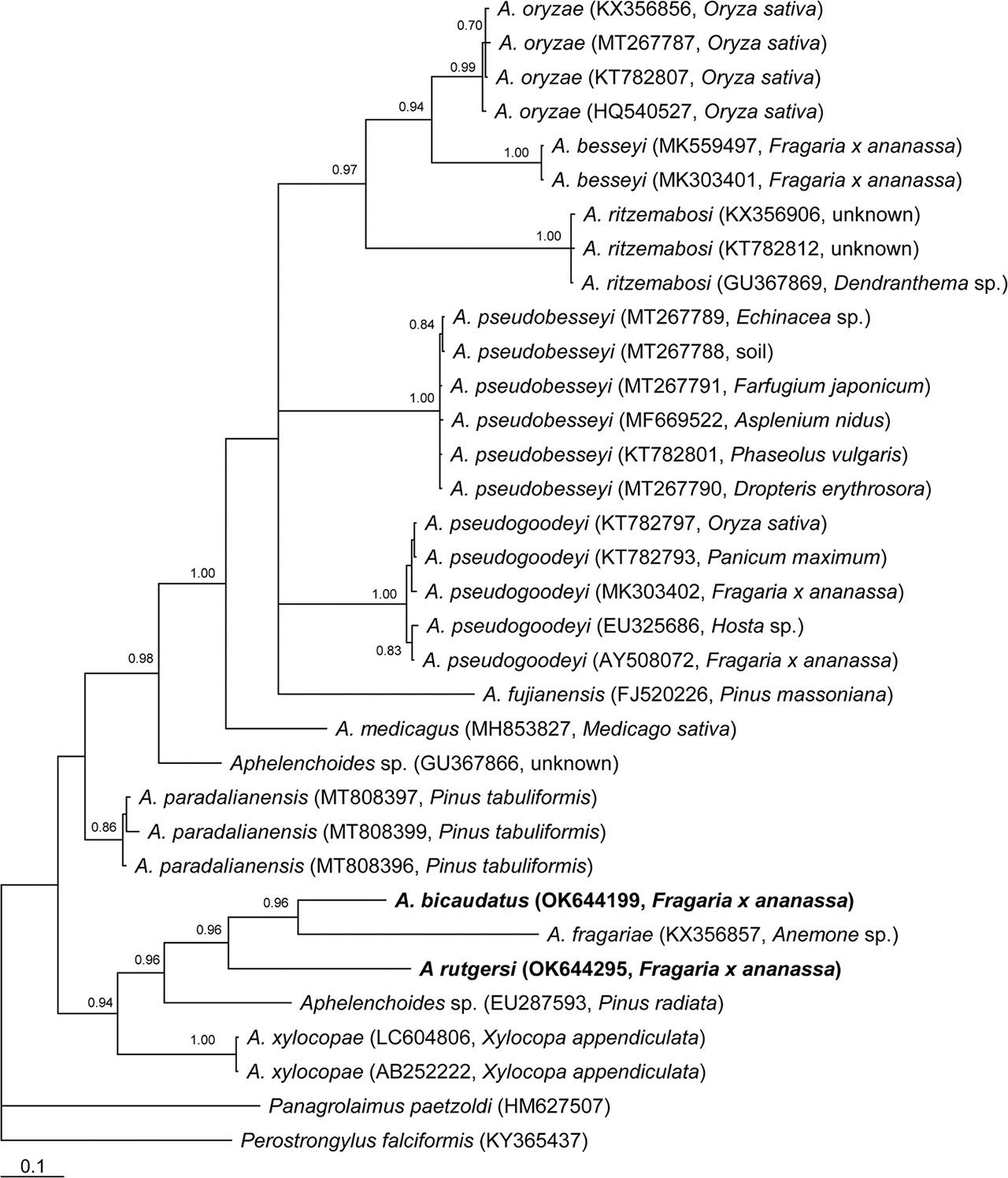
Figure 6.
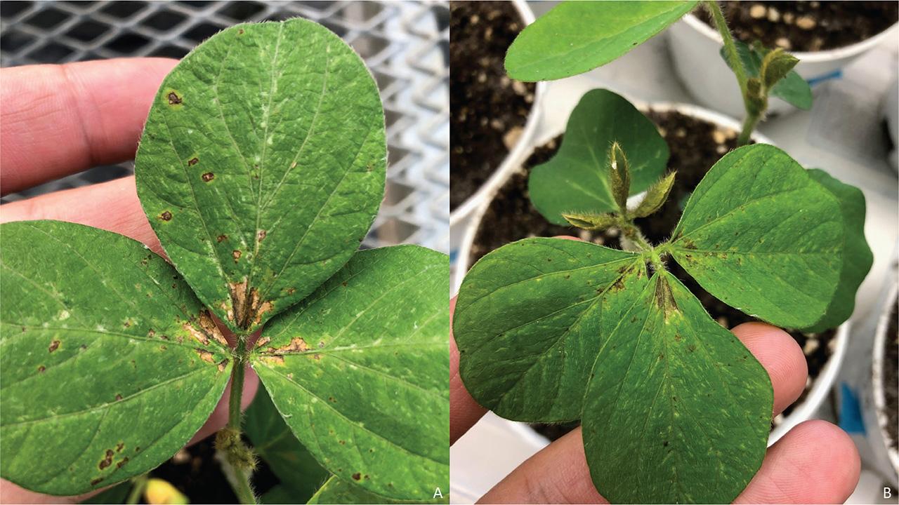
Figure 7.
Populations of Aphelenchoides characterized in the present study_
| Species | Location | Host | Sample code | GenBank accession number | Source | ||
|---|---|---|---|---|---|---|---|
| SSU | LSU | COI | |||||
| A. bicaudatus | Wimauma, Florida, USA | Fragaria × ananassa | N18-1001-3 | OK644201 | OK644281 | OK644199 | C. Oliveira |
| OK644282 | |||||||
| A. rutgersi | Wimauma, Florida, USA | Fragaria × ananassa | N18-00206 | OK644202 | OK644283 | OK644295 | C. Oliveira |
| OK644203 | |||||||
Morphometrics of live females of a Florida population of Aphelenchoides bicaudatus from strawberry reared on Monilinia fructicola compared to those in the original (Imamura, 1931) and subsequent re-descriptions by Siddiqui & Taylor (1967), Jen et al_ (2012), Kim et al_ (2016), Israr et al_ (2017) and Shokoohi and Moyo (2022)_a
| Population Substrate | Florida | Japan | Illinois | Taiwan | South Korea | Pakistan | South Africa |
|---|---|---|---|---|---|---|---|
| Reference | Present paper N18-01001-3 | Imamura (1931) | Siddiqui and Taylor (1967) | Jen et al. (2012) | Kim et al. (2016) | Israr et al. (2017) | Shokoohi and Moyo (2022) |
| Character n | 10♀♀ | 18♀♀ | 50♀♀ | 50♀♀ | 7♀♀ | 2♀♀ | 8♀♀ |
| L | 494.1 ± 32.0 | 499 ± 67.9 | 517.9 ± 3.8 | 455.0 ± 64.5 | |||
| (454.8–552.4) | (380.0–470.0) | (410.0–550.0) | (376–637) | (513.6–522.6) | (360.0–360.0) | (409.0–529.0) | |
| A | 31.2 ± 3.6 | 33.0 ± 2.4 | 28.3 ± 0.5 | 29.1 ± 2.0 | |||
| (27.6–41.1) | (31.3–31.7) | (25–31) | (27.0–38.6) | (27.7–28.8) | (30.1–32.7) | (27.1–31.1) | |
| B | 8.0 ± 0.5 | 9.0 ± 0.7 | 7.3 ± 0.0 | 4.6 ± 0.5 | |||
| (7.2–9.0) | (6.8–8.4) | (7.3–9.6) | (7.5–10.0) | (7.3–7.4) | (8.8–7.2) | (4.1–5.1) | |
| b’ | 4.1 ± 0.2 | - | - | 5.1 ± 0.7 | - | - | |
| (3.7–4.6) | (3.6–7.9) | (5.6–5.8) | |||||
| C | 13.2 ± 0.9 | 11.9 ± 0.9 | 11.3 ± 0.5 | 16.3 ± 2.0 | |||
| (11.9–14.8) | (9.4–12.6) | (9.8–13.7) | (10.1–14.8) | (10.7–11.9) | (11.3–12.0) | (14.1–17.8) | |
| c’ | 4.4 ± 0.5 | - | - | 5.4 ± 0.5 | 4.6 ± 0.1 | 3.2 ± 0.6 | |
| (3.7–5.3) | (4.1–7.1) | (4.4–4.8) | (2.9–3.7) | (2.6–3.6) | |||
| V | 68.7 ± 1.5 | 68.5 ± 1.2 | 66.0 ± 0.2 | 68.8 ± 1.6 | |||
| (65.8–71.1) | (61.7–90.2) | (65.0–70.0) | (64.9–71.8) | (65.7–66.4) | (66.8–67.2) | (67.0–71.0) | |
| OV | 27.8 ± 2.0 | - | - | 15.2 ± 2.7 | - | - | |
| (23.9–30.9) | (11.0–22.0) | (12.0–12.5) | |||||
| Max. body diameter | 15.9 ± 1.3 | - | - | - | - | 15.6 ± 1.5 | |
| (13.5–17.8) | (25.0–26.2) | (14.0–17.0) | |||||
| Body diameter at anus | 8.6 ± 0.8 | - | - | - | - | 8.8 ± 0.7 | |
| (7.2–9.9) | (11.8) | (8.0–9.0) | |||||
| Anterior genital tract length | 136.9 ± 10.4 | - | - | - | - | 179.3 ± 18.0 | |
| (121.7–150.1) | (84.0–95.0) | (167.0–200.0) | |||||
| Lip region width | 4.9 ± 0.3 | - | - | - | 5.7 ± 0.1 | ||
| (4.5–5.5) | (5.0–5.2) | (4.0–4.3) | (5.6–5.8) | ||||
| Lip region height | 2.8 ± 0.2 | - | - | - | 2.8 ± 0.9 | ||
| (2.4–3.0) | (2.5–2.9) | (2.0–2.0) | (2.0–4.0) | ||||
| Stylet length | 10.9 ± 0.4 | - | 10.4 ± 0.6 | 11.2 ± 0.5 | 11.1 ± 1.7 | ||
| (10.3–11.8) | (10–12) | (9.0–12.0) | (10.4–11.7) | (10.0–11.0) | (10.0–13.0) | ||
| Stylet cone | 5.1 ± 0.3 | - | - | - | - | - | 4.7 ±0.3 |
| (4.5–5.8) | (4.5–5.0) | ||||||
| Stylet knob height | 1.4 ± 0.2 | - | - | - | - | - | - |
| (1.2-1.7) | |||||||
| Stylet knob width | 1.9 ± 0.3 | - | - | - | - | - | - |
| (1.2–2.3) | |||||||
| Median bulb length | 12.1 ± 0.7 | - | - | - | 11.2 ± 0.7 | ||
| (10.9–13.3) | (12.8–12.9) | (10.0–10.0) | (11.0–12.0) | ||||
| Median bulb width | 9.6 ± 0.8 | - | - | - | 9.0 ±0.8 | ||
| (7.9–11.1) | (8.6–8.9) | (7.0–8.0) | (8.5–10.0) | ||||
| Median bulb valve length | 4.1 ± 0.4 | - | - | - | - | - | - |
| (3.6–4.9) | |||||||
| Median bulb valve width | 2.9 ± 0.2 | - | - | - | - | - | - |
| (2.5–3.2) | |||||||
| Pharynx length | 62.1 ± 2.7 | - | - | - | - | 88.3 ± 11.2 | |
| (59.0–68.7) | (90.0–92.0) | (76.0–98.0) | |||||
| Pharyngeal overlap | 57.0 ± 2.1 | - | - | - | - | - | |
| (53.0–59.7) | (50.0–75.0) | ||||||
| Ant. end to pharyngeal gland lobe | 118.8 ± 3.3 | - | - | - | - | - | - |
| (4.5–5.8) | |||||||
| Anterior end to excretory pore | 66.9 ± 3.0 | - | - | - | - | 61.3 ± 3.2 | |
| (62.3–71.2) | (50.0–51.0) | (60.0–65.0) | |||||
| Postuterine sac (PUS) | 22.3 ± 1.9 | - | - | - | - | 47.0 ± 2.6 | |
| (18.0–24.2) | (22.0–24.0) | (45.0–50.0) | |||||
| Vulva anus distance (VA) | 123.5 ± 10.3 | - | - | - | - | - | |
| (105.9–137.6) | (84.0–85.0) | ||||||
| Ant. end to vulva | 349.3 ± 22.3 | - | - | - | - | - | |
| (311.0–392.7) | (242.0–248.0) | ||||||
| Post end to vulva | 144.8 ± 26.5 | - | - | - | - | - | - |
| (89.3–170.4) | |||||||
| Tail length | 37.4 ± 0.8 | - | 45.9 ± 2.5 | 28.0 ± 3.6 | |||
| (36.6–38.6) | (41.8) | (34–42) | (43.0–48.8) | (30.0–31.0) | (24.0–31.0) | ||
| Body width at vulva (BWV) | 14.5 ± 0.8 | - | - | - | - | - | - |
| (13.1–15.8) | |||||||
| PUS/L | 4.5 ± 0.4 | - | - | - | - | - | |
| (3.7–5.2) | (6.1–6.6) | ||||||
| PUS/BWV | 1.6 ± 0.2 | - | - | - | - | - | - |
| (1.1–1.8) | |||||||
| PUS/VA | 18.1 ± 1.1 | - | - | 18.9 ± 4.5 | - | - | |
| (16.0–20.5) | (9.2–33.8) | (22.4–24.0) | |||||
| Lateral field width | 2.7 ± 0.4 | - | - | - | - | - | |
| (2.1–3.4) | (2.2) | ||||||
Morphometrics of live females of a Florida populations of Aphelenchoides rutgersi from strawberry and reared on Monilinia fructicola compared to those in the original description by Hooper and Myers (1971) and populations from Pakistan and South Africa reported by Erum and Shahina (2010) and Girgan et al_ (2018), respectively_a
| Population Substrate | Florida | Florida | Florida | Florida | Pakistan | South Africa |
|---|---|---|---|---|---|---|
| Reference | Present study (N18-00206) | Hooper and Myers (1971) | Hooper and Myers (1971) | Hooper and Myers (1971) | Erum and Shahina (2010) | Girgan at al. (2018) |
| Character n | 20♀♀ | 20♀♀ | 20♀♀ | 20♀♀ | 12♀♀ | 3♀♀ |
| L | 507.6 ± 71.0 | 535.0 ± 20 | 480.0 ± 25 | 360.0 ± 20 | 440 ± 30 | 556.0 ± 22.6 |
| (371.2–614.7) | (500.0–570.0) | (430.0–530.0) | (320.0–405.0) | (370.0–500.0) | (542.0–582.0) | |
| A | 29 ± 2 | 27.0 ± 1.8 | 25.0 ± 1.7 | 27.0 ± 1.7 | 26.7 ± 1.4 | 34.1 ± 0.8 |
| (25.8–33.5) | (23–30) | (23–29) | (24–31) | (24.0–31.7) | (33.5–34.9) | |
| B | 8.4 ± 1.0 | 10.7 ± 0.4 | 9.3 ± 0.4 | 7.60 ± 0.16 | 5.7 ± 1.3 | 4.6 ± 0.2 |
| (6.5–10.0) | (9.7–11.6) | (8.3–10.1) | (6.7–8.9) | (4.4–8.4) | (4.4–4.8) | |
| b’ | 4.0 ± 0.6 | 5.0 ± 0.5 | 4.1 ± 0.4 | 3.5 ± 0.3 | 4.4 ± 0.8 | - |
| (3.0–5.2) | (4.2–6.2) | (3.6–4.6) | (3.1–4.0) | (4.1–5.0) | ||
| C | 15.4 ± 1.7 | 16.9 ± 0.6 | 16.3 ± 1.0 | 14.1 ± 0.8 | 8.0 ±0.4 | 15.8 ± 0.5 |
| (12.9–18.9) | (15.9–18.1) | (15.0–18.7) | (12.4–15.5) | (7.2–8.6) | (15.5–16.4) | |
| c’ | 3.4 ± 0.4 | 3.0 ± 0.2 | 2.9 ± 0.1 | 3.3 ± 0.2 | 5.7 ± 0.4 | 3.6 ± 0.3 |
| (2.5–4.0) | (2.6–3.4) | (2.6–3.2) | (2.8–4.0) | (4.8–6.4) | (3.3–3.9) | |
| V | 70.3 ± 1.0 | 71.0 ± 1.0 | 72.0 ± 0.9 | 70.0 ± 0.7 | 64.5 ± 0.9 | 69.0 ± 0.8 |
| (68.6–72.0) | (69.0–72.0) | (70.0–74.0) | (69.0–72.0) | (63.3–65.5) | (69.0–70.0) | |
| Max. body diameter | 17.6 ± 3.1 | - | - | - | - | 16.0 ± 0.9 |
| (12.8–22.7) | (16.0–17.0) | |||||
| Body diameter at anus or cloacal opening | 10.0 ± 1.4 | - | - | - | - | 10.0 ± 0.9 |
| (7.4–13.3) | (9.0–11.0) | |||||
| Body diameter at vulva (BDV) | 18.1 ± 2.3 | - | - | - | - | 16.0 ± 0.8 |
| (15–23) | (15.017.0) | |||||
| OV or Testis/L | 24.8 ± 6.4 | 60.0 ± 7.1 | 50.0 ± 4.6 | 24.0 ± 2.8 | - | - |
| (15.4–40.9) | (50–77) | (42–60) | (18–29) | |||
| Anterior genital tract length | 127.2 ± 44.0 | - | - | - | - | 220.0 ± 5.6 |
| (70.0–242.5) | (215.0–226.0) | |||||
| Lip region width | 6.3 ± 0.5 | - | - | - | 5.6 ± 0.4 | |
| (5.0–6.9) | (4.8–5.6) | (5.0–6.0) | ||||
| Lip region height | 2.9 ± 0.2 | - | - | - | 3.2 ± 0.5 | |
| (2.5–3.0) | (2.4–3.2) | (3.0–4.0) | ||||
| Stylet length | 11.0 ± 0.5 | - | - | 10.7 ± 0.6 | 11.0 ± 0.4 | |
| (10.1–11.9) | (10.0) | (10.0–12.0) | (11.0–12.0) | |||
| Stylet cone | 5.4 ± 0.4 | - | - | - | - | - |
| (4.8–6.0) | ||||||
| Stylet knob height | 1.5 ± 0.2 | - | - | - | - | - |
| (1.2–1.9) | ||||||
| Stylet knob width | 2.3 ± 0.3 | - | - | - | - | - |
| (1.9-2.8) | ||||||
| Median bulb length | 13.7 ± 0.7 | - | - | - | 12.0 ±0.5 | |
| (12.3–14.9) | (12.8–13.6) | (12.0–13.0) | ||||
| Median bulb width | 10.3 ± 0.7 | - | - | - | 10.0 ± 0.5 | |
| (9.0-11.9) | (9.6–10.4) | (9.0–10.0) | ||||
| Median bulb valve length | 4.0 ± 0.4 | - | - | - | - | - |
| (3.1–4.5) | ||||||
| Median bulb valve width | 3.0 ± 0.2 | - | - | - | - | - |
| (2.5–3.4) | ||||||
| Pharynx length | 60.4 ± 2.1 | 44 ± 2 | 46 ± 2 | 27.0 ± 1.7 | - | |
| (54.4–66.3) | (42–47) | (42–49) | (24–31) | (58.0) | ||
| Pharyngeal overlap | 67.5 ± 12.5 | - | - | - | - | - |
| (45.5–95.8) | ||||||
| Ant. end to pharyngeal gland lobe | 127.8 ± 13.7 | - | - | - | - | 122.0 ± 1.5 |
| (99.9–162.1) | (121.0–124.0) | |||||
| Anterior end to excretory pore | 66.6 ± 5.4 | - | - | - | - | 76.0 ±1.9 |
| (58.4–76.2) | (75.0–76.0) | |||||
| Postuterine sac | 26.1 ± 4.5 | - | - | - | 42.6 ±2.2 | 40.0 ± 4.1 |
| (15.8–41.0) | (36.0–55.0) | (37.0–43.0) | ||||
| Vulva anus distance | 117.7 ± 19.2 | - | - | - | - | - |
| (76.2–149.4) | ||||||
| Ant. end to vulva | 356.9 ± 50.3 | - | - | - | 386.0 ± 13.6 | - |
| (266.3–437.5) | (374.0 401.0) | |||||
| Post end to vulva | 150.7 ± 21.7 | - | - | - | - | - |
| (104.9–191.9) | ||||||
| Tail length | 33.0 ± 3.6 | - | - | 35.0 ± 2.1 | - | |
| (26.7–42.5) | (30.5) | (33.0–37.0) | ||||
| Spermatheca length | 14.1 ± 3.9 | - | - | - | - | - |
| (9–21) | ||||||
| Spermatheca width | 9.9 ± 1.6 | - | - | - | - | - |
| (8–13) | ||||||
| PUS/VA | 22.1 ± 3.8 | - | - | - | - | - |
| (16.1–27.5) | ||||||
| Lateral field width | 3.0 ± 0.2 | - | - | - | - | - |
| (2.5–3.4) | ||||||
| PUS/L | 5.1 ± 0.8 | - | - | - | - | - |
| (3.8–6.4) | ||||||
| PUS/BDV | 1.5 ± 0.2 | - | - | - | - | - |
| (1.0–1.9) | ||||||
| Spikes | 1 | 1 | 1 | - | 1 | - |
Morphometrics of live males of a Florida population of Aphelenchoides rutgersi from strawberry reared on Monilinia fructicola compared to those in the original description by Hooper and Myers (1971) and a population from Pakistan described by Erum and Shahina (2010)_a
| Population Substrate | Florida | Florida | Pakistan |
|---|---|---|---|
| Reference | Present study N18-00206 | Hooper and Myers (1971) | Erum and Shahina (2010) |
| Character n | 2♂♂ | 20♂♂ | 7♂♂ |
| L | 538.8 ± 20.5 | 440.0 ± 35 | 400.0 ± 30.0 |
| (518.3−559.2) | (380.0−490.0) | (360.0−470.0) | |
| A | 29.7 ± 0.3 | 29.0 ± 2.8 | 28.7 ± 2.5 |
| (29.4−30.0) | (21−32) | (24.0−31.7) | |
| B | 8.9 ± 0.4 | 8.7 ± 0.9 | 5.3 ± 1.3 |
| (8.5−9.3) | (7.1−11.0) | (3.9−7.2) | |
| b’ | 4.0 ± 0.1 | 4.0 ± 0.1 | 4.5 ± 1.0 |
| (4.0−4.1) | (3.6−4.4) | (4.2−5.5) | |
| C | 15.9 ± 1.2 | 14.3 ± 0.8 | 13.7 ± 1.6 |
| (14.7−17.2) | (13.0−15.4) | (10.6−16.0) | |
| c’ | 2.6 ± 0.3 | 2.9 ± 0.2 | 2.7 ± 0.3 |
| (2.3−2.9) | (2.4−3.3) | (2.4−3.1) | |
| Max. body diameter | 18.2 ± 0.9 | - | - |
| (17.3−19.0) | |||
| Body diameter at cloacal opening | 13 ± 1 | - | - |
| (12−14) | |||
| Testis | 240.5 ± 8.5 | - | - |
| (232−249) | |||
| Testis/L% | 44.7 ± 0.1 | 72.0 ± 6.2 | - |
| (44.5−44.8) | (58.0−78.0) | ||
| Lip region width | 6.5 ± 0.2 | - | - |
| (6.3−6.6) | |||
| Lip region height | 3 | - | - |
| Stylet length | 11.9 | 10.0 ± 1 (9.5−11.0) | 10.6 ± 0.4 |
| (10.0−11.2) | |||
| Stylet cone | 6 | - | - |
| (5.9−6.0) | |||
| Stylet knob width | 2.4 ± 0.1 | - | - |
| (2.3−2.4) | |||
| Stylet knob height | 1.8 ± 0.1 | - | - |
| (1.7−1.8) | |||
| Median bulb width | 10.0 ± 0.5 | - | - |
| (9.5−10.5) | |||
| Median bulb length | 14.2 ± 0.7 | - | - |
| (13.5−14.8) | |||
| Median bulb valve length | 4.5 | - | - |
| Median bulb s valve width | 3 | - | - |
| Pharynx length | 60.6 ± 0.2 | 45 ± 3 | - |
| (60.4−60.8) | (38.0−51.0) | ||
| Pharyngeal overlap | 72.8 ± 3.5 | - | - |
| (69.3−76.2) | |||
| Ant. end to pharyngeal gland lobe | 133.7 ± 2.9 | - | - |
| (130.8−136.6) | |||
| Anterior end to excretory pore | 67.8 ± 3.5 | - | - |
| (64.3−71.2) | |||
| Tail length | 33.9 ± 1.3 | - | - |
| (32.6−35.2) | |||
| Spicule length | 18.2 ± 0.6 | 15.5 ± 1.0 | 14.2 ± 1.7 |
| (16.6−19.8) | (14−17) | (13.0−17.0) | |
| Lateral field width | 3 | - | - |