
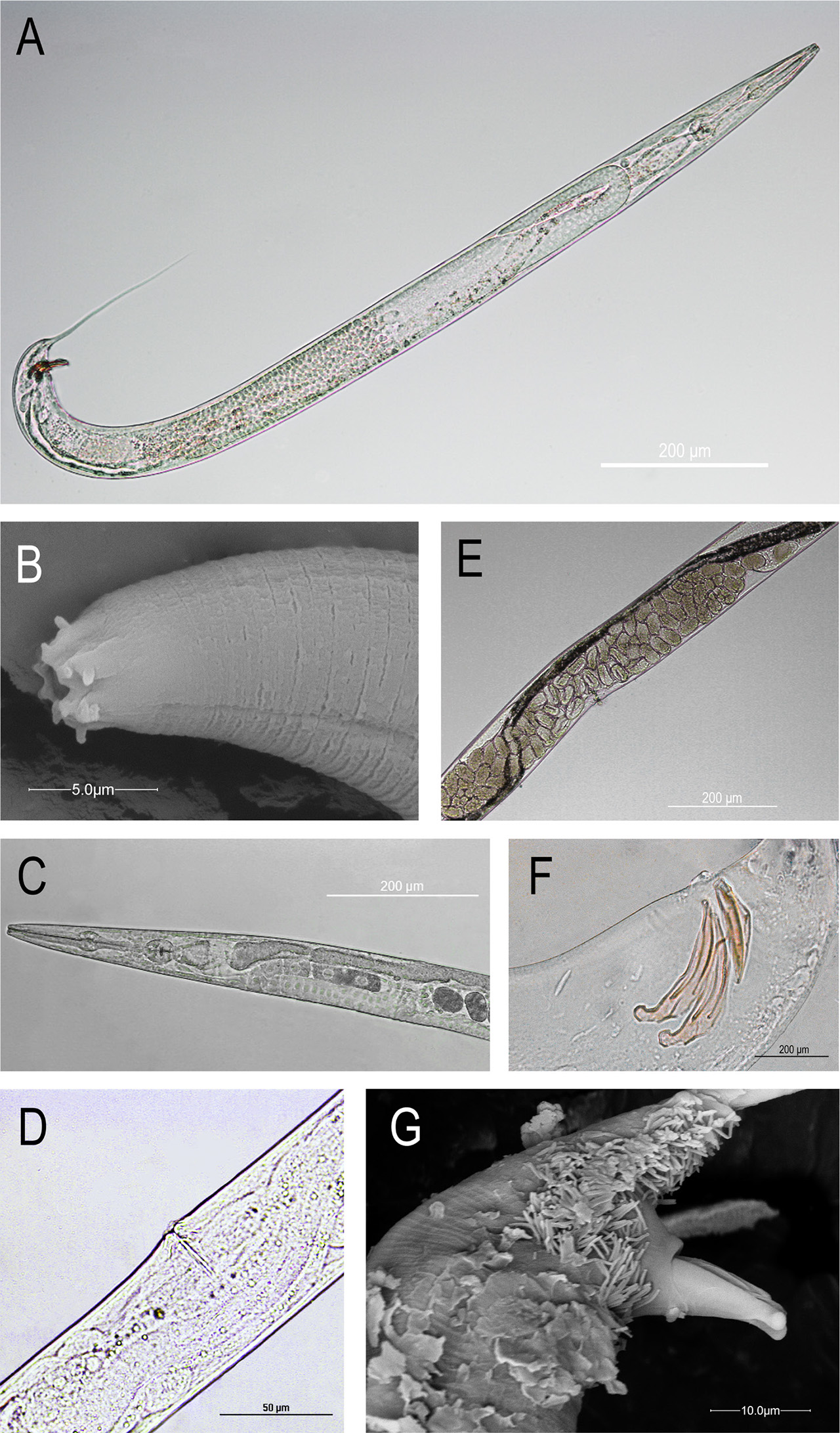
Figure 1:
Rhabditis axei: (A) male, entire body; (B) head region at SEM; (C) pharyngeal region; (D) vulva; (E) eggs at mid-body; (F) spicules and gubernaculum; (G) spicules at SEM.
Table 1.
Measurements Morphometrics of Rhabditis axei (14 females and 21 males). All measurements are in μm: mean ± s.d. (range). Comparison with measurements given by Goodey (1963) and Meamar et al. (2007). L: body length; MBD: maximum body diameter; ES: distance of the base of the pharynx from head end; B: buccal cavity length; T: tail length; SP: spicule length; GU: gubernaculum length; V: distance of vulva from head end; a: L/MBD; b: L/ES; c: L/T; V%: V/L*100.
| Character | Current study | According to Goodey, 1963 | According to Meamar et al., 2007 | |||
|---|---|---|---|---|---|---|
| Female (μm) | Male (μm) | Female (μm) | Male (μm) | Female (μm) | Male (μm) | |
| L | 1369.1 ± 196.1 (1128.2–1862.4) | 1236.5 ± 194.9 (890.9–1705.0) | (760.0–1180.0) | (740.0–950.0) | 1135.7 ± 88.3 (910.0–1374.1) | 894.3 ± 62.7 (673.4–1137.5) |
| MBD | 63.4 ± 14.9 (42.5–92.6) | 47.2 ± 13.9 (25.4–75.9) | - | - | 53.7 ± 5.3 (45.5–68.2) | 39.9 ± 3.3 (27.3–54.6) |
| ES | 213.2 ± 17.1 (191.8–242.8) | 205.2 ± 11.8 (183.8–233.5) | - | - | - | - |
| B | 26.5 ± 3.1 (23.4–34.4) | 24.6 ± 2.1 (21.9–30.9) | - | - | 22.5 ± 1.4 (19.9–26.4) | 22.9 ± 1.2 (19.8–26.4) |
| T | 301.7 ± 56.3 (221.5–434.4) | 265.8 ± 38.6 (177.1–332.8) | - | - | 307.6 ± 25.8 (235.4–363.0) | 189.1 ± 12.8 (154.0–228.8) |
| SP | - | 44.5 ± 5.0 (37.7–53.9) | - | - | - | 38.1 ± 1.3 (33.0–44.0) |
| GU | - | 30.2 ± 3.1 (26.0–35.4) | - | - | - | 27.3 ± 0.9 (24.2–30.8) |
| V | 664.9 ± 99.1 (524.6 – 912.4) | - | - | - | 490.9 ± 39.6 451.4–530.5 | - |
| a | 22.2 ± 3.5 (16.8–29.4) | 27.4 ± 5.4 (18.7–39.6) | (20.0–32.0) | - | - | (21.0–29.0) |
| b | 6.4 ± 0.8 (4.8–8.2) | 6.0 ± 0.8 (4.5–7.6) | (4.1–6.5) | - | - | (4.0–5.4) |
| c | 4.6 ± 0.5 (3.8–5.4) | 4.6 ± 0.6 (3.8–7.0) | (3.6–5.0) | - | - | (3.5–5.0) |
| V% | 48.6 ± 2.8 (44.8–53.8) | - | (38.0–50.0) | - | - | - |
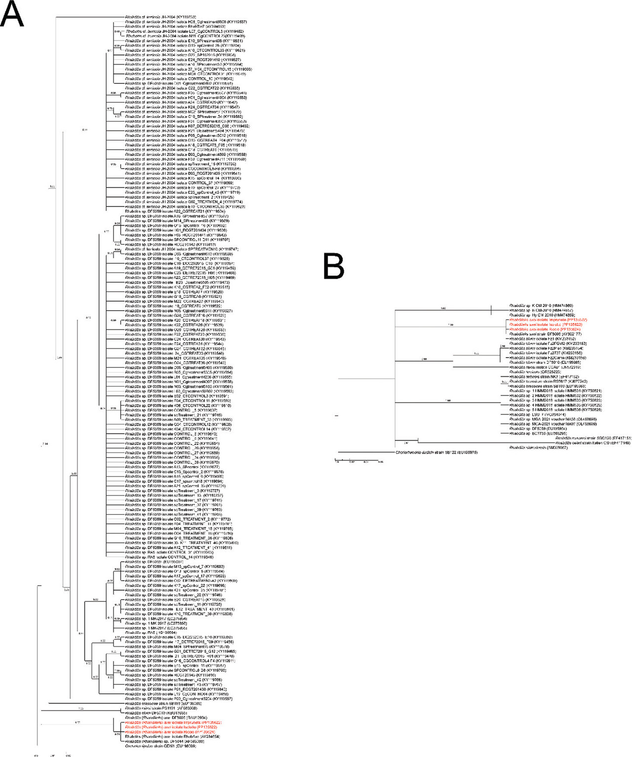
Figure S1:
Bayesian trees of the genus Rhabditis based on A) SSU and B) LSU gene fragments. Accession numbers of sequences were reported in brackets and posterior probability was annotated near the branches. The strains isolated during this work were highlighted in red.