
Table 1.
Pratylenchus delattrei and Quinisulcius capitatus recovered from chickpea roots and soil, districts, sampling locality, laboratory codes, altitude, GPS coordinators and GenBank accession numbers.
| Districts | Sampling locality | Codes | Species | Altitude (m) | Longitude(°) | Latitude(°) | GenBank accession number | ||
|---|---|---|---|---|---|---|---|---|---|
| 28S–rDNA | ITS–rDNA | COI gene | |||||||
| Minjar | Kitecha | Mki-5 | P. delattrei | 1600-1800 | 8°52′58.16″N | 39°29′46.01″E | OP646170 | - | - |
| Mki-8 | OP646169 | - | - | ||||||
| Mki-12 | - | - | OP730534 | ||||||
| Adea’a | Gollodhertu | AG-1 | P. delattrei | 1800-1900 | 8°38′55.54″N | 38°55′0.94″E | - | OP646171 | - |
| AG-2 | OP646168 | OP646172 | - | ||||||
| AG-3 | OP646167 | - | OP730535 | ||||||
| Mesekan | Jolle-2 | JO2-3 | Q. capitatus | 1900-1950 | 8°11′49.79″N | 38°27′51.24″E | OP626319 | OP646173 | OP627909 |
| JO2-5 | OP626320 | OP646174 | OP627910 | ||||||
| JO2-7 | OP626321 | OP646175 | OP627911 | ||||||
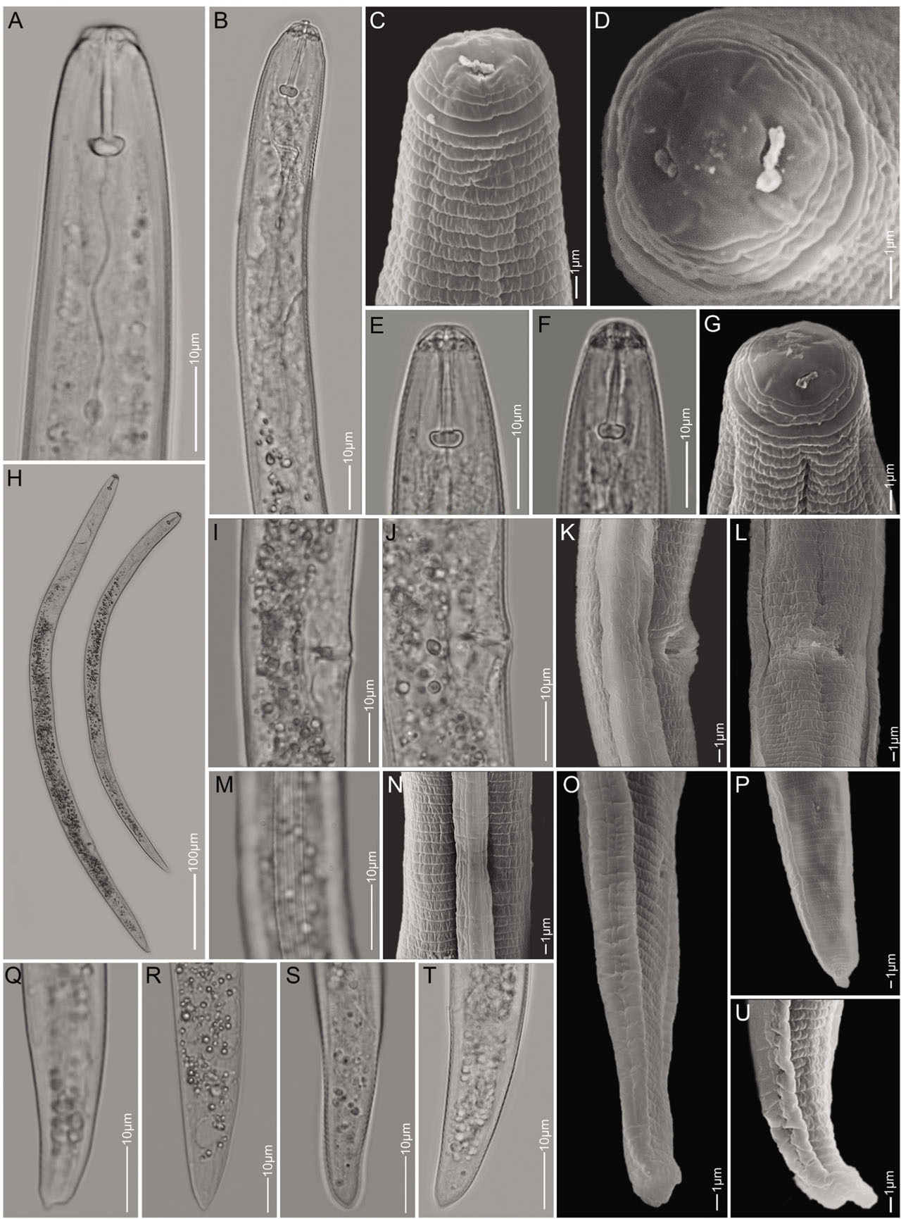
Figure 1:
Light microscopy and scanning electron microscopy images of Pratylenchus delattrei. A–C, E–G: Anterior part of the body showing lip and neck region; D: En face view; H: Whole female's body; I–L: Vulva region (L, ventral view); M,N: Lateral fields at mid-body; O–U: Tail region.
Table 2.
Comparison of morphometrics of the Ethiopian Pratylenchus delattrei from chickpea in Ethiopia with the original description from Madagascar (Luc, 1958), and two other P. delattrei populations from Cape Verde and Iran. All measurements are in μm and in the form: mean ± s.d. (range).
| Character | P. delattrei from chickpea in Ethiopia (present study) | P. delattrei from cotton in Madagascar (Luc, 1958) | P. delattrei from tomato in Cape Verde (Flis et al., 2018) | P. delattrei from vegetables in Iran (Majd Taheri et al., 2013) | |
|---|---|---|---|---|---|
| Hormozgan 1 | Hormozgan 2 | ||||
| n | 10 | 13 | 20 | 7 | 12 |
| L | 475 ± 47.1 (410 – 560) | 390 – 470 | 532 ± 33 (498 – 586) | 543 ± 55 (467 – 616) | 508 ± 49.2 (434 – 576) |
| a | 25.2 ± 2.2 (22.8 – 29) | 20.4 – 25.8 | 26.6 ± 2.2 (22.1 – 31.3) | 23.8 ± 2.1 (21.2 – 26.9) | 22.6 ± 1.0 (21.1 – 25) |
| b | 5.6 ± 0.6 (4.8 – 6.6) | 3.7 – 4.8 | 6.6 ± 0.5 (6.1 – 7.7) | 6.1 ± 0.7 (5.0 – 7.2) | 5.9 ± 0.6 (5.2 – 6.9) |
| b’ | 4.7 ± 0.5 (4.1 – 5.6) | – | 4.5 ± 0.4 (4.0 – 5.3) | 4.3 ± 0.2 (4.0 – 4.6) | 4.1 ± 0.4 (3.6 – 4.9) |
| c | 14.6 ± 1.9 (13.7 – 20.3) | 18 – 22.3 | 21.9 ± 2.1 (18.5 – 25.1) | 20 ± 2 (18.1 – 23.1) | 19.7 ± 2.6 (16.7 – 24.1) |
| c’ | 2.3 ± 0.2 (2.0 – 2.8) | – | 2.2 ± 0.2 (1.9 – 2.8) | 2.2 ± 0.3 (1.9 – 2.6) | 1.9 ± 0.2 (1.6 – 2.2) |
| V | 72.2 ± 7.6 (62.4 – 86.9) | 73 – 81 | 76 ± 1 (75 – 78) | 75.1 ± 1.9 (71.4 – 77.1) | 75.9 ± 1.3 (74 – 78.7) |
| Stylet length | 16.7 ± 0.7 (15.8 – 17.8) | 16.5 – 18.0 | 16.4 ± 0.4 (15.4 – 16.9) | 16.3 ± 0.8 (15 – 17) | 16.0 ± 0.6 (15 – 17) |
| Dorsal gland opening from stylet base | 3.0 ± 0.5 (2.3 – 3.5) | – | 2.9 ± 0.3 (2.4 – 3.1) | – | – |
| O | 17.7 ± 2.9 (13.6 – 22.2) | – | 17.4 ± 1.7 (14.3 – 21.3) | – | – |
| Pharynx length | 85 ± 1.0 (82.5 – 85.9) | – | 80.1 ± 3.2 (73.1 – 84.2) | 89 ± 10.4 (76 – 105) | 87 ± 4 (82 – 94) |
| Pharyngeal overlap | – | – | 39.0 ± 7.2 (29.8 – 49.0) | – | |
| Anterior end to end of pharyngeal gland lobe | 101 ± 1.4 (98 – 102) | – | – | 125 ± 7.3 (116 – 135) | 123 ± 8.3 (113 – 138) |
| Maximal body diameter | 22.7 ± 0.5 (21.6 – 23.1) | – | 20.2 ± 2.2 (16.8 – 23.7) | 23 ± 3.3 (20 – 29) | 22.5 ± 2.1 (19 – 26) |
| Anal body diameter | 13.0 ± 1.2 (10.9 – 14.1) | – | 11.2 ± 1.2 (8.9 – 13.2) | 12.4 ± 1.5 (11 – 14) | 13 ± 0.9 (12 – 14) |
| Tail length | 29.0 ± 0.9 (27.5 – 30.1) | – | 24.8 ± 2.3 (21.0 – 27.1) | 26.6 ± 2.3 (23 – 29) | 24.8 ± 2.3 (23 – 29) |
| Tail annuli | 20 ± 2 (18 – 23) | – | 19 ± 2 (16 – 24) | 20 ± 2.1 (18 – 23) | 19 ± 1.5 (17 – 21) |
| Phasmid to terminus | 14.1 ± 0.4 (13.5 – 14.5) | – | 10.6 ± 2.7 (6.0 – 14.9) | – | – |
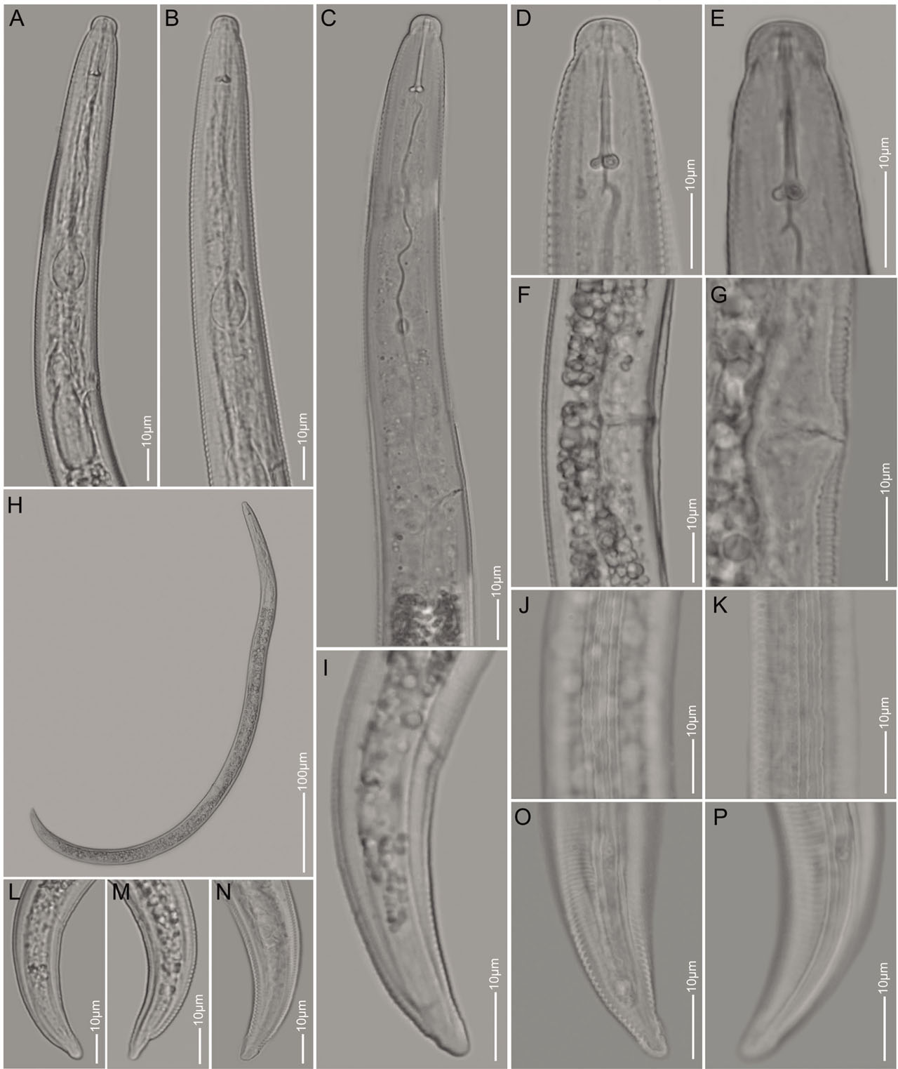
Figure 2:
Light microscopy images of Quinisulcius capitatus. A–E: Anterior part of the body showing lip and neck regions; F,G: Vulval regions (lateral view); H: Whole female body; J,K: Lateral field showing five distinct incisures; I, L–P: Tail region lateral view.
Table 3.
Comparison of important morphological character and morphometrics of the Ethiopian Quinisulcius capitatus, found from chickpea in Ethiopia, with original description from USA (Allen, 1955), and two other Q. capitatus from Ethiopia and Canada. All measurements are in μm and in the form: mean ± s.d. (range).
| Character | Q. capitatus from chickpea in Ethiopia (present study) | Q. capitatus from pear in USA (Allen, 1955) | Q. capitatus from coffee in Ethiopia (Mekete et al., 2008) | “Q. capitatus”* from grass in Canada (Munawar et al., 2021) |
|---|---|---|---|---|
| n | 10 | 13 | 10 | 20 |
| L | 699 ± 11.6 (667 – 707) | 630 – 850 | 630 – 790 | 810.3 ± 44.6 (744.0 – 911.0) |
| a | 31.2 ± 1.3 (29.7 – 34.4) | 30 – 38 | 30.9 – 38.6 | 41.4 ± 1.8 (38.6 – 43.7) |
| b | 5.4 ± 0.2 (5.1 – 5.7) | 5.0 – 5.8 | – | 5.5 ± 0.3 (5.0 – 6.3) |
| c | 14.7 ± 0.7 (14.1 – 16.3) | 12 – 17 | 15.3 – 17.6 | 22.4 ± 1.1 (19.9 – 23.8) |
| c’ | 1.9 ± 0.2 (1.5 – 2.1) | – | – | 2.6 ± 0.2 (2.2 – 3.2) |
| V | 56.6 ± 2.2 (52.7 – 59.0) | 51 – 58 | 54.7 – 63.6 | 57.4 ± 1.5 (53.4 – 59.8) |
| Stylet length | 18.8 ± 0.6 (17.8 – 19.7) | 16 – 18 | 15 – 18 | 18.3 ± 1.0 (15.5 – 20.4) |
| Lip height | 4.0 ± 0.5 (3.2 – 4.5) | – | – | 4.0 ± 0.2 (3.7 – 4.4) |
| Lip width | 7.6 ± 0.5 (7.0 – 8.3) | – | – | 7.6 ± 0.4 (6.9 – 8.3) |
| Median bulb length | 16.1 ± 0.3 (15.7 – 16.6) | – | – | 14.0 ± 1.6 (11.3 – 16.9) |
| Median bulb width | 13.1 ± 0.6 (12.2 – 13.9) | – | – | 10.4 ± 1.4 8.4 – 14.2 |
| Pharyngeal length | 129 ± 5.1 (123 – 138) | – | – | 147.8 ± 5.8 (140.2 – 159.0) |
| SE pore from anterior end | 121 ± 3.3 (113 – 123) | – | 121 | 128.6 ± 5.3 (121.0 – 139.0) |
| Midbody diameter | 22.4 ± 1.0 (20.5 – 23.5) | 21 – 27 | – | – |
| Tail length | 47.8 ± 3.0 (41.0 – 50.0) | – | – | 35.8 ± 2.4 (31.3 – 40.4) |
| Anal body diameter | 25.3 ± 3.2 (23.0 – 34.2) | – | – | – |
| Phasmid position | Middle of tail | Middle of tail | – | Middle of tail |
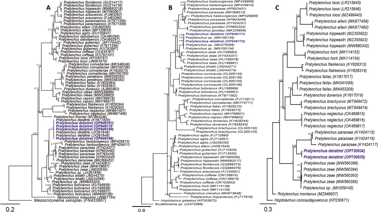
Figure 3:
Bayesian 50% majority rule consensus phylogeny of Pratylenchus delattrei from Ethiopia and related species based on 28S (A) and (B) ITS of rDNA genes and (C) COI of mtDNA using a GTR model. Branch support is indicated with PP. The sequences from this study were marked by blue color and bold font.
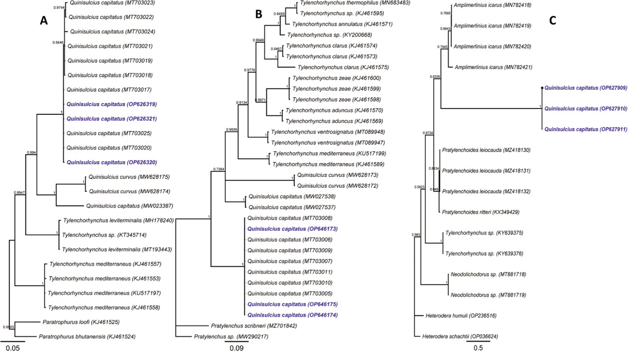
Figure 4:
Bayesian 50% majority rule consensus phylogeny of Quinisulcius capitatus from Ethiopia and related species on 28S (A) and (B) ITS of rDNA genes and (C) COI of mtDNA using a GTR model. Branch support is indicated with PP. The sequences from this study were marked by blue color and bold font.