
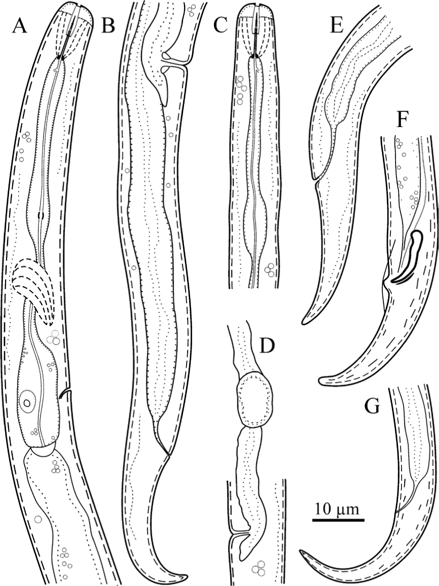
Figure 1
Line drawings of Boleodorus bushehresnsis n. sp. (A, B, D, E, G: female; C, F: male). (A) Pharynx. (B) Posterior body region. (C) Anterior body region. (D) Vulval region, showing offset spermatheca. (E–G) Tail.
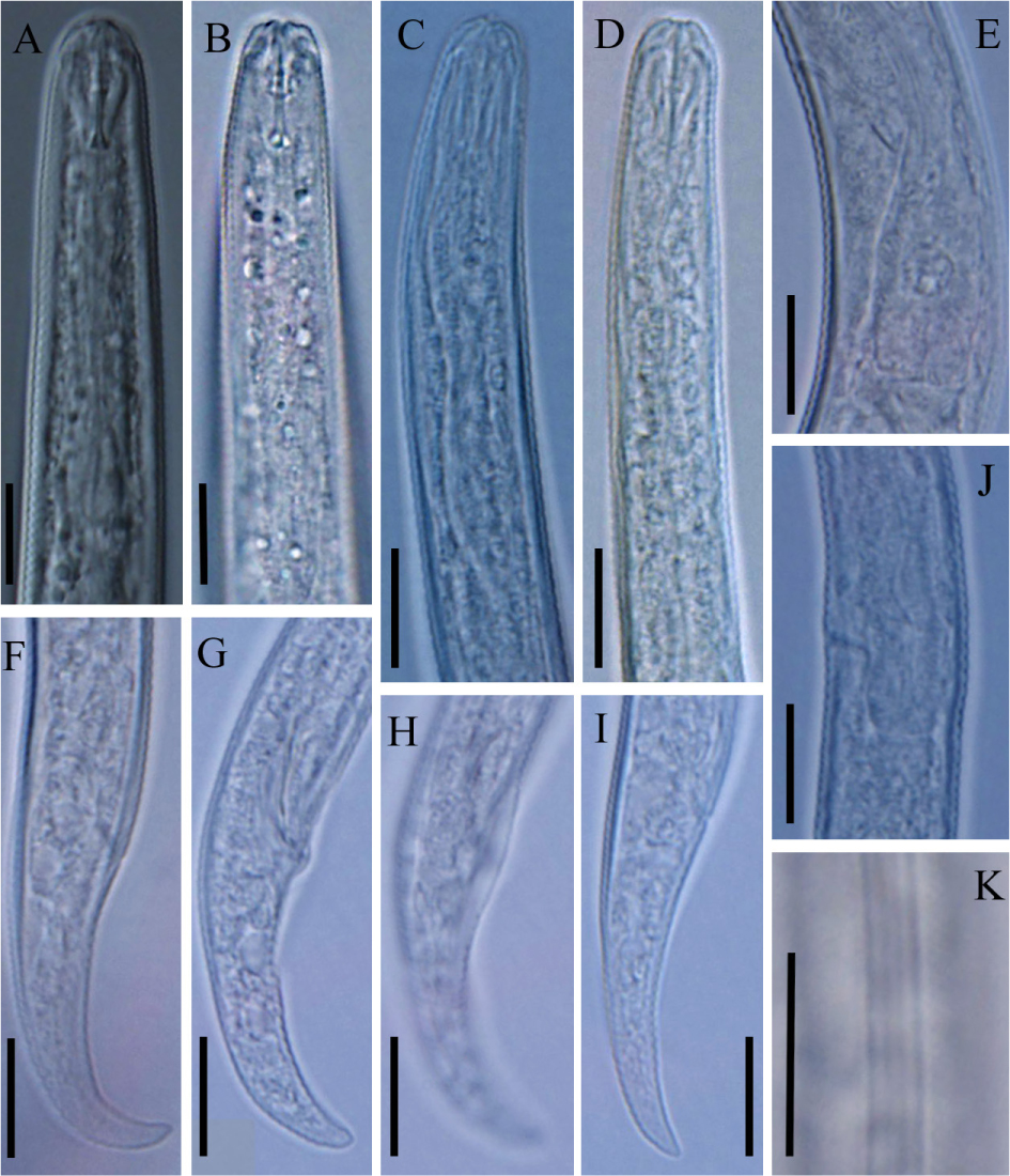
Figure 2
Light microphotographs of Boleodorus bushehrensis n. sp. (A, B, C, E, F, I, J, K: female; D, G, H: male). (A, B) Anterior region showing cephalic region and stylet, respectively. (C) Pharyngeal metacorpus. (D) Anterior body region showing the oral aperture in a depression. (E, J) Pharyngeal bulb region showing excretory pore. (F, G, I) Tail tip. (H) Bursa. (K) Lateral field at midbody. (All scale bars = 10 μm).
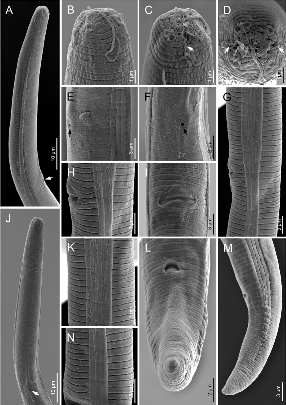
Figure 3
Scanning electron microphotographs of Boleodorus bushehrensis n. sp. (female). (A) Anterior body region showing beginning of lateral field (arrow showing the excretory pore). (B–D) Anterior end in ventrolateral, ventral, and en face views, respectively (arrows pointing to the amphidial openings). (E, F) Excretory pore in lateral and ventral views, respectively (arrow). (G) Lateral field at vulva. (H, I) Vulva in lateral and ventral views, respectively. (J) Anterior body region in ventral view showing excretory pore. (K,N) Lateral field at midbody showing unusual division and four incisures, respectively. (L,M) Anus in ventral and lateral views, respectively.
Table 1
Morphometrics of Boleodorus bushehrensis n. sp.
| Characteristics | Holotype | Paratypes | |
|---|---|---|---|
| Females | Males | ||
| n | 13 | 3 | |
| L | 449 | 412 ± 35.5 (334–464) | 432 ± 17 (417–450) |
| L’ | 413 | 379 ± 34 (298–432) | 399 ± 16 (385–416) |
| a | 34 | 32.5 ± 3.0 (27.8–37.4) | 39.8 ± 2.5 (37.2–42.0) |
| b | 4.8 | 4.8 ± 0.5 (4–6) | 5.0 ± 0.5 (4.3–5.4) |
| c | 12 | 12.6 ± 1.5 (9.3–15.8) | 13.1 ± 0.1 (12.9–13.0) |
| c’ | 4.9 | 3.9 ± 0.5 (2.9–4.9) | 4.1 ± 0.1 (4.0–4.3) |
| V or T | 79 | 74.8 ± 1.9 (70.8–79.5) | – |
| V’ | 80.7 | 80.9 ± 1.2 (77.5–82.0) | – |
| Cephalic region height | 2.3 | 2.1 ± 0.3 (1.7–2.6) | 2.1 ± 0.4 (1.8–2.7) |
| Cephalic region width | 6.5 | 5.9 ± 0.6 (5.0–6.7) | 6.1 ± 0.4 (5.6–6.5) |
| Stylet length | 8 | 8.7 ± 0.4 (8.0–9.3) | 9.7 ± 0.3 (9.5–10.0) |
| Conus length | 3 | 3.2 ± 0.2 (3.0–3.5) | 3.3 ± 0.3 (3.0–3.5) |
| m | 37.5 | 36.8 ± 3.4 (32.3–42.7) | 33.6 ± 2.3 (31.5–36.0) |
| DGO | 3 | 3.1 ± 0.2 (2.8–3.5) | 3.0 ± 0.07 (2.9–3.0) |
| Distance from excretory pore to anterior end | 78.5 | 69.5 ± 5.2 (64–79) | 63.9 ± 1.6 (62–65) |
| MB | 48.2 | 55.8 ± 12.1 (43.0–82.3) | 57.8 (n=1) |
| Pharynx length | 91 | 87 ± 7 (73–99) | 87.7 ± 7.5 (80–95) |
| Distance from anterior end to vulva | 333 | 307 ± 28 (245–347) | – |
| BW | 12 | 12.7 ± 1.3 (12–15) | 10.8 ± 0.3 (10.6–11.0) |
| Anal BW | 9 | 8.6 ± 0.6 (7–9) | 8.7 ± 0.9 (8–9) |
| Vulva–anus (V–A) distance | 80 | 72.2 ± 8.5 (54–84) | – |
| PUS length | 11.5 | 10.6 ± 1.3 (8.2–12.0) | – |
| Tail/V–A | 0.5 | 0.5 ± 0.07 (0.4–0.7) | – |
| PUS/BW | 1 | 0.8 ± 0.07 (0.7–1.0) | – |
| Tail length | 36 | 33 ± 3 (26–38) | 32.5 ± 0.7 (32–33) |
| Spicules length | – | – | 11.5 ± 0.4 (11.5–12.0) |
| Gubernaculum length | – | – | 4.5 ± 0.2 (4–5) |
| Bursa length | – | – | 6.3 ± 0.5 (6–7) |
[i] All measurements are in micrometers and in the form: mean ± SD (range).
[ii] BW, body width; DGO, dorsal gland opening; PUS, postvulval uterine sac; SD, standard deviation.
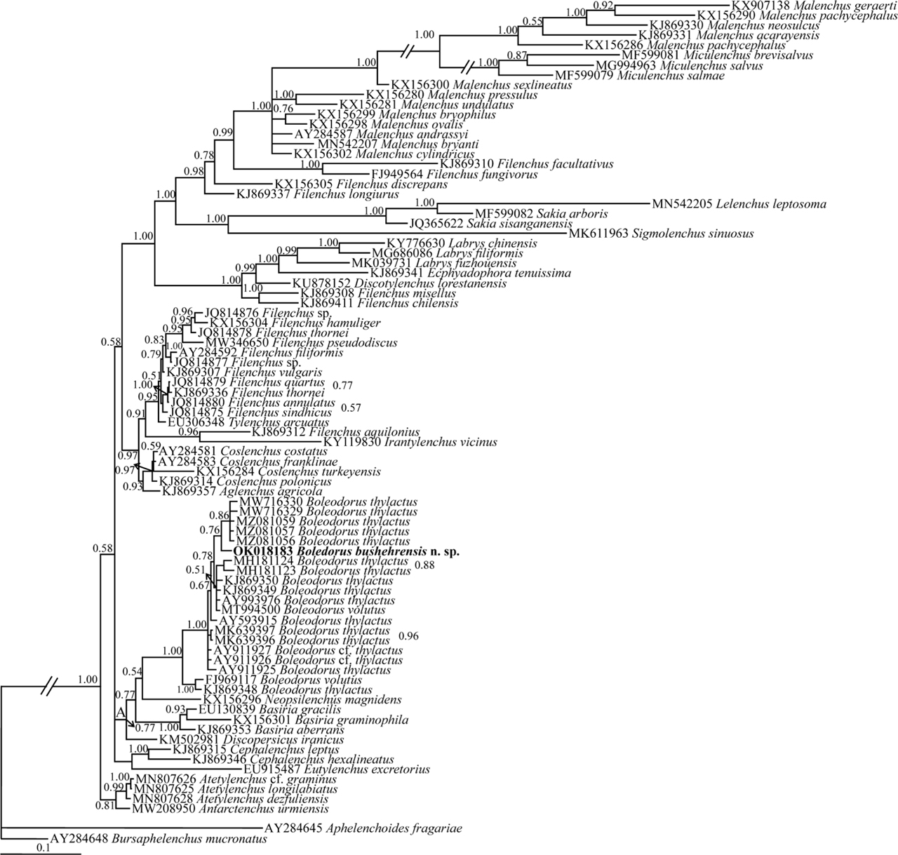
Figure 4
Bayesian 50% majority rule consensus tree inferred from the SSU rDNA of Boleodorus bushehrensis n. sp. under the GTR + G + I model. Bayesian posterior probability values are given for corresponding clades. The new species is in bold font. GTR, general time-reversible; G, gamma; I, invariant; rDNA, ribosomal DNA; SSU, small subunit.
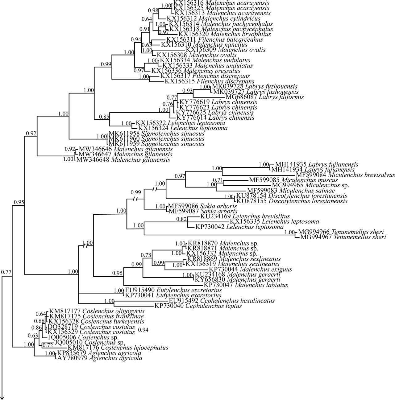
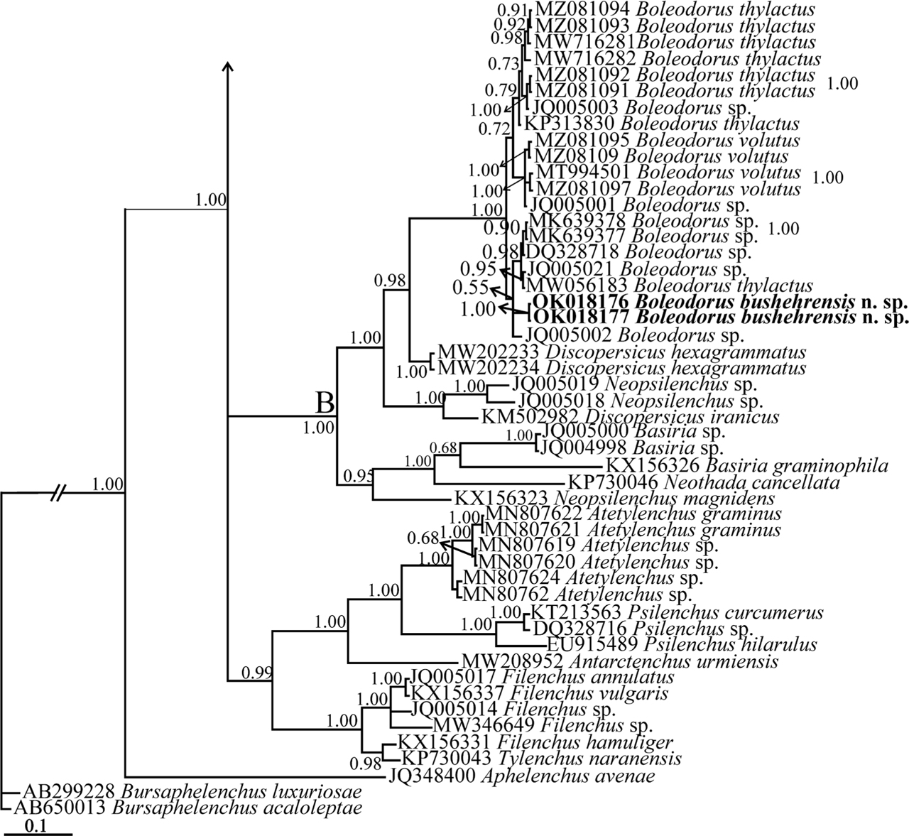
Figure 5
Bayesian 50% majority rule consensus tree inferred from the LSU rDNA D2–D3 sequences of Boleodorus bushehrensis n. sp. under the GTR + G + I model. Bayesian posterior probability values are given for the corresponding clades. The new species is in bold font. GTR, general time-reversible; G, gamma; I, invariant; LSU, large subunit; rDNA, ribosomal DNA.
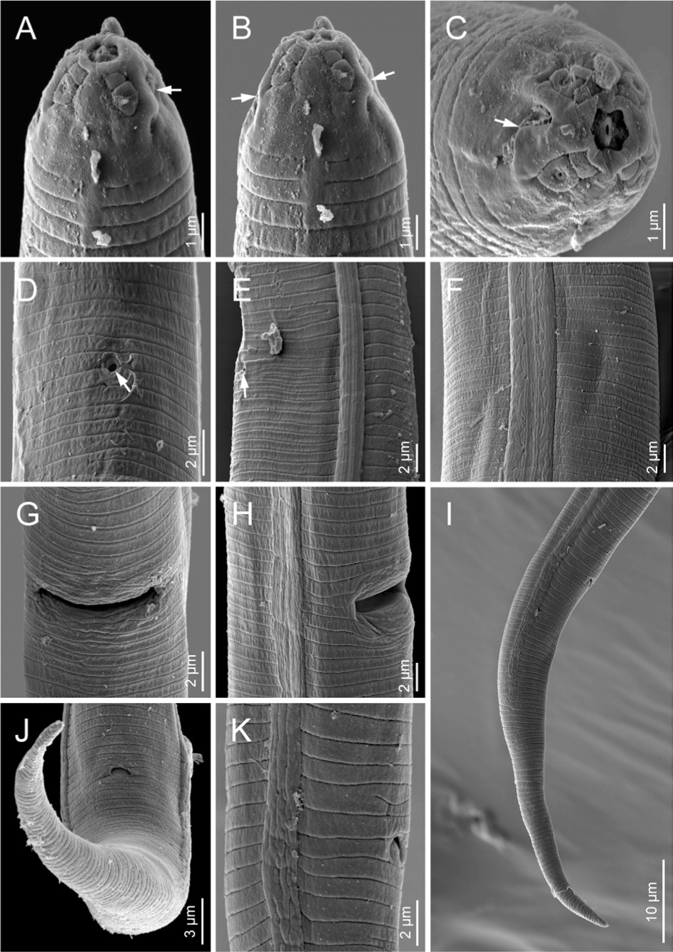
Figure 6
Scanning electron microphotographs of Boleodorus thylactus Thorne, 1941 (female). (A–C) Anterior end in ventral and frontal views, respectively (arrows pointing to the amphidial openings). (D,E) Excretory pore in ventral and lateral views (arrow). (F, K) Lateral field at midbody showing four incisures. (G,H) Vulva in lateral and ventral views, respectively. (I) Posterior end in lateral view. (J) Anus in ventral view.