
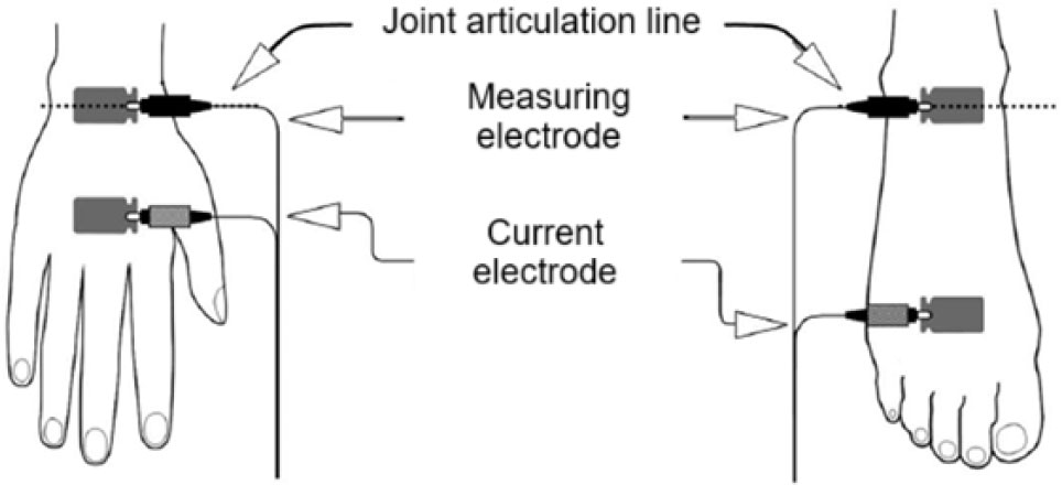
Fig. 1:
Diagram of the location of measuring (potential) and current electrodes on the patient's hand and foot.
Table 1:
Study participant data.
| Parameters | M M ± SD | F M ± SD |
|---|---|---|
| Height, cm | 182 ± 12 | 164 ± 6 |
| Body mass, kg | 61.4 ± 11.7 | 78.9 ± 15.5 |
| BMI, kg/m2 | 22.3 ± 2.8 | 24.2 ± 3.9 |
| Age, years | 18.1 ± 1.3 | 18.4 ± 0.9 |
| Shoulder girth, cm | 27.3 ± 3.26 | 34.6 ± 3.7 |
| Calf circumference, cm | 34.8 ± 2.3 | 36.6 ± 3.3 |
| Hip circumference, cm | 69.7 ± 8.5 | 78.6 ± 7.9 |
| Diameter of the distal epiphysis of the humerus, cm | 5.3 ± 0.5 | 6.7 ± 0.6 |
| Diameter of the distal femoral epiphysis, cm | 8.1 ± 1.0 | 9.4 ± 0.8 |
| Subscapular, mm | 16.3 ± 7.0 | 14.3 ± 8.8 |
| Triceps, mm | 16.0 ± 5.6 | 9.9 ± 4.6 |
| Iliac crest, mm | 16.1 ± 8.0 | 13.8 ± 10.3 |
| Medial calf, mm | 19.5 ± 8.3 | 11.9 ± 5.4 |
[i] Notes: M – Male, F – Female, Subscapular – fold of skin and fat under the lower angle of the scapula in an oblique direction at an angle of 45° to the line of the spine, Triceps – vertical fold of skin and fat, on the back of the shoulder, in the middle between the olecranon and acromion process, on a freely lowered arm, Iliac crest – a diagonal fold passing directly above the iliac crest in a vertical line from the mid-axillary line, Medial calf – vertical skinfold, on the medial side of the leg at the level of the maximum circumference.
Table 2:
Results of a comparative analysis of somatotype assessment using anthropometric measurements and bioelectrical impedance analysis.
| Female | |||||
| Somatotype | BIA | Anthropometry | p | pc | r |
| M ± SD | M ± SD | ||||
| Endo | 4.2±1.1 | 4.6±1,3 | 0.028* | 0.84 (0.62; 0.94) | 0.89 |
| Meso | 4.9±1.0 | 4.1±0.3 | 0.001* | 0.31 (0.12; 0.48) | 0.81 |
| Ekto | 2.3±1.2 | 2.3±1.4 | 0.886 | 0.96 (0.90; 0.99) | 0.97 |
| Male | |||||
| Somatotype | BIA | Anthropometry | p | pc | r |
| M ± SD | M ± SD | ||||
| Endo | 2.1±1.3 | 3.1±1.1 | 0.432 | 0.21 (−0.40; 0.69) | 0.23 |
| Meso | 5.6±1.6 | 3.8±1.4 | 0.001* | 0.42 (0.10; 0.66) | 0.84 |
| Ecto | 2.4±1.3 | 2.4±1.2 | 0.167 | 0.98 (0.94; 0.99) | 0.99 |
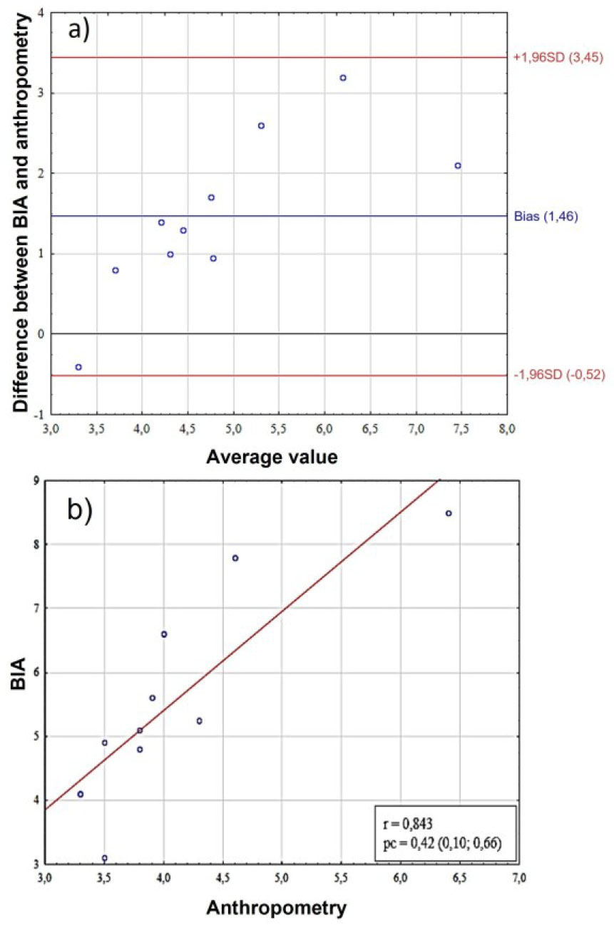
Fig. 2:
Comparative analysis of endomorphy scores in female. A – assessment of bias using the Bland-Altman method, the Y-axis shows the “BIA-Anthro” difference in endomorphy scores, B – scatter plot between two methods for assessing somatotype. BIA – bioelectrical impedance analysis, SD (standard deviation), r – Pearson correlation coefficient, pc – Lin correlation concordance coefficient.
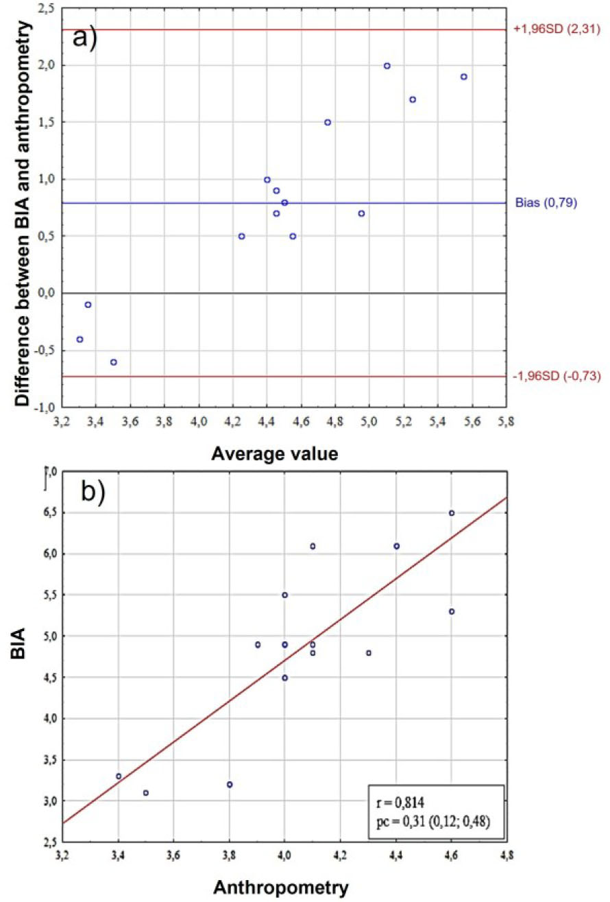
Fig. 3:
Comparative analysis of mesomorphy scores in females. A– assessment of bias using the Bland-Altman method, the Y-axis shows the “BIA-Anthro” difference in mesomorphy scores, B – scatter plot between two methods for assessing somatotype. BIA – bioelectrical impedance analysis, SD (standard deviation), r – Pearson correlation coefficient, pc – Lin correlation concordance coefficient.
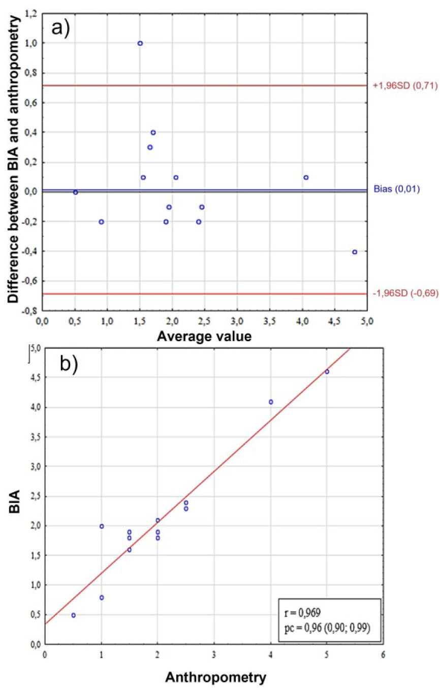
Fig. 4:
Comparative analysis of ectomorphy scores in females. A – assessment of bias using the Bland-Altman method, the Y-axis shows the “BIA-Anthro” difference in ectomorphy scores, B – scatter plot between two methods for assessing somatotype. BIA – bioelectrical impedance analysis, SD (standard deviation), r – Pearson correlation coefficient, pc – Lin correlation concordance coefficient.
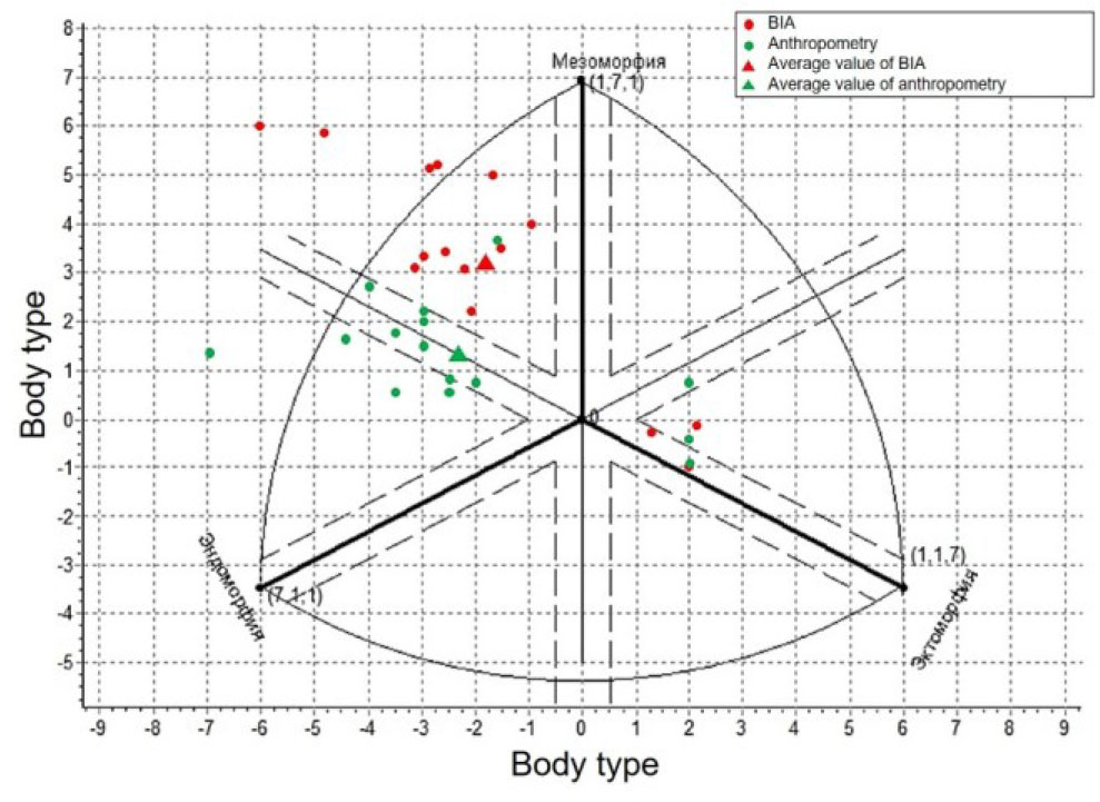
Fig. 5:
Comparison of female somatotyping results by the Heath-Carter method using BIA and anthropometry. BIA – bioelectrical impedance analysis.
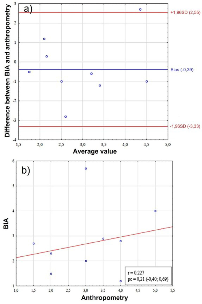
Fig. 6:
Comparative analysis of endomorphy scores in young men. A – assessment of bias using the Bland-Altman method, the Y-axis shows the “BIA-Anthro” difference in endomorphy scores, B – scatter plot between two methods for assessing somatotype. BIA – bioelectrical impedance analysis, SD (standard deviation), r – Pearson correlation coefficient, pc – Lin correlation concordance coefficient.
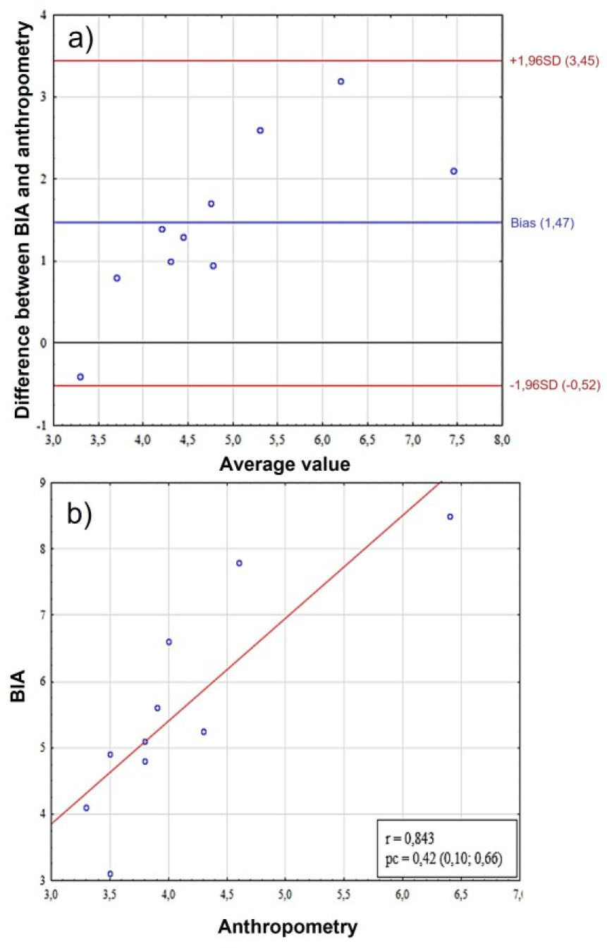
Fig. 7:
Comparative analysis of mesomorphy scores in male. A – assessment of bias using the Bland-Altman method, the Y-axis shows the “BIA-Anthro” difference in mesomorphy scores, B – scatter plot between two methods for assessing somatotype. BIA – bioelectrical impedance analysis, SD (standard deviation), r – Pearson correlation coefficient, pc – Lin correlation concordance coefficient.
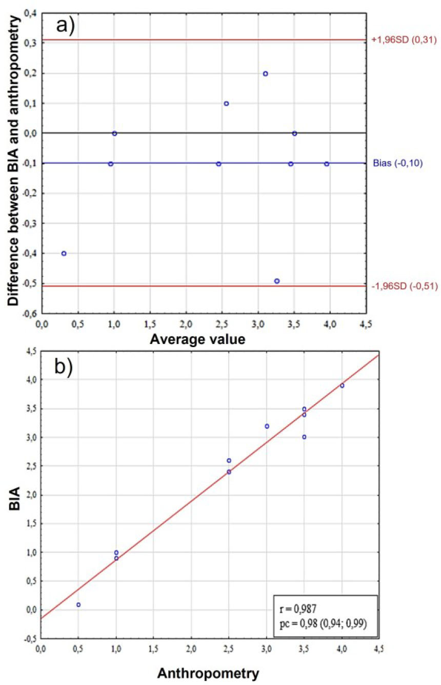
Fig. 8:
Comparative analysis of ectomorphy scores in male. A – assessment of bias using the Bland-Altman method, the Y-axis shows the “BIA-Anthro” difference in ectomorphy scores, B – scatter plot between two methods for assessing somatotype. BIA – bioelectrical impedance analysis, SD (standard deviation), r – Pearson correlation coefficient, pc – Lin correlation concordance coefficient.
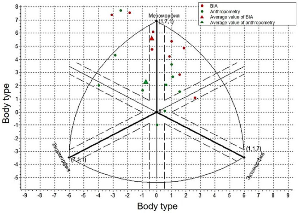
Fig. 9:
Comparison of somatotyping results of male by Heath-Carter method using BIA and anthropometry.