
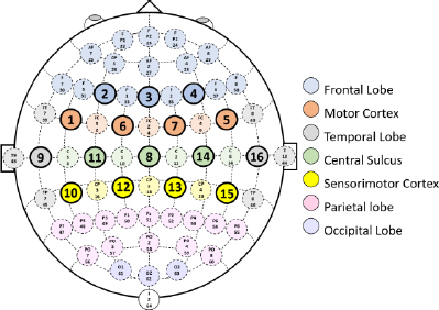
Fig.1:
A schematic representation of the brain regions selected for EEG signal recording [13].
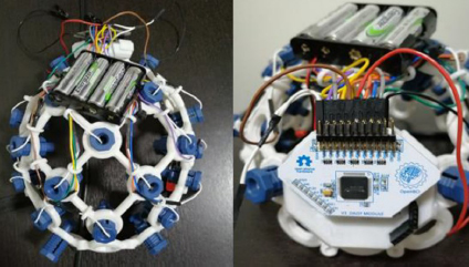
Fig.2:
The OpenBCI Cyton with Daisy Biosensing Board and associated EEG headgear, detailing the hardware setup used in signal collection [13].
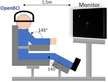
Fig.3:
The reclining chair with delineated limb angles and the monitor placement at a 1.5-meter distance, ensuring direct alignment with the participant’s field of vision [13].
Tab.1:
Task abbreviations assigned to different limb movements.
| Task | Description |
|---|---|
| BEO | Recording a Baseline with Eyes Open |
| CLH | Closing Left Hand: five times per run |
| CRH | Closing Right Hand: five times per run |
| DLF | Dorsal flexion of Left Foot: five times per run |
| PLF | Plantar flexion of Left Foot: five times per run |
| DRF | Dorsal flexion of Right Foot: five times per run |
| PRF | Plantar flexion of Right Foot: five times per run |
| Rest | Resting in between tasks: after each task |
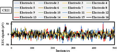
Fig.4:
EEG signals over a 4-second interval with 500 instances for Subject 10 in CRH phase.
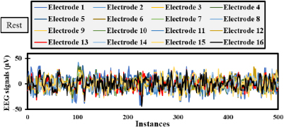
Fig.5:
EEG signals over a 4-second interval with 500 instances for Subject 10 in Resting phase.
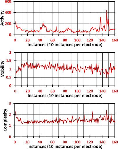
Fig.6:
Hjorth parameters of the Resting movement, namely activity, mobility, and complexity for all electrodes.
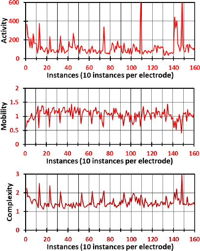
Fig.7:
Hjorth parameters of the CRH movement, namely activity, mobility, and complexity for all electrodes.
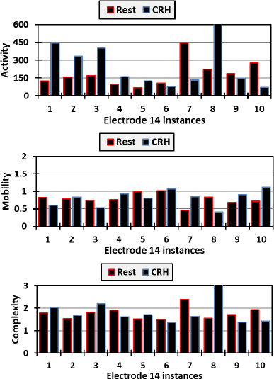
Fig.8:
Activity, mobility, and complexity of electrode 14 readings for both resting and closing right hand.
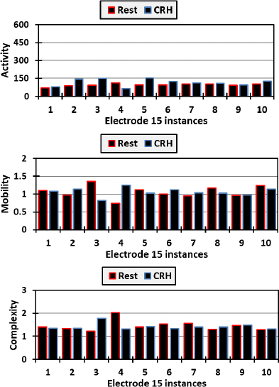
Fig.9:
Activity, mobility, and complexity of electrode 15 readings for both resting and closing right hand.