
Table 1.
Demographics, FFR, OCT and angiographic findings
| n | Min | Max | Mean | s.d. | FFR ≤ 0.8 | s.d. | FFR > 0.8 | s.d. | p value | |
|---|---|---|---|---|---|---|---|---|---|---|
| FFR | 151 | 0.41 | 0.98 | 0.81 | 0.10 | 0.72 | 0.07 | 0.88 | 0.04 | <0.001 |
| Age | 151 | 36.00 | 81.00 | 60.41 | 10.42 | 61.00 | 10.19 | 59.97 | 10.63 | 0.55 |
| OCT proximal area | 151 | 2.83 | 20.56 | 8.84 | 3.04 | 8.45 | 2.95 | 9.13 | 3.09 | 0.17 |
| OCT distal area | 151 | 1.79 | 18.38 | 6.79 | 2.77 | 6.23 | 2.66 | 7.22 | 2.80 | 0.03 |
| Average area | 151 | 2.68 | 19.47 | 7.82 | 2.55 | 7.34 | 2.37 | 8.17 | 2.64 | 0.04 |
| Lesion length | 151 | 6.20 | 65.60 | 31.33 | 13.20 | 35.55 | 14.52 | 28.14 | 11.18 | 0.001 |
| MLA | 151 | 0.75 | 5.51 | 2.21 | 1.06 | 1.61 | 0.62 | 2.67 | 1.09 | <0.001 |
| %AS average reference | 21.51 | 92.00 | 70.97 | 11.80 | 76.97 | 8.04 | 66.44 | 12.19 | <0.001 | |
| Proximal reference diameter | 1.90 | 5.11 | 3.27 | 0.57 | 1.25 | 0.28 | 1.59 | 0.36 | <0.001 | |
| Distal reference diameter | 1.51 | 4.84 | 2.86 | 0.58 | 2.76 | 0.56 | 2.98 | 0.58 | 0.02 | |
| Average reference diameter | 1.84 | 4.97 | 3.08 | 0.50 | 3.00 | 0.48 | 3.17 | 0.51 | 0.03 | |
| Minimum diameter | 151 | 0.84 | 2.53 | 1.44 | 0.37 | 1.25 | 0.28 | 1.59 | 0.36 | <0.001 |
| %Diameter stenosis (vs. average reference diameter) | 26.18 | 83.69 | 46.78 | 10.19 | 50.35 | 10.17 | 42.05 | 8.15 | <0.001 | |
| FFR ≤ 0.8 | 65 | 43% | 65 | 86 | ||||||
| Vessel | LAD | 80 | 53.30% | 47 (72.3%) | 33 (38.4%) | <0.001 | ||||
| RCA | 38 | 25.30% | ||||||||
| LCX | 33 | 21.85% | ||||||||
| Male | 127 | 84.10% | 56 (86.2%) | 71 (82.6%) | 0.55 | |||||
| Smoker | 46 | 30.50% | 23 (35.4%) | 23 (26.7%) | 0.253 | |||||
| Hypertension | 116 | 76.80% | 45 (69.2%) | 71 (82.6%) | 0.055 | |||||
| Dyslipidemia | 121 | 80310% | 50 (76.9%) | 71 (82.6%) | 0.341 | |||||
| Diabetes mellitus | 47 | 31.10% | 20 (30.8%) | 27 (31.4%) | 0.93 |
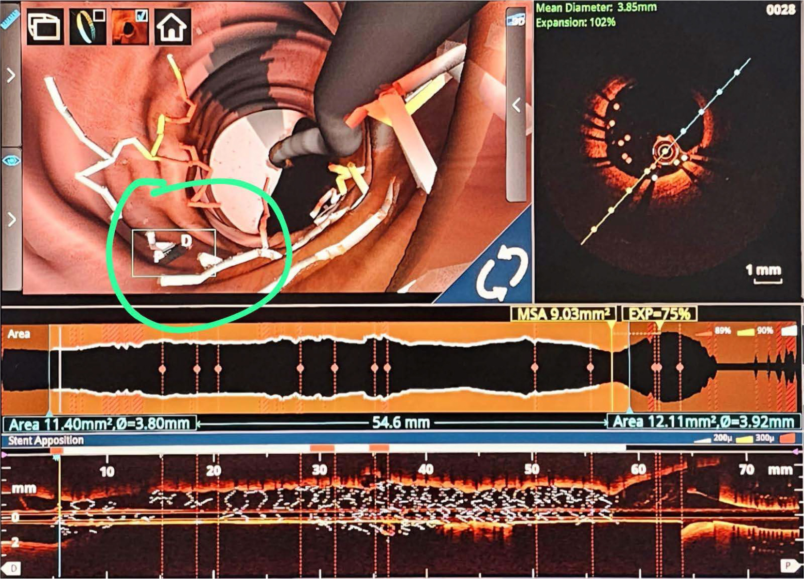
Figure 1.
OCT with 3D reconstruction. Severe malposition of the stent requiring 1:1 noncompliant balloon post dilatation.
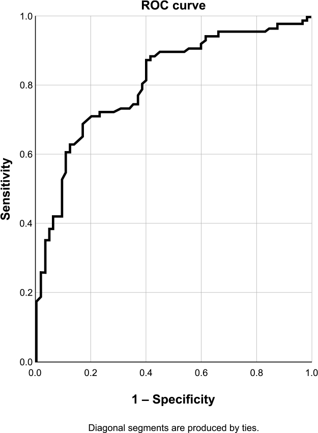
Figure 2.
ROC curve for OCT-derived MLA to predict FFR < 0.8. Sensibility 83.1%, specificity 68.6%, accuracy 74.8%.
| AUC, Test result variable(s): MLA | |||
|---|---|---|---|
| Area | Std. Errora | Asymptotic Sig.b | Asymptotic 95% CI |
| 0.810 | 0.035 | 0.000 | 0.741–0.879 |
| AUC Test result variable(s): PAS | |||
|---|---|---|---|
| Area | Std. Errora | Asymptotic Sig.b | Asymptotic 95% CI |
| 0.761 | 0.039 | 0.000 | 0.685–0.836 |
| Area under the ROC curve | ||||
|---|---|---|---|---|
| Test result variable(s) | Area | Std. Errora | Asymptotic Sig.b | Asymptotic 95% CI |
| %RMA | 0.761 | 0.039 | 0.000 | 0.685–0.837 |
| MLA x | 0.810 | 0.035 | 0.000 | 0.741–0.879 |
| Paired-sample area difference under the ROC curves | |||||
|---|---|---|---|---|---|
| Test result pair(s) | Asymptotic | AUC difference | Std. error difference | Asymptotic 95% CI | |
| RMA | -1.572 | 0.116 | -0.049 | 0.269 | -0.111–0.012 |
| MLA x | |||||
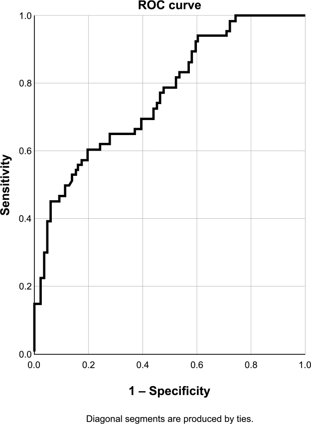
Figure 3.
ROC curve for PAS to predict FFR < 0.8. Sensibility 60.0%, specificity 80.2%, accuracy 71.5%.
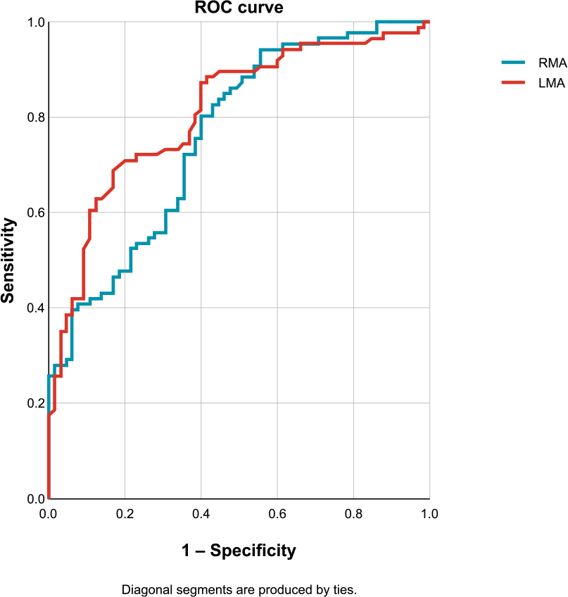
Figure 4.
Comparative analysis of RMA and LMA.
| AUC Test result variable(s): %PAS | |||
|---|---|---|---|
| Area | Std. Errora | Asymptotic Sig.b | Asymptotic 95% CI |
| 0.752 | 0.055 | 0.000 | 0.644–0.861 |
| AUC Test result variable(s): %AS average reference | |||
|---|---|---|---|
| Area | Std. Errora | Asymptotic Sig.b | Asymptotic 95% CI |
| 0.926 | 0.052 | 0.000 | 0.823–1.000 |
| AUC Test result variable(s): MLA x | |||
|---|---|---|---|
| Area | Std. Errora | Asymptotic Sig.b | Asymptotic 95% CI |
| 0.916 | 0.046 | 0.000 | 0.826–1.000 |
| AUC Test result variable(s): MLA x | |||
|---|---|---|---|
| Area | Std. Errora | Asymptotic Sig.b | Asymptotic 95% CI |
| 0.866 | 0.069 | 0.004 | 0.731–1.000 |
| AUC Test result variable(s): %AS average reference | |||
|---|---|---|---|
| Area | Std. Errora | Asymptotic Sig.b | Asymptotic 95% CI |
| 0.823 | 0.094 | 0.010 | 0.638–1.000 |
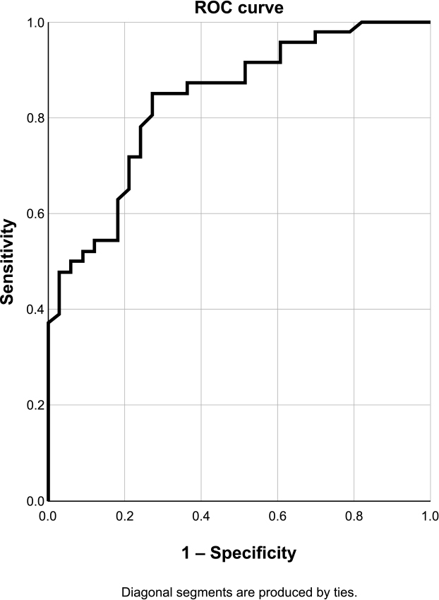
Figure 5.
ROC curve for MLA in the LAD. Sensibility 78.3%, specificity 75.8%, accuracy 77.2%.
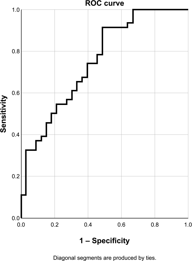
Figure 6.
ROC curve for PAS in the LAD. Sensibility 91.3%, specificity 51.5%, accuracy 74.7%.
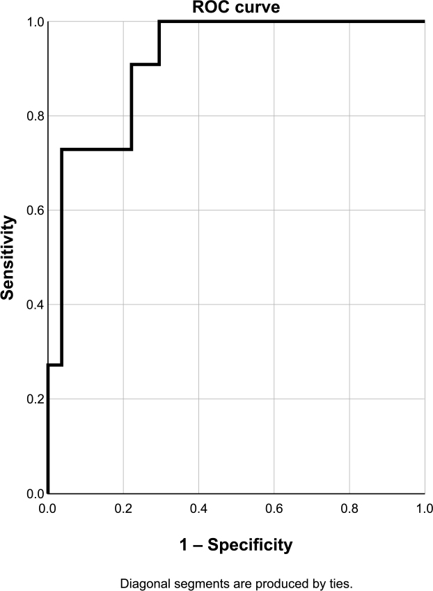
Figure 7.
ROC curve for MLA in the RCA. Sensibility 90.1%, specificity 77.8%, accuracy 81.6%.
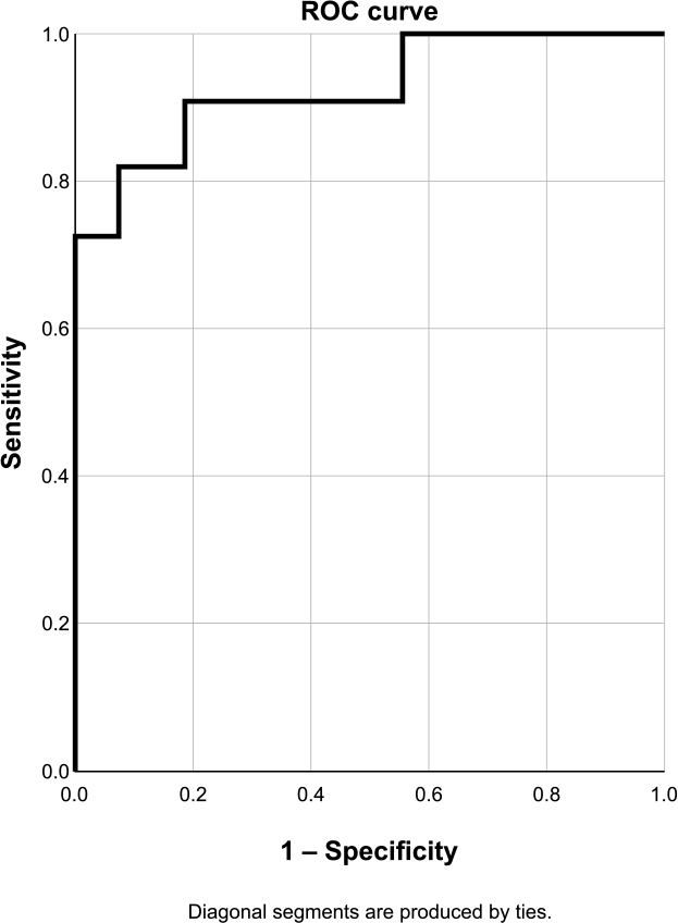
Figure 8.
ROC curve for PAS in the RCA. Sensibility 100.0%, specificity 33.3%, accuracy 52.6%.
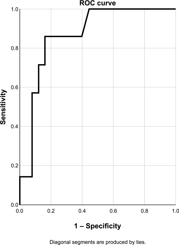
Figure 9.
ROC curve for MLA in the CX. Sensibility 100.0%, specificity 48.0%, accuracy 59.4%.
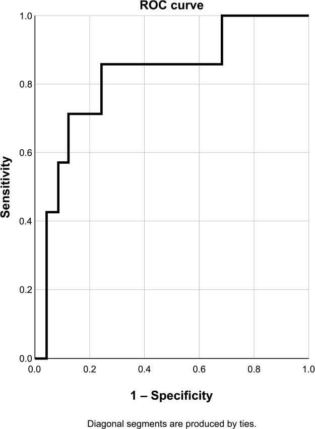
Figure 10.
ROC curve for PAS in the CX. Sensibility 85.7%, specificity 76.0%, accuracy 78.1%.
TABLE 2.
Univariate predictors for FFR ≤ 0.8
| B | p value | Exp(B) (95% CI) | |
|---|---|---|---|
| LAD location | 1.434 | 0.000 | 4.19 (2.09-8.41) |
| OCT proximal reference area | 0.077 | 0.173 | 0.93 (0.83-1.03 |
| OCT distal reference area | 0.140 | 0.034 | 0.87 (0.76-0.99) |
| Average reference area | 0.138 | 0.050 | 0.87 (0.76-1) |
| Lesion length | 0.045 | 0.001 | 1.05 (1.02-1.07 |
| MLA | 1.538 | 0.000 | 0.21 (0.12-0.38) |
| PAS vs. RA | 0.110 | 0.000 | 1.12 (1.07-1.17) |
| PAS vs. proximal reference | 0.103 | 0.000 | 1.11 (1.06-1.16) |
| PAS vs. distal reference | 0.071 | 0.000 | 1.07 (1.04-1.11) |