FIGURE 1.
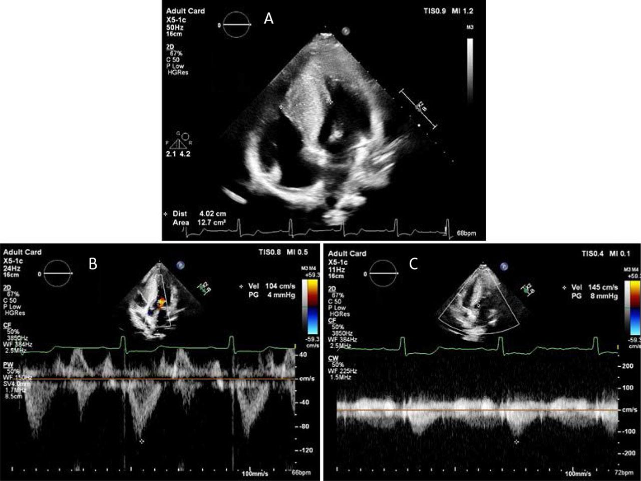
FIGURE 2.
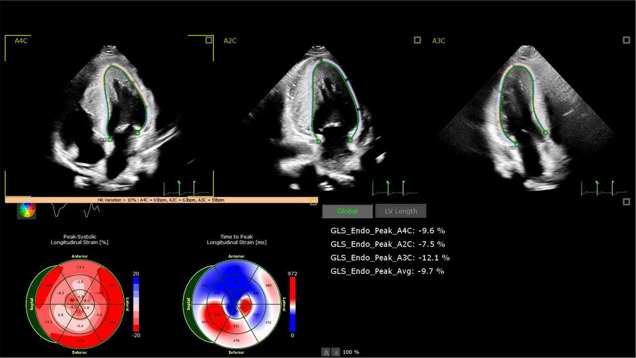
FIGURE 3.
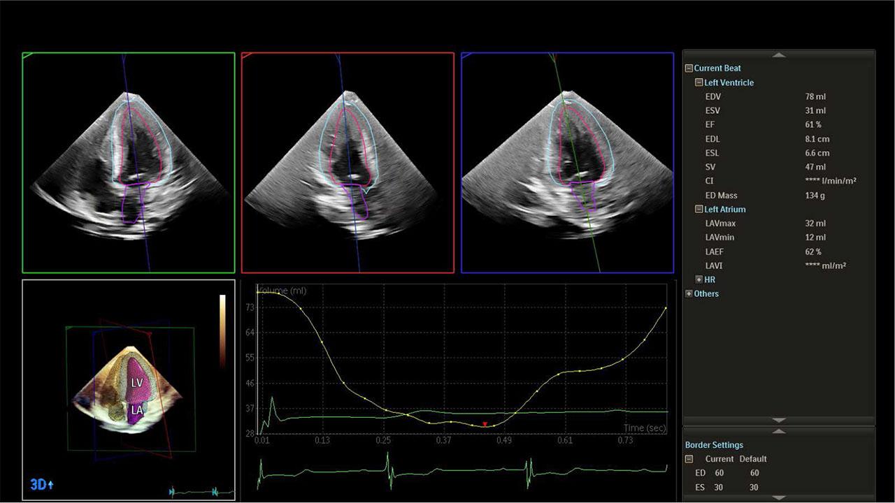
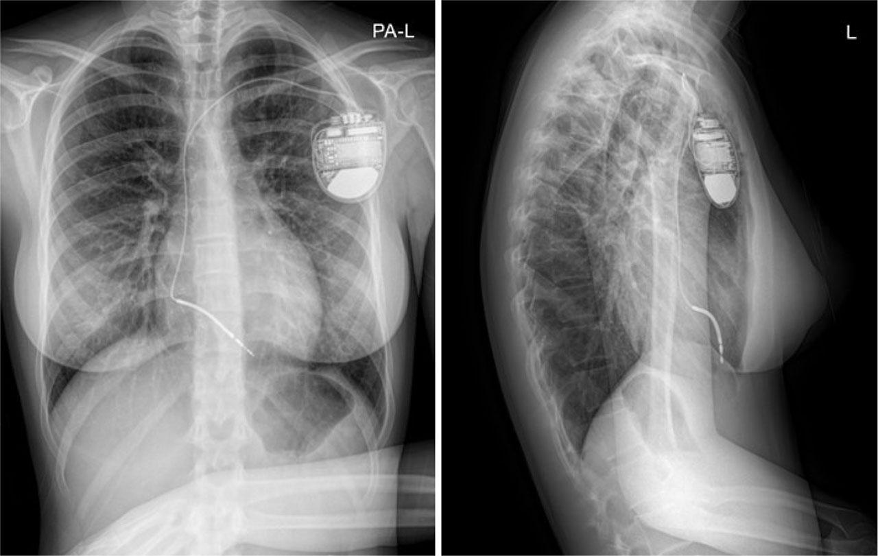
FIGURE 4.
© 2025 Beatrix-Julia Hack, Ioana Sus, Diana-Ramona Iurian, Liliana Gozar, Anca Voichita Popoiu, Iolanda Muntean, published by Asociatia Transilvana de Terapie Transvasculara si Transplant KARDIOMED
This work is licensed under the Creative Commons Attribution-NonCommercial-NoDerivatives 3.0 License.