
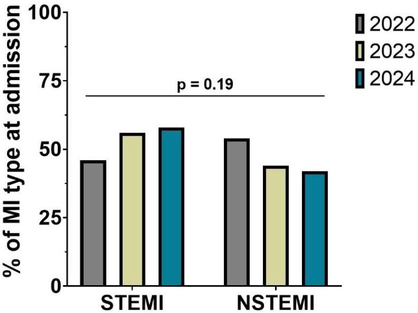
FIGURE 1.
Trends in the type of AMI presentation over the three timespans
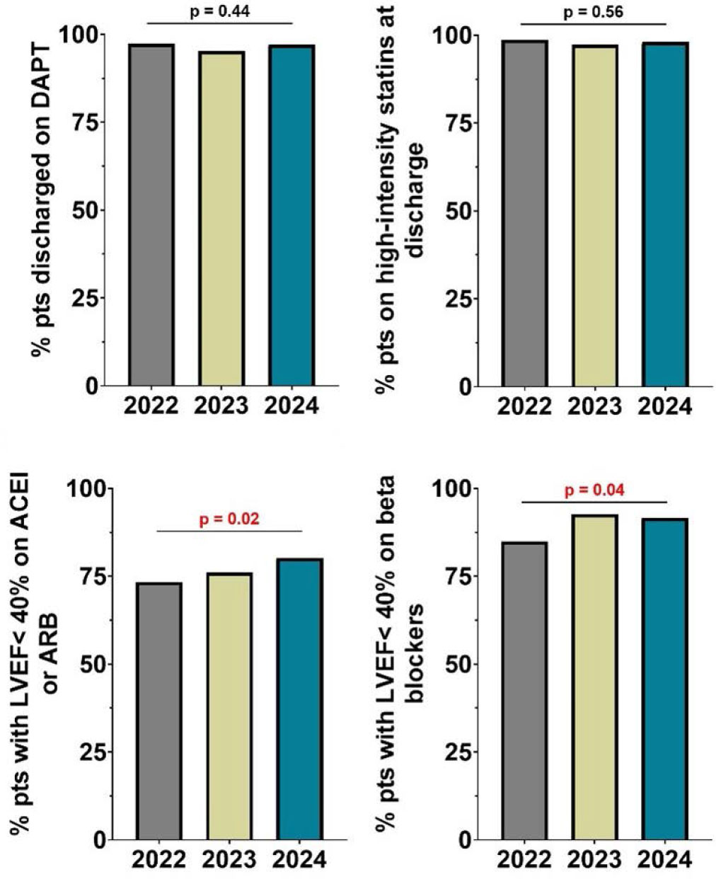
FIGURE 2.
Differences in the management of patients with AMI over the three timespans. A. Percentage of patients discharged on dual antiplatelet therapy. B. Percentage of patients discharged on high intensity statin. C. Percentage of patients with LVEF <40% discharged on ACEI or ARB. D. Percentage of patients with LVEF <40% discharged on beta-blockers.
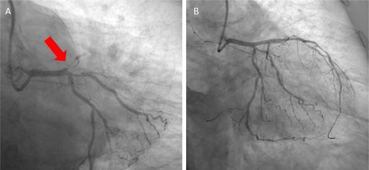
FIGURE 3.
Left coronary artery angiography. A. Acute thrombosis on the left descending anterior artery. B. Post drug eluting stent implantation.
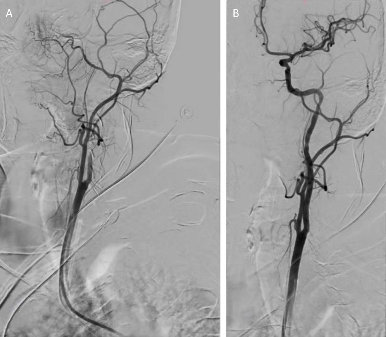
FIGURE 4.
Emergency thrombectomy procedure
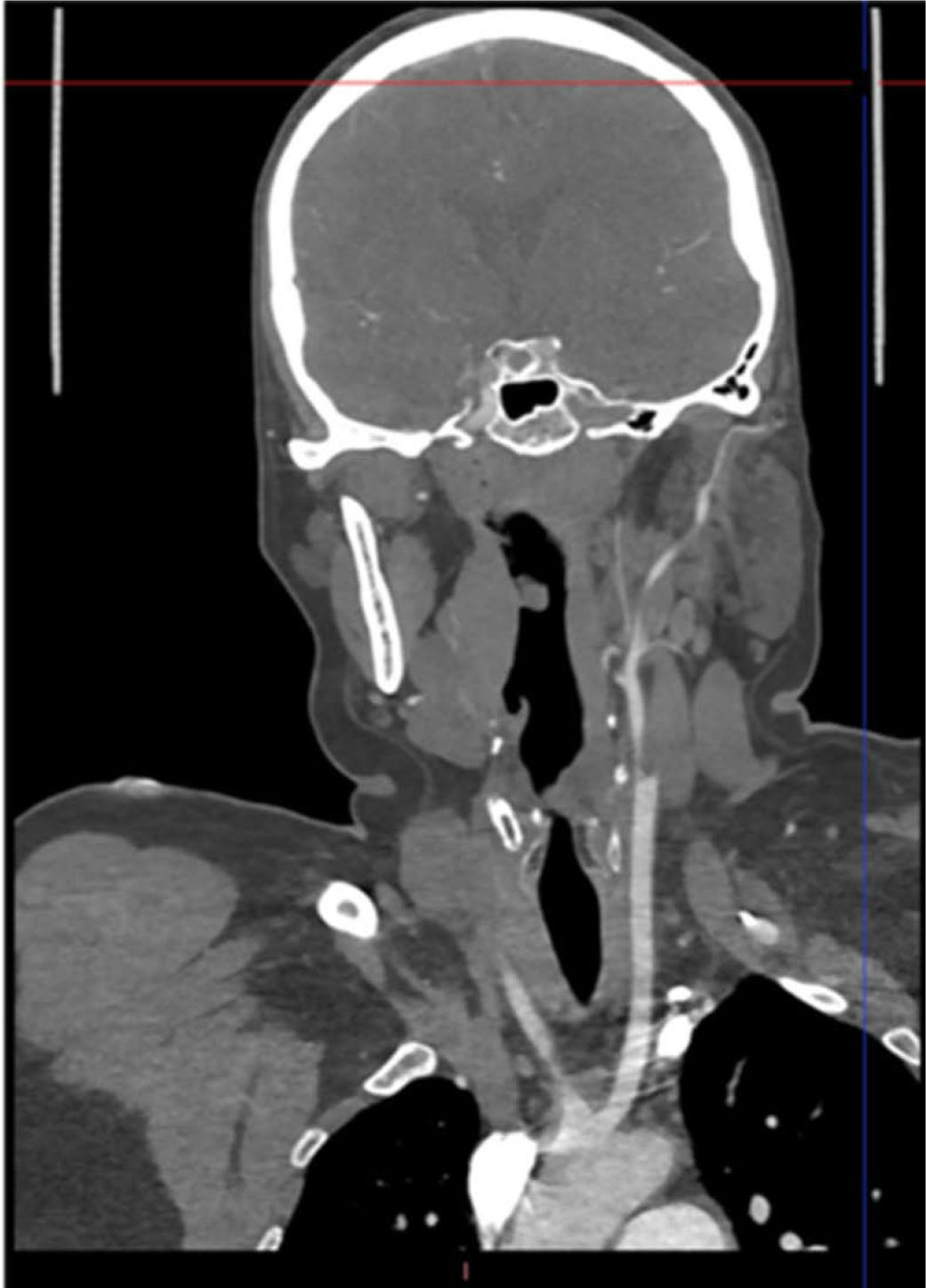
FIGURE 5.
CT scan performed 24 hours post thrombectomy showing no evidence of cerebral ischemic lesions