
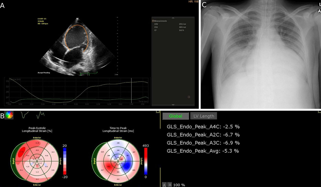
FIGURE 1.
Severe left ventricular dysfunction. A. Severely dilated LV with impaired systolic function (auto 2D quantification). B. Decreased LV global longitudinal strain (GLS) C. Chest radiograph revealing pulmonary edema.
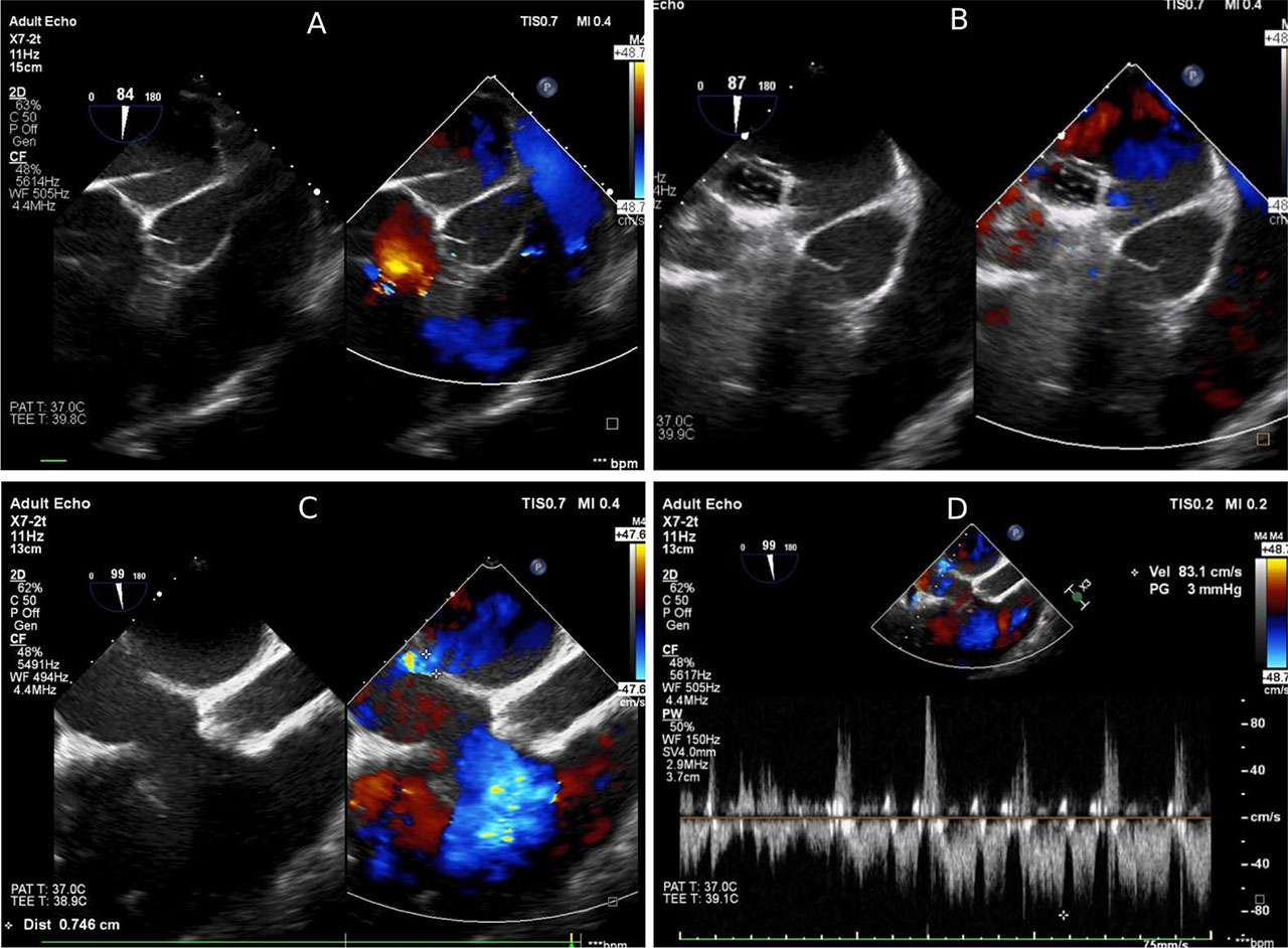
FIGURE 2.
Echocardiography-guided transseptal puncture through the interatrial septum. A. Transseptal needle. B. Balloon dilation of the interatrial communication. C. Doppler color-flow in atrial septal defect. D. Pulsed-wave Doppler revealing unrestrictive atrial septal defect.
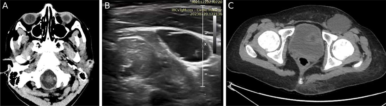
FIGURE 3.
Complications of VA-ECMO. A. Thromboembolic event: head CT scan revealing a bulbar ischemic stroke. B. Ultrasonic image of left inguinal seroma. C. Pelvic CT scan revealing a large, thin-walled left inguinal collection.
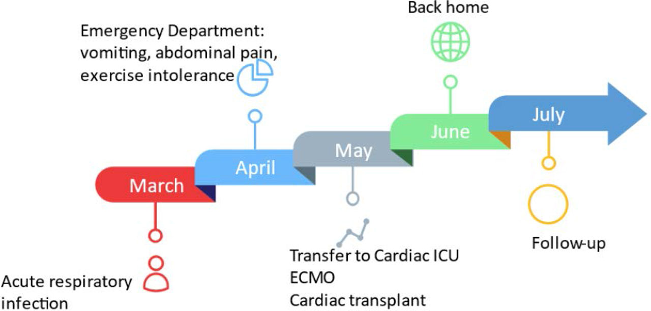
FIGURE 4.
Medical history timeline of the patient