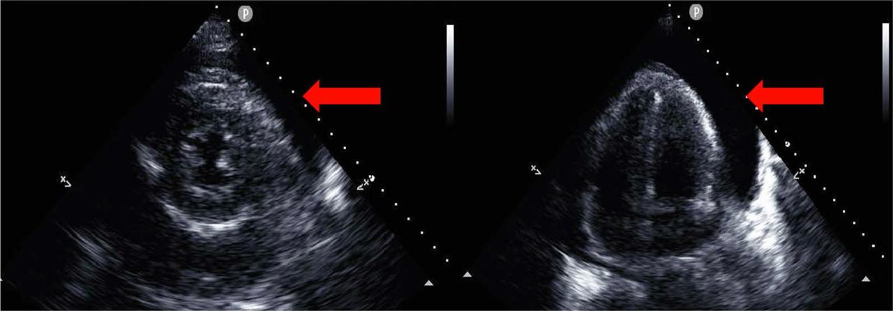
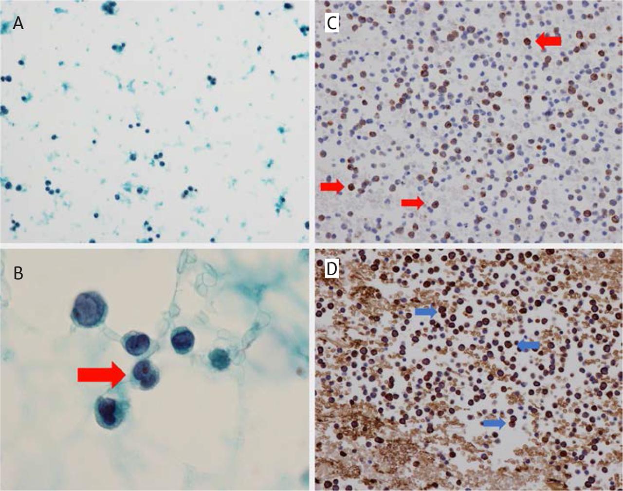
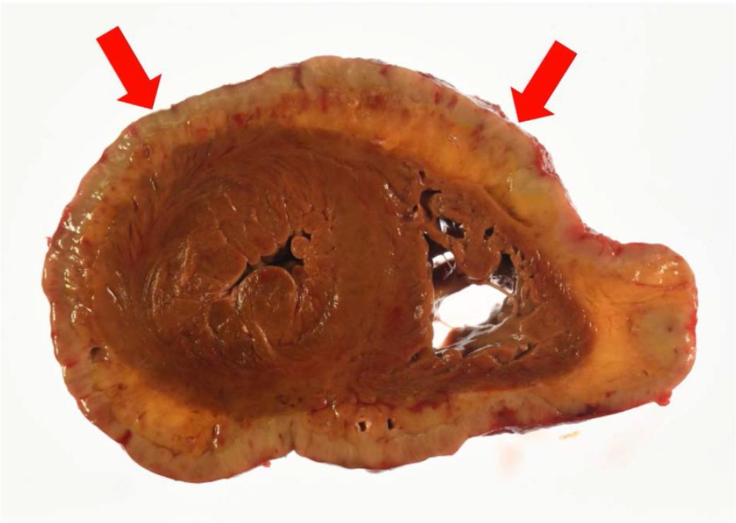
Have a personal or library account? Click to login
An Autopsy Case of Acute Transformation of Myelodysplastic Syndrome Leading to Carcinomatous Cardiac Tamponade Cover

An Autopsy Case of Acute Transformation of Myelodysplastic Syndrome Leading to Carcinomatous Cardiac Tamponade

Open Access
|Jun 2024

Figures & Tables

FIGURE 1.

A,B, Echocardiography on arrival to the hospital. A, Parasternal left border left ventricular short-axis image showing pericardial effusion (red arrow). B, The apical tetralogy of the pericardium also shows pericardial effusion (red arrow).

FIGURE 2.

A–D, Cytology of pericardial effusion and cell block at the time of presentation. A,B, Atypical cells are scattered, and the possibility of myelomonocytic blasts (red arrows) is suspected, suggesting the possibility of acute MDS transformation. C,D, In the cell block, numerous blast-like cells with irregular nuclei are seen. Many are CD34 positive (stained brown, red arrow) and MPO positive (stained brown, blue arrow), consistent with acute myeloid leukemia.

FIGURE 3.

Cross-section of the heart at autopsy. Diffuse greenish-white neoplastic lesions extending circumferentially and diffusely around the pericardial epicardium (red arrow).

Laboratory findings at admission

WBC2,100/μlT-Bil3.0 U/LpH7.21
Seg28%AST1,440 U/LPaCO219 mmHg
Band8.0%ALT1,391 U/LPaO2169 mmHg
Lymph58%LDH2,076 U/LHCO37.9 mmol/L
Mono2.5%ALP276 U/LLactate140 mg/dL
Blast1.0%GGT41 U/L
Hb8.0 g/dlBUN57.5 mg/dl
Plt8.2 × 104/μlCr2.9 mg/dl
APTT34.2 sNa142 mEq/L
PT-INR1.98K5.2 mEq/L
D-dimer8.9 μg/dlCRP5.09 mg/dlL
CK166 U/L
CK-MB106 U/L
TropT0.08 ng/ml
BNP153 pg/ml

Reported cases of myeloid sarcoma with MDS

Author, yearBase diseaseAge/SexSymptomClinical findingsTreatmentPericardiocentesisOutcome
Mateen et al., 20066MDS, RAEB64/FDyspneaEjection fraction decrease Pericardial effusionBlood transfusionNADied
Matkowskyj et al., 20105t-MDS, RAEB59/MDyspneaAcute heart failure Pericardial tamponadeDiuretic Intravenous dobutamine Pericardial drainageNo appearance of blastsDied
Present case, 2020MDS80/MLoss of consciousnessPericardial tamponadePericardial drainageHigher blasts ratio compared to peripheral bloodDied
DOI: https://doi.org/10.2478/jce-2024-0013 | Journal eISSN: 2457-5518 | Journal ISSN: 2457-550X
Language: English
Page range: 83 - 88
Submitted on: May 30, 2024
Accepted on: Jun 17, 2024
Published on: Jun 27, 2024
Published by: Asociatia Transilvana de Terapie Transvasculara si Transplant KARDIOMED
In partnership with: Paradigm Publishing Services
Publication frequency: 4 issues per year

© 2024 Junji Hatakeyama, Ryo Ageishi, Fumito Kato, published by Asociatia Transilvana de Terapie Transvasculara si Transplant KARDIOMED
This work is licensed under the Creative Commons Attribution-NonCommercial-NoDerivatives 3.0 License.