FIGURE 1.
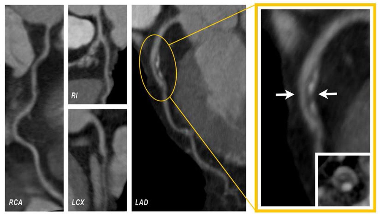
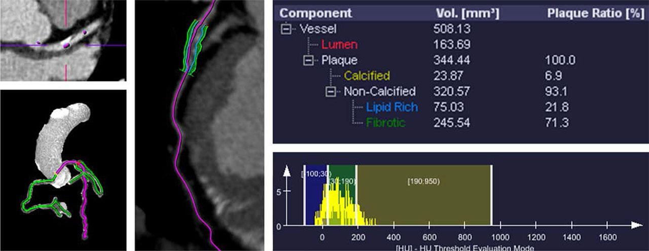
FIGURE 2.
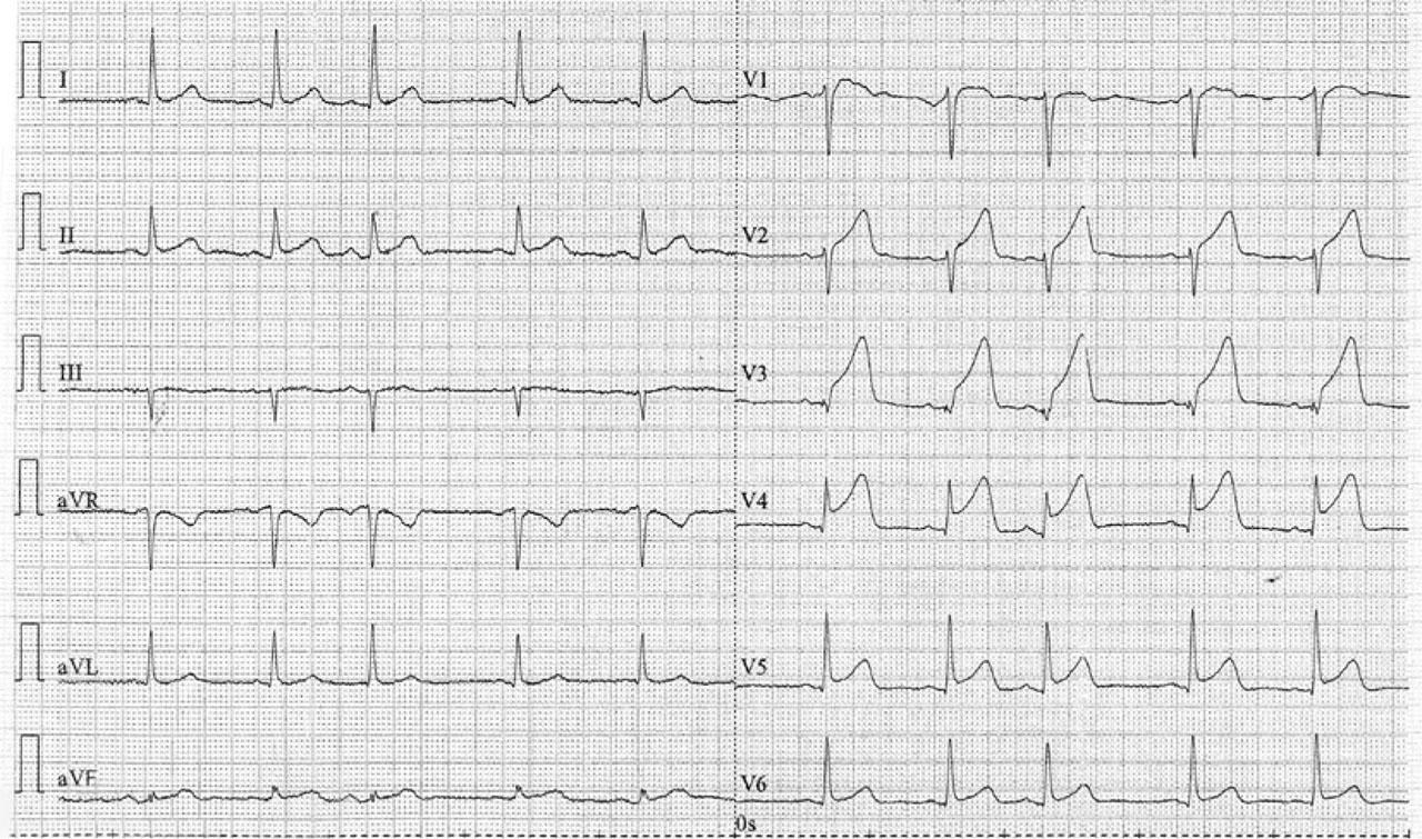
FIGURE 3.
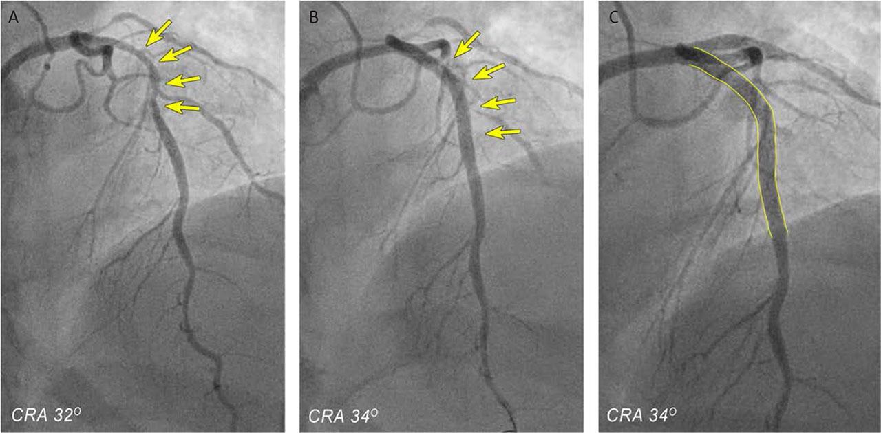
FIGURE 4.
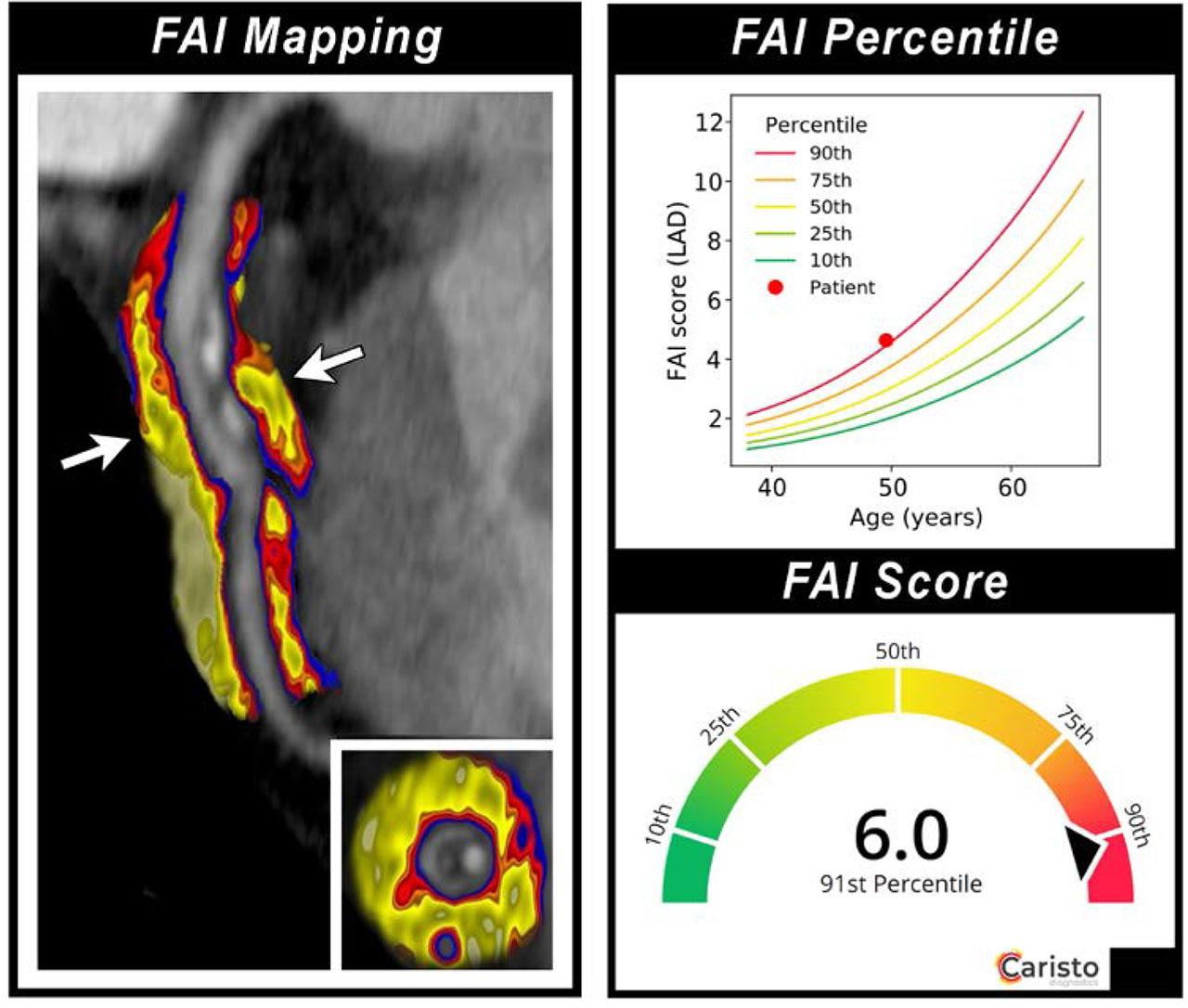
FIGURE 5.
Baseline, risk factors, laboratory and PVAT inflammation parameters for each case_ Increased values are marked with bold_
| Parameters | Normal values | Patient's values |
|---|---|---|
| White blood cells (× 109/L) | 4.5–11 | 30.48 |
| Platelets (× 109/L) | 150–400 | 486 |
| Hemoglobin (g/dL) | 13.8–17.2 | 15.9 |
| Hematocrit (%) | 41–50 | 47.2 |
| Creatinine (mg/dL) | 0.74–1.35 | 1.29 |
| Urea (mg/dL) | 6–24 | 43.80 |
| Glucose (mg/dL) | < 99 | 201 |
| K+ (mmol/L) | 3.6–5.2 | 6.60 |
| Na+ (mmol/L) | 135–145 | 140 |
| CK (U/L) | 55–170 | 813 |
| Total cholesterol (mg/dL) | < 200 | 209.3 |
| HDL cholesterol (mg/dL) | > 60 | 58.2 |
| LDL cholesterol (mg/dL) | < 100 | 129.7 |
| Triglycerides (mg/dL) | < 150 | 97.5 |
| Uric acid (mg/dL) | 3.5–7.2 | 6.8 |
| AST (GOT) (U/L) | 8–33 | 468 |
| ALT (GPT) (U/L) | 4–36 | 208 |
| aPTT (s) | 21–35 | 117.2 |
| CK-MB (ng/mL) | 5–25 | 125.7 |
| hs-cTnI (μg/L) | < 14 | 4.710 |