
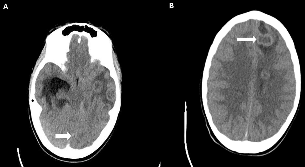
Fig. 1.
A: Axial head CT image showing the midline shift (white arrow); B: Axial head CT image showing multiple brain abscesses.
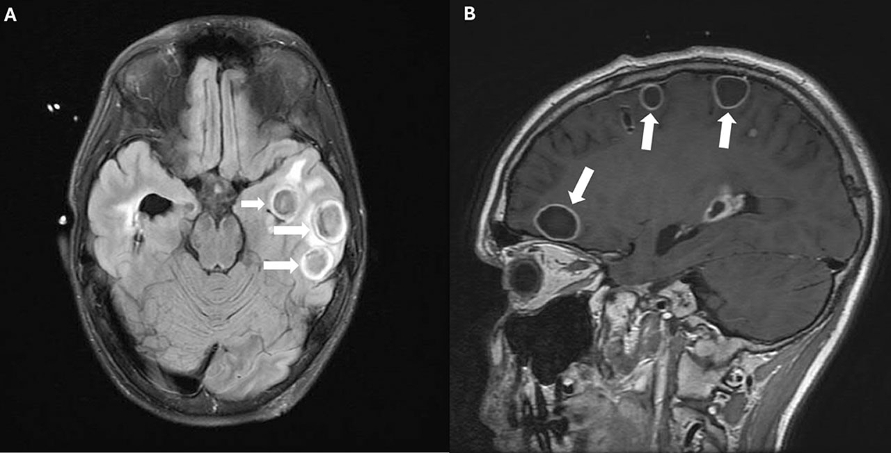
Fig. 2.
A and B Axial MRI images demonstrating resolving brain abscesses. White arrows highlight the reduced size of abscesses