
Introduction
The ISAPS (International Society of Aesthetic Plastic Surgery) 2019 report demonstrated that there was a 10.4 % global increase in noninvasive aesthetic treatments, such as fillers, from the previous year. Silicone (polydimethylsiloxane) injections were originally thought to be ideal for cosmetic augmentation given its durability, stability, and lack of immunogenicity. Therefore, it has been used for cosmetic procedures by both medical and nonmedical personnel. However, the use of these treatments has been associated with several life-threatening complications such as acute pneumonitis, alveolar hemorrhage, and acute respiratory distress, among others [1,2].
Respiratory symptoms are the most predominant and usually manifest within several days from initial exposure but reactions have been seen up to a year following injection [3,4]. The pathophysiological mechanism is thought to be due to an inflammatory response and cellular damage in response to systemic spread of silicone emboli [5]. Treatment is supportive but often requires a steroid regimen [4]. The use of clandestine silicone injections therefore, creates potential risks for susceptible populations.
Case Presentation
A 44-year-old Hispanic female with a history of anxiety presented to the Emergency Department with a two-week history of exertional dyspnea. She also had associated dry cough, chills, pleurisy, lightheadedness, and fatigue. At the time of presentation to the emergency department, her vitals were notable for tachypnea and hypoxia, requiring a high-flow nasal cannula. In addition, a chest x-ray revealed patchy bibasilar airspace opacities with peripheral involvement (see appendix, figure 1). A CT pulmonary angiography (CTPA) revealed bibasilar ground glass airspace disease without evidence of filling defects (see appendix, figure 2). Laboratory tests on arrival were notable for leukocytosis, elevated prothrombin time (PT), D-dimer, lactate dehydrogenase (LD), and fibrinogen (see appendix, table 1).
On day of admission to the general wards, the patient’s presentation was concerning for COVID-19 and/or community-associated pneumonia, therefore, she was started empirically on Dexamethasone (6 mg, intravenously, daily [Mylan Laboratories Ltd., Hyderabad, IN], Low Molecular Weight Heparin (LMWH; enoxaparin; 40 mg, subcutaneously, daily [Meitheal Pharmaceuticals Inc., Chicago, USA]), Ceftriaxone (1 g, intravenously, daily [Sandoz GmbH, Kundl, AUT]) and Doxycycline hyclate (100 mg, oral, twice daily [Amneal Pharmaceuticals Pvt. Ltd., Ahmedabad, IND]). However, point of care (POC) and multiple SARS-CoV2 RNA tests returned negative on day 2 of admission.
On day 2 of admission, the patient became increasingly encephalopathic and hypoxic, and was subsequently intubated, placed on high ventilator settings, and transferred to the ICU for higher level of care (see appendix, table 2). Her hypoxia improved minimally with inhaled nitrous oxide (iNO) and proning on day 3 of admission. On day 4 of admission, she was found to have tracheal bloody secretions, therefore, bronchoscopy with bronchoalveolar lavage (BAL) was performed on day 5 of admission and revealed moderate erythema with residual blood seen throughout. Sequential BALs were consistent with diffuse alveolar hemorrhage with negative cultures (see appendix, table 3). An extensive autoimmune and infectious work-up was also performed on day 5 of admission and was unremarkable (see appendix, table(s) 4 & 5).
Further history was obtained from the patient’s family members on day 5 of admission, who stated that the patient had received bilateral gluteal and lip injections with silicone from an unlicensed professional. She had received these routinely over several years and a few days prior to her onset of symptoms. On receiving this information, it was concluded that the patient was experiencing diffuse alveolar hemorrhage secondary to suspected silicone embolism syndrome.
Following review of the literature on day 6 of admission, the patient was initiated on high-dose methylprednisolone (60 mg, intravenously, every 6 hours [Pharmacia & Upjohn Co, Division of Pfizer Inc., Kalamazoo, USA]) for management [1]. Initially, a lung biopsy was considered to confirm the diagnosis of silicone embolism syndrome. However, due to the patient’s high oxygen requirements, this was not carried out due to safety concerns and was postponed.
After ten days of high-dose steroids, from day 6 to day 15 of admission, the patient’s clinical status improved, and therefore, biopsy was deferred knowing that it would not change the overall outcome. The patient was eventually discharged home on a prednisone taper (Deltasone®; 40 mg, orally, daily for 7 days and then subsequent halving of dosage over the next 3 weeks [Oculus Innovative Sciences, Petaluma, USA]).
Discussion
Over the past two decades, social media has increasingly glorified body image through the use of filters and Photoshop that alter the perceived physical appearance, resulting in more and more people turning toward augmentation as a way to achieve the ‘ideal’ body type. Buttock augmentation, in particular, has become very popular among women. Coincidentally, the number of legal buttock augmentation procedures in the United States has gone up 90% since 2015 [6].
However, the cost of these procedures ranges from several thousands of dollars (from $4,459 to $5,352) making it difficult for individuals from lower socioeconomic status to afford them [7]. Therefore, many turn to illegal plastic surgery clinics which perform the same procedures at a fraction of the cost. As a result, these patients put themselves at serious risk of health complications that may lead to life-threatening outcomes as has been reported by the media over the past several years [8]. Many of these illegal clinics do not use the proper equipment for administering these injections (eg, ultrasound guidance) which can lead to accidental puncture of a gluteal vessel or increased perivascular pressure leading to the development of silicone emboli [9]. Past studies have discovered that anti-silicone IgG antibodies can form immune complexes within the vasculature leading to an amplified inflammatory response [10]. As silicone invades the vasculature and forms immune complexes, these complexes can cause intravascular damage which can ultimately lead to the activation of the coagulation cascade.
The coagulopathy and elevated inflammatory markers observed in our case led to confusion early on when attempting to make a diagnosis because initial laboratory findings suggested a severe COVID-19 infection [11]. Additionally, the radiographic findings (eg, CTPA and CXR) of our patient were similar to those found in COVID-19 in which there is bilateral airspace disease with tendency to peripheral distribution [12]. The culmination of these findings made it difficult to distinguish between COVID-19 pneumonia versus SES. Therefore, it is important for physicians, regardless of the underlying cause, to utilize anticoagulation therapy when either SES or COVID-19 is suspected, as the use of LMWH can prevent the progression to disseminated intravascular coagulation (DIC); a lethal thrombotic event that may occur in either COVID-19 or SES [6]. High dose steroids should also be considered to reduce the inflammation that results from SES. Initially, we utilized the COVID-19 protocol steroid regimen (eg, Dexamethasone 6 mg, oral, daily), but did not see clinical improvement until high dose steroids were initiated, suggesting the need for a more aggressive regimen when treating SES.
Conclusion
With the COVID-19 pandemic fresh in the mind of most physicians, recency bias played a role early in the clinical course of this patient, leading to increased costs and a delay in optimized treatment. Therefore, it is of the utmost importance for clinicians to obtain a thorough history when attempting to diagnose the cause of a patient’s respiratory failure so that the proper treatment measures can be initiated as soon as possible.
Notes
Appendices
Appendix
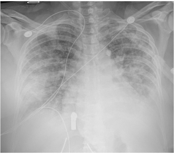
Fig. 1
Chest X-Ray (Single View) on Day 3 of Hospitalization
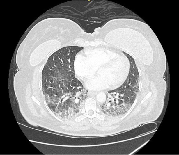
Fig. 2
Chest CT Angiography on Day 2 Of Hospitalization
Table 1
Example of Inflammatory Markers Trended Early in Patient’s Hospital Course
| Day 1 | Day 2 | Day 3 | Day 4 | Day 5 | Day 6 | |
|---|---|---|---|---|---|---|
| PT | 12.8 | N/A | 15.3 | N/A | 15.0 | N/A |
| D-dimer | 1.6 | 1.3 | 1.6 | 1.3 | 4.4 | > 21 |
| LD | 202 | 202 | 392 | 500 | 691 | 1,175 |
| Fibrinogen | 492 | 555 | 891 | 827 | N/A | 570 |
Table 2
Ventilation Settings and Corresponding Arterial Blood Gas (ABG) Values
| Hospital Day # | Ventilation Mode | PEEP (cm H2O) | Respiratory Rate (bpm) | FiO2 (%) | Tidal Volume (Vt; mL) | Arterial Blood Gas Values (pH/ paCO2/paO2/HCO3) | Ventilator Changes2 |
|---|---|---|---|---|---|---|---|
| 31 | AC/VC | 12 | 16 | 100 | 400 | 7.33/39/184/19.7 | ↓ FiO2 to 50% |
| 4 | AC/VC | 12 | 20 | 60 | 400 | 7.28/46/86/21.5 | ↓ FiO2 to 50%; ↑ RR to 26 bpm |
| 5 | AC/VC | 16 | 26 | 100 | 390 | 7.32/43/133/21.7 | ↓ FiO2 to 90%; ↑ Vt to 400 mL |
| 6 | AC/VC | 18 | 23 | 90 | 400 | 7.37/41/118/23.2 | ↓ FiO2 to 80%; ↑ Vt to 300 mL |
| 73 | AC/VC | 16 | 28 | 65 | 300 | 7.07/99/162/27.6 | ↓ FiO2 to 60%; ↑ RR to 32 bpm; ↑ Vt to 320 mL |
| 8 | AC/VC | 10 | 38 | 70 | 320 | 7.30/62/117/29.6 | ↓ FiO2 to 60% |
| 9 | AC/VC | 12 | 38 | 50 | 320 | 7.36/65/68/35.1 | None |
| 10 | AC/VC | 10 | 38 | 45 | 320 | 7.43/61/91/39.1 | None |
| 11 | AC/VC | 6 | 32 | 40 | 350 | 7.46/49/58/35.1 | ↑ FiO2 to 60%; ↑ PEEP to 8 cmH2O |
| 12 | AC/VC | 8 | 32 | 45 | 350 | 7.44/48/110/32.0 | ↓ FiO2 to 40%; ↓ PEEP to 6 cmH2O |
| 13 | PS | 6 | N/A | 30 | N/A | 7.46/42/71/29.0 | Extubated successfully to NC, 6 LPM |
[i] Abbreviations: paCO2 = partial pressure of arterial carbon dioxide (mmHg); paO2 = partial pressure of arterial oxygen (mmHg); HCO3 = concentration of bicarbonate within arterial blood sample (mMol/L); PEEP = positive end expiratory pressure (mmHg); bpm = breathes per minute; FiO2 = fraction of inspired oxygen; AC/VC = assist-control/volume-control; PS = pressure support; NC = nasal cannula; LPM = Liters per minute
1First day of intubation
2Ventilator changes made following corresponding ABG values. Repeat ABGs were not obtained because pulse oximeter readings revealed stable oxygen saturations following adjustments.
3First day proning was initiated to improve oxygenation & ventilation. Proning occurred in 16-hour intervals.
Table 3
Broncheoalveolar Lavage (BAL) Samples Collected on Day 5 of Hospital Course
| RBC | Nucleated Cells | Cell Differentials | |
|---|---|---|---|
| BAL #1 | 28000 | 388 | - Segs: 86% |
| - Lymphs: 1% | |||
| - Mono/Mcrphg: 8% | |||
| - Var Lymph: 1% | |||
| - Other: 4% | |||
| BAL #2 | 71000 | 406 | - Segs: 72% |
| - Bands: 3% | |||
| - Lymphs: 15% | |||
| - Mono/Mcrphg: 1% | |||
| - Eosins: 1% | |||
| - Other: 4% | |||
| BAL #3 | 76000 | 511 | - Segs: 70% |
| - Lymphs: 2% | |||
| - Mono/Mcrphg: 23% | |||
| - Other: 5% |
[i] Abbreviations: BAL = Bronchoalveolar Lavage; RBC = Red Blood Cells; Segs = Segmented Cells; Lymphs = Lymphocytes; Mono/Mcrphg = Monocytes/Macrophages; Var Lymph = Various Lymphocytes; Eosins = Eosinophils
Of Note: Trend of RBCs on sequential BALs led to the diagnosis of diffuse alveolar hemorrhage (DAH).
Table 4
Various Lab Studies Performed to Rule Out Infectious Etiology
| Infectious Agent | Type of Test | Source | Results |
|---|---|---|---|
| HIV 1,2 Ag/Ab | Polymerase Chain Reaction (PCR) | Serum | Non-reactive |
| Respiratory Viral Panel (RVP)1 | Polymerase Chain Reaction (PCR) | Nasal Swab | Not Detected |
| Legionella Antigen | Enzyme Immunoassay (EIA) | Urine | Negative |
| Q Fever Ab IgM/IgG (Phase I-Ii) | Indirect Immunofluorescence Antibody (IFA) | Serum | <1:16 |
| Coccidiodes Antibody | Immunodiffusion (ID) & Complement Fixation (CF) | Serum | Negative |
| Cocci Immunodiffusion IgM/IgG | Immunodiffusion (ID) | Serum | Negative |
| Quantiferon TB Gold | Interferon-Gamma (IFN-γ) Release Assay (IGFRA) | Serum | Negative |
| Hepatitis Panel2 | Polymerase Chain Reaction (PCR) | Serum | Non-reactive |
| Acid Fast | Culture | Sputum | No Growth Detected |
| Gram Stain | Culture | Blood & BAL3 | No Growth Detected |
| Viral | Culture | BAL | No Growth Detected |
| Respiratory | Culture | Sputum | No Growth Detected |
| Fungal Stain | Culture | Blood & BAL3 | No Growth Detected |
[i] Abbreviations: HIV = Human Immunodeficiency Virus; Ag = Antigen; Ab = Antibody; IgM = Immunoglobulin M; IgG = Immunoglobulin G; TB = Tuberculosis; BAL = Bronchoalveolar Lavage
1RVP consists of the following viral PCR test(s): Adenovirus, Metapneumovirus, Rhinovirus/Enterovirus, Influenza A&B, Parainfluenza 1-4, RSV, Bordetella parapertusis, RP, Bordetella pertussis, RP, Chlamydophila pneumoniae, Mycoplasma pneumoniae, RP, Coronavirus HKV1/NL63/229E/OC43
2Hepatitis panel consists of the following antibody (AB) and antigen (Ag) test(s): HAV AB IgM, HBC AB IgM, HBS AB, HBS Ag, & HCV AB
3Bronchoalveolar Lavage (BAL) samples as listed above under Table 2
Table 5
Studies Performed to Rule Out Autoimmune Cause
| Autoimmunity Tested | Type of Test | Source | Results |
|---|---|---|---|
| C-ANCA/P-ANCA AB1 | Enzyme Immunoassay (EIA) | Serum | Negative |
| Antinuclear Antibody (ANA) Panel2 | Indirect Immunofluorescence Antibody (IFA) | Serum | Negative |
| Angiotensin Converting Enzyme | Enzyme-Linked Immunosorbent Assay (ELISA) | Serum | 20 U/L (ref. range: 16-85 U/L) |
| C1 Esterase Inhibitor | Enzyme Immunoassay (EIA) | Serum | 60 mg/dL (ref. range 19 - 37 mg/dL |
| C1 Esterase Inhibitor, Functional | Enzyme Immunoassay (EIA) | Serum | > 90 (ref. range: > 67 (Normal) 41-67 (Equivocal) <41 (Abnormal)) |
| C2 Complement | Immunodiffusion (ID) | Serum | 63 u/mL (ref. range: 25 - 47 u/mL) |
| C5 Complement | Immunodiffusion (ID) | Serum | 34 mg/dL (ref. range: 10.6 - 26.3 mg/dL) |
[i] Abbreviations: AB = Antibody
1Antineutrophil Cytoplasmic Antibody (ANCA) testing includes: Anti-myeloperoxidase (MPO) antibodies & anti-proteinase 3 (PR3) antibodies
2ANA panel consists of the following antibody (AB) tests: ANA AB Quant, anti-CCP3 AB, anti-Centromere AB, anti-Chromatin AB, anti-RNP AB, anti-SCL-70 AB, anti-Sm AB, anti-SS-A AB, and anti-SS-B AB