
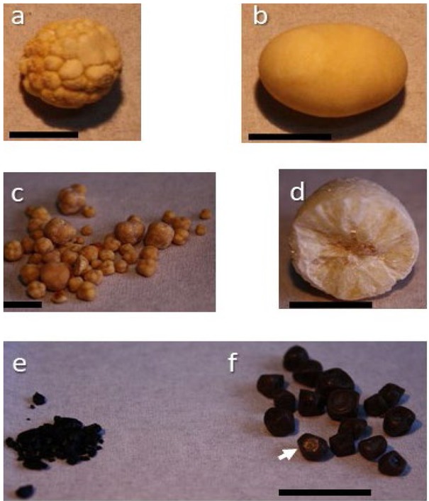
Fig. 1
Human gallbladder stones. Pure cholesterol stones with spherical shape and morular surface (a), oval shape and smooth surface (b), multiple stones (c). The cut surface is shown with a brownish centre and radial disposition of cholesterol monohydrate crystals (d). Pure pigment stones are shown as debris (e). Mixed stones are shown as multifaceted concernments (f). Note that the black pigment surface and the inner centre are made of a small amount of solid cholesterol crystals (arrow) Black horizontal line=1 cm.
Table 1
Complications of gallstones
| Complications |
|---|
| Acute cholecystitis* |
| Acute biliary pancreatitis* |
| Acute cholangitis* |
| Acute acalculous cholecystitis* |
| Biliary enteric fistula and gallstone ileus* |
| Choledocholithiasis |
| Cholestatic jaundice* |
| Cholesterolosis and gallbladder polyps |
| Chronic cholecystitis |
| Gallbladder carcinoma and porcelain gallbladder |
| Recurrent pyogenic cholangitis* |
[i] *Conditions characterized by critical care aspects.
Table 2
Conditions predisposing to increased risk of acute cholecystitis
| Acute non-biliary diseases | Immunocompromised illness |
|---|---|
| Acute renal failure | Infections (hepatitis B virus, ascariasis in developing countries) |
| Age > 60 years | Major surgery |
| Biliary sludge | Female gender |
| Cardiovascular disease (history of ischemic stroke, cerebral hemorrhage) | Multiple trauma |
| Diabetes mellitus | Sepsis |
| Gallstones | Severe burns |
| Hormonal replacement therapy | Systemic vasculitis |
| Hypertriglyceridemia | Total parenteral nutrition, long term fasting |
[i] Adapted from Portincasa P, Wang DQH. Gallstones. In: Podolsky KD, Camilleri M, Fitz JG, Kalloo AN, Shanahan F, Wang TC, eds. Yamada’s Textbook of Gastroenterology. 6th ed. Hoboken, New Jersey (USA): Wiley-Blackwell, 2015:1808-1834 [56].
Table 3
Imaging techniques for acute cholecystitis
| Technique | Major findings | Notes |
|---|---|---|
| Abdominal ultrasonography | Enlarged gallbladder size | First choice |
| Gallbladder wall thickening (>4mm) Incarcerated gallstone(s) Intraluminal debris echoes Pericholecystic fluid collection or abscess Positive “sonographic” Murphy sign Sonolucent “double wall sign” | Moderate sensitivity (88%90%) and specificity (80%) [63, 122-124] | |
| Hepatobiliary scintigraphy (technetium iminodiacetic | Compound given intravenously and excreted by the liver Test is positive if the gallbladder is not visualized (i.e., cystic | Highest sensitivity and specificity (≈90-95%) [123] |
| acid or hydroxyiminodiacetic acid) | duct obstruction due to edema by acute cholecystitis) [63] | Not easily available |
| Computed Tomography (CT) | Gallbladder wall edema | High sensitivity 94% |
| Pericholecystic fluid Other complications [125] | Low specificity 59% [123, 126] | |
| Magnetic Resonance | Gallstones are likely passed in the common bile duct [127] | Moderate accuracy [123] |
| Cholangio-Pancreatography | Not easily available | |
| (MRCP) | Under evaluation |
[i] Adapted from Portincasa P, Wang DQH. Gallstones. In: Podolsky KD, Camilleri M, Fitz JG, Kalloo AN, Shanahan F, Wang TC, eds. Yamada’s Textbook of Gastroenterology. 6th ed. Hoboken, New Jersey (USA): Wiley-Blackwell, 2015:1808-1834 [56].
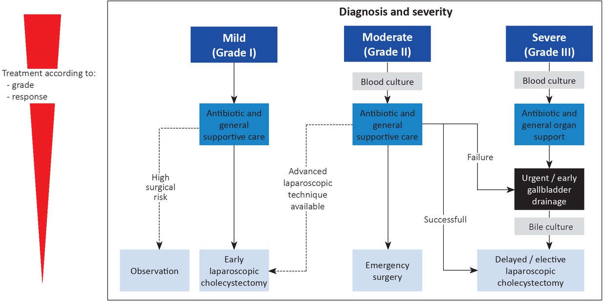
Fig. 2
Algorithm of the management of acute cholecystitis
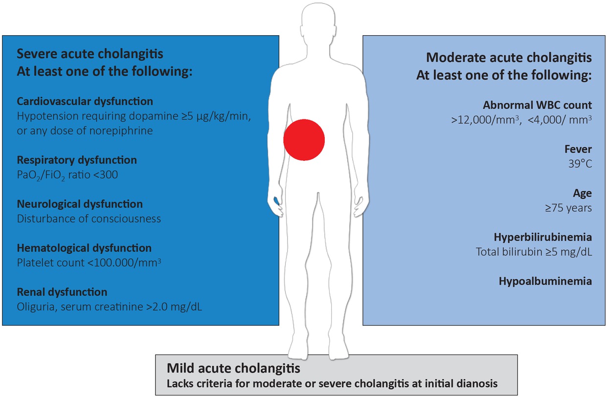
Fig. 3
Definition of severity of acute cholangitis.
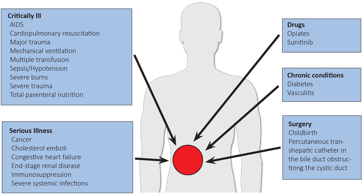
Fig. 4
Factors predisposing to acute acalculous cholecystitis.