
Introduction
B-cell non-Hodgkin lymphomas (NHL) in childhood and adolescence are the fifth most common pediatric malignancy worldwide (1). Among malignant lymphomas, B-cell NHL is the most common type, accounting for approximately 7% of all childhood cancers (2).
The cure rate for B-cell non-Hodgkin lymphoma in high-income countries exceeds 90%, largely due to robust supportive care and the addition of targeted therapy with dose-intense chemotherapy. However, in low- and middle-income countries (LMICs), survival outcomes remain below 50% because of inadequate supportive care and limited access to intensive chemotherapy and targeted therapies. Reported event-free survival (EFS) rates for B-cell NHL from India and other LMICs range from 20% to 72% (3).
In LMICs such as Pakistan, pediatric B-cell non-Hodgkin lymphoma presents additional challenges because of limited diagnostic resources, inadequate supportive care, and high rates of treatment-related mortality (3,4). Furthermore, treatment at centers lacking the capacity to monitor and manage tumor lysis syndrome (TLS) has been associated with increased early mortality. Addressing these challenges requires optimized risk stratification, early intervention strategies, and improved access to essential supportive care measures.
We conducted a retrospective study and evaluated risk factors influencing long term survival outcomes in patients with B-cell non-Hodgkin lymphoma treated at a tertiary cancer care hospital.
Methods and Material
This retrospective descriptive study was conducted at Shaukat Khanum Memorial Cancer Hospital and Research Centre (SKMCH&RC), Lahore, Pakistan, and included data from patients aged 1–18 years, following approval from the institutional review board. Newly diagnosed patients with CD20-positive B-cell non-Hodgkin lymphoma—including Burkitt lymphoma (BL), high-grade B-cell non-Hodgkin lymphoma not otherwise specified (BNHL-NOS), and diffuse large B-cell lymphoma (DLBCL)—who were diagnosed between January 2016 and December 2020 were included. All patients had a five-year post-treatment follow-up period.
Patients with T-cell lymphoblastic lymphoma (TCL), precursor B-cell lymphoblastic lymphoma, anaplastic large cell lymphoma (ALCL), and those with missing data were excluded. Data were retrieved from the electronic database. The diagnosis of B-cell non-Hodgkin lymphoma was confirmed by tissue biopsy, histopathological examination, and immunohistochemistry.
Data were collected for age, gender, residence, diagnosis, site, stage, lactate dehydrogenase levels, treatment group, tumor lysis syndrome, diagnostic procedure (biopsy vs laparotomy), and outcomes. Staging was performed according to the St. Jude classification, using computed tomography (CT) scans, bone marrow aspiration and biopsy, and cell count and cytology of cerebrospinal fluid (5). MRI of the spine was performed in some patients to evaluate spinal cord involvement. Risk stratification and the treatment protocol used at the center were based on the United Kingdom Children’s Cancer Study Group (UKCCSG) NHL 2003, which follows the FAB/LMB (French-American-British/Lymphome Malins De Burkitt) protocol (6).
In our study, tumor lysis syndrome was identified based on laboratory abnormalities, including elevated serum potassium, uric acid, creatinine, calcium, and phosphate levels.
Group A treatment consisted of cyclophosphamide 250 mg/m2 every 12 hours for the first 3 days, vincristine 2 mg/m2 on days 1 and 6, prednisolone 60 mg/m2 in two divided doses for 5 days (then tapered over 3 days), and doxorubicin 60 mg/m2 (COPAD regimen).
Groups B and C received initial cytoreductive chemotherapy with vincristine 1 mg/m2, cyclophosphamide 300 mg/m2, prednisolone 60 mg/m2 in two divided doses for 5 days, and intrathecal chemotherapy with methotrexate (MTX) and hydrocortisone (COP regimen).
Group B patients received two cycles of induction chemotherapy: cyclophosphamide 250 mg/m2 every 12 hours for 3 days, vincristine 2 mg/m2 on day 1, prednisolone 60 mg/m2 in two divided doses for 5 days (then tapered over 3 days), methotrexate (MTX) 3 g/m2 on day 1 with folinic acid rescue, doxorubicin 60 mg/m2 on day 1, and two doses of intrathecal MTX and hydrocortisone (COPADM regimen). This was followed by two cycles of consolidation chemotherapy: cytarabine 100 mg/m2 as a 24-hour infusion for 5 days, high-dose MTX 3 g/m2 with folinic acid rescue, and intrathecal MTX, hydrocortisone, and cytarabine for 2 days (CYM regimen).
Group C patients received very high-dose MTX 8,000 mg/m2 on day 1 followed by folinic acid rescue and cyclophosphamide 1,000 mg/m2/day for 3 days during induction, along with vincristine, doxorubicin, and prednisolone. In the consolidation phase, they received high-dose cytarabine 3,000 mg/m2/day for 4 days and etoposide 200 mg/m2/day for 4 days (CYVE regimen). CNS-positive patients also received high-dose MTX 8,000 mg/m2 with folinic acid rescue and intrathecal methotrexate, followed by four cycles of maintenance chemotherapy. Rituximab was introduced in January 2020 for patients with stage IV disease; it was not available earlier due to resource limitations.
Response criteria on reassessment imaging followed the United Kingdom Children’s Cancer Study Group (UKCCSG) NHL 2003 guidelines. Complete remission was defined as the disappearance of all measurable or evaluable lesions, with no L3 blast cells in the CSF or bone marrow. Partial response was defined as the existence of histologically proven residual disease.
Disease progression was defined as an increase of more than 25% in the product of the two largest diameters of measurable lesions, the appearance of new lesions, or the reappearance of L3 blasts in the bone marrow or cerebrospinal fluid. Disease response was also assessed at the end of therapy. Patients were followed for five years after completing therapy.
Reassessment CT scans for treatment group B were performed after the first cycle of CYM chemotherapy. If histologically proven residual disease was present, chemotherapy was intensified from Group B to Group C. No centralized radiology review was available at our center.
Statistical analysis was performed using SPSS software (version 26.0; SPSS, Chicago, IL, USA). Continuous variables were presented as mean ± SD, and categorical variables were summarized as frequencies and percentages. Categorical variables were compared using the chi-square test or Fisher’s exact test, as appropriate.
Continuous variables were compared using the independent t-test. A multivariable logistic regression (MLR) model was used to identify independent risk factors associated with mortality. Statistical significance was defined as a two-tailed p-value < 0.05. Overall survival (OS) and event-free survival (EFS) were calculated using the Kaplan-Meier method. EFS was calculated from the date of diagnosis until the first event, including death from any cause, disease progression, relapse, second malignancy, or abandonment of treatment. OS was calculated as the percentage of patients alive at last follow-up.
Results
A total of 276 patients with B-cell non-Hodgkin lymphoma were registered between January 2016 and December 2020. The median age was 5 years (IQR: 2), with a male predominance (76.8%). Most patients were from Khyber Pakhtunkhwa (57.2%), followed by Punjab (23.2%). Burkitt lymphoma was the most common diagnosis (89.1%), and the diagnosis was mainly established by biopsy (54.3%). Most children presented with advanced disease (stage III–IV: 95.3%), and the abdomen was the most common primary site (83.7%). Tumor lysis syndrome occurred in 24.6% of patients. On reassessment scan, complete response was achieved in 63%, while 3.3% had a partial response (Table 1). The median follow-up duration was 53 months. At the end of treatment, 93 patients died (30 [28.5%] due to disease burden and 63 [60%] due to sepsis), 7 (6.5%) had disease progression, 2 (2%) relapsed, and 3 (2%) were lost to follow-up.
Table 1.
Baseline Demographic and Clinicopathological Characteristics of Children with B-Cell Non-Hodgkin’s Lymphoma.
| Variables | Characteristics | Frequency n (%) |
|---|---|---|
| Age (years) | Median (IQR) | 5.0 (2.0) |
| Gender | Male | 212 (76.8) |
| Female | 64 (23.2) | |
| Area of Residence | Punjab | 64 (23.2) |
| Khyber Pakhtunkhwa | 158 (57.2) | |
| Afghanistan | 39 (14.1) | |
| Balochistan | 13 (4.7) | |
| Sindh | 1 (0.4) | |
| Others | 3 (1.1) | |
| Diagnosis | Burkitt’s lymphoma | 246 (89.1) |
| DLBCL | 26 (9.4) | |
| B-cell NHL, NOS | 4 (1.4) | |
| Diagnostic Procedure | Biopsy | 150 (54.3) |
| Laparotomy | 126 (45.7) | |
| Tumor Lysis Syndrome | Yes | 68 (24.6) |
| No | 208 (75.4) | |
| Clinical Stage | Low stage (I–II) | 13 (4.7) |
| Advanced stage (III–IV) | 263 (95.3) | |
| Primary Site of Disease | Abdomen | 231 (83.7) |
| Face | 28 (10.1) | |
| Neck | 6 (2.2) | |
| Paraspinal | 8 (2.9) | |
| Others | 3 (1.1) | |
| Stage IV Disease | None | 203 (73.6) |
| Bone marrow involvement | 28 (10.1) | |
| CNS disease (cranial mass or nerve palsy) | 28 (10.1) | |
| CSF positive | 3 (1.1) | |
| Both BM & CNS involvement | 14 (5.1) | |
| Reassessment Scan | Not done | 88 (31.9) |
| Complete response | 174 (63.0) | |
| Partial treatment response | 9 (3.3) | |
| Disease progression | 5 (1.8) |
Of the 276 patients, 176 (63.7%) were alive (censored) and 100 (36.3%) died. Gender was not significantly associated with outcome (p=0.182). Baseline LDH was strongly associated with mortality, with a greater proportion of deaths occurring in patients with LDH ≥2× the upper limit of normal (54.1% vs 19.6%; p<0.01). Advanced stage, tumor lysis syndrome, and poor reassessment response were also significantly associated with mortality (p<0.001) (Table 2).
Table 2.
Association of Baseline Clinical Characteristics with Mortality Among Children with B-cell Non-Hodgkin’s Lymphoma.
| Variables | Characteristics | Alive: n (%) | Death: n (%) | P-value |
|---|---|---|---|---|
| Sex | Male | 140 (66.0) | 72 (34.0) | 0.182 |
| Female | 36 (56.3) | 28 (43.8) | ||
| Baseline LDH | ≤2× upper limit of normal | 115 (80.4) | 28 (19.6) | <0.001 |
| >2× upper limit of normal | 61 (45.9) | 72 (54.1) | ||
| Primary Disease Site | Abdomen | 151 (65.4) | 80 (34.6) | 0.051 |
| Face | 15 (53.6) | 13 (46.4) | ||
| Paraspinal | 2 (25.0) | 6 (75.0) | ||
| Clinical Stage | Low stage (I–II) | 12 (92.0) | 1 (8.0) | <0.001 |
| Advanced stage (III–IV) | 163 (63.0) | 99 (37.0) | ||
| Surgery Type at Diagnosis | Biopsy | 82 (54.7) | 68 (45.3) | <0.001 |
| Laparotomy | 94 (74.6) | 32 (25.4) | ||
| Tumour Lysis Syndrome | No | 157 (75.5) | 51 (24.5) | <0.001 |
| Yes | 19 (27.9) | 49 (72.1) | ||
| Reassessment Scan | Complete response | 165 (94.8) | 9 (5.2) | <0.001 |
| Partial response | 7 (77.8) | 2 (22.2) | ||
| Disease progression | 1 (20.0) | 4 (80.0) |
In univariate regression analysis, patients with baseline LDH >2× normal had significantly higher odds of death (OR: 3.31, 95% CI: 2.13–5.13; p<0.001). Patients with tumor lysis syndrome had a fourfold higher risk of mortality (OR: 4.04, 95% CI: 2.71–6.02; p<0.001). Biopsy was associated with lower odds of death (OR: 0.50, 95% CI: 0.33–0.77; p=0.002). In multivariate analysis, patients with LDH >2× normal had a twofold higher risk of death compared to those with LDH <2× normal (OR: 2.03, 95% CI: 1.25–3.31; p=0.004), indicating that elevated LDH is independently associated with worse survival. Patients with tumor lysis syndrome had a 2.9-fold higher risk of death compared to those without TLS (OR: 2.86, 95% CI: 1.84–4.46; p<0.001) (Table 3).
Table 3.
Predictors of Mortality in B-cell Non-Hodgkin Lymphoma (Unadjusted and Adjusted).
| Analysis | Variables | Characteristics | Unadjusted OR (95% CI) | p-Value |
|---|---|---|---|---|
| Univariate | Gender | Male | Reference | |
| Female | 1.31 (0.85–2.04) | 0.216 | ||
| Baseline LDH | <2× upper limit of normal | Reference | ||
| >2× upper limit of normal | 3.31 (2.13–5.13) | <0.001 | ||
| Tumor Lysis Syndrome | No | Reference | ||
| Yes | 4.04 (2.71–6.02) | <0.001 | ||
| Surgery at Diagnosis | Laparotomy | Reference | ||
| Biopsy | 0.50 (0.33–0.77) | 0.002 | ||
| Multivariate | Gender | Male | Reference | |
| Female | 1.41 (0.91–2.20) | 0.120 | ||
| Baseline LDH | <2× upper limit of normal | Reference | ||
| >2× upper limit of normal | 2.03 (1.24–3.30) | 0.004 | ||
| Tumour Lysis Syndrome | No | Reference | ||
| Yes | 2.86 (1.83–4.45) | <0.001 | ||
| Surgery at Diagnosis | Laparotomy | Reference | ||
| Biopsy | 0.86 (0.54–1.37) | 0.530 |
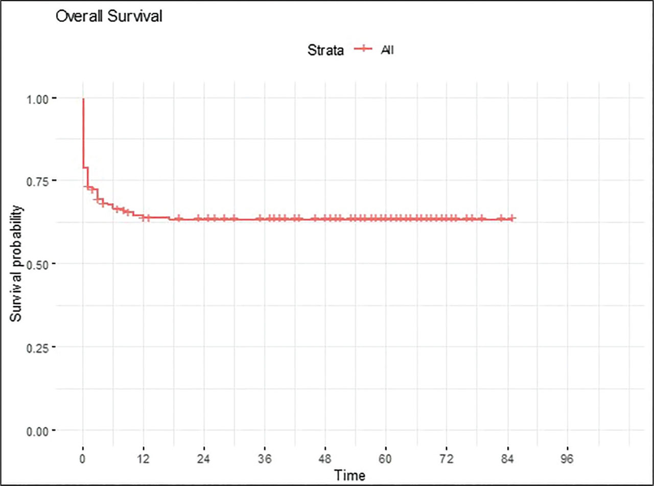
Figure 1.
Kaplan–Meier Curve of Overall Survival in Patients with B-cell Non-Hodgkin Lymphoma.
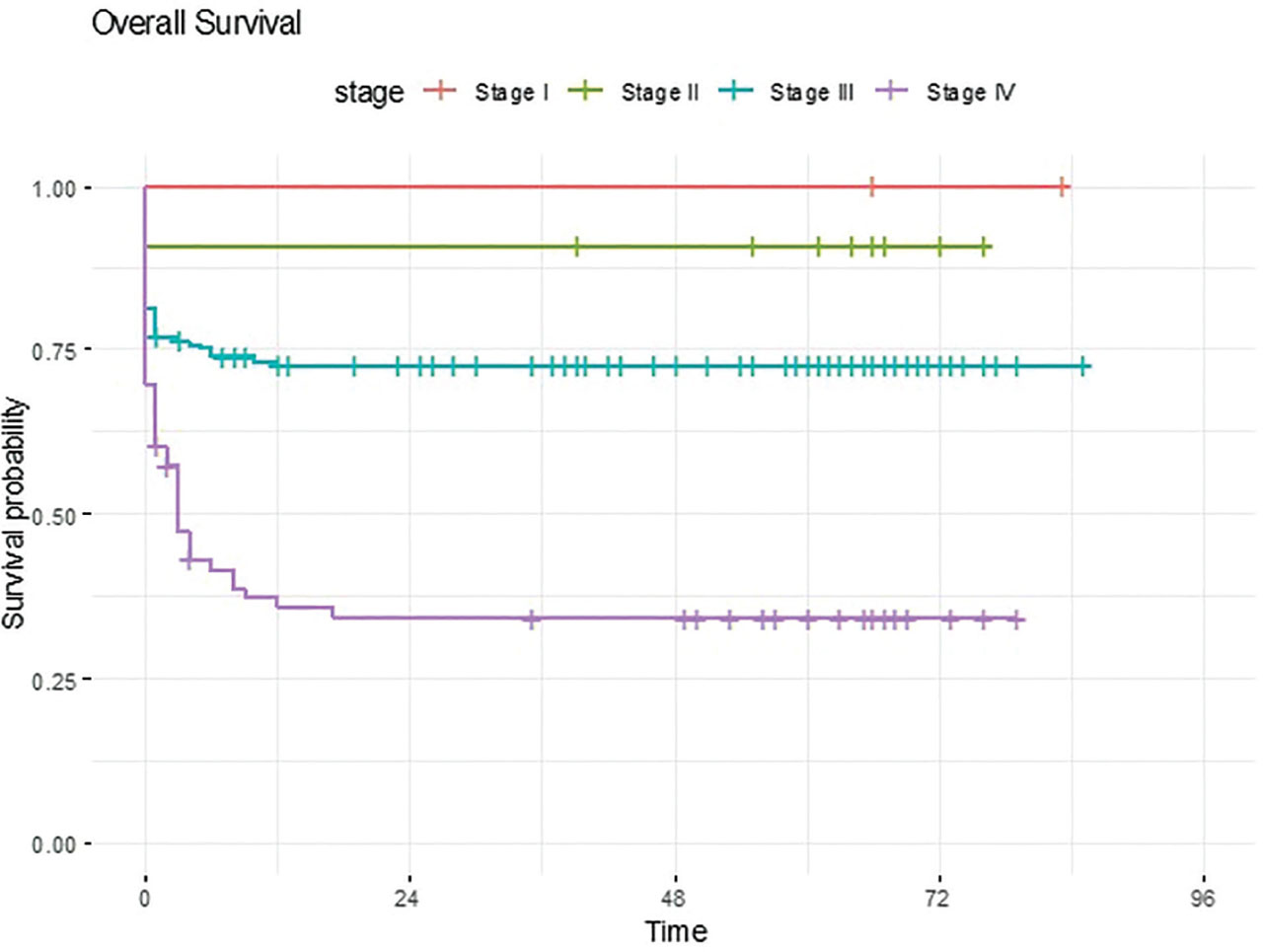
Figure 2.
Kaplan–Meier Curve of Overall Survival Stratified by Clinical Stage in Patients with B-cell Non-Hodgkin Lymphoma.
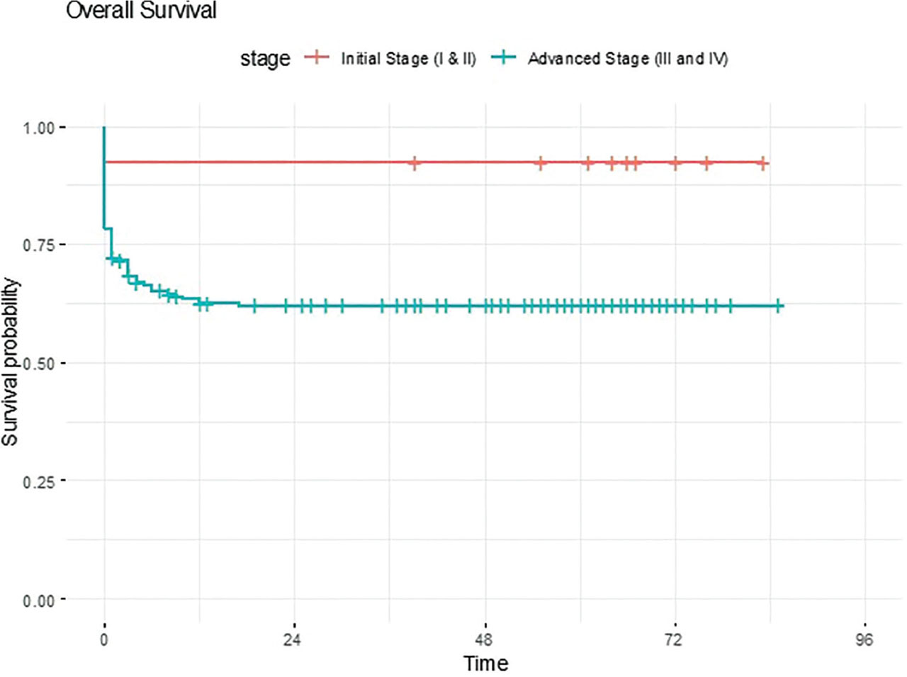
Figure 3.
Kaplan–Meier Curve of Overall Survival Stratified by Disease Burden in Patients with B-cell Non-Hodgkin Lymphoma.
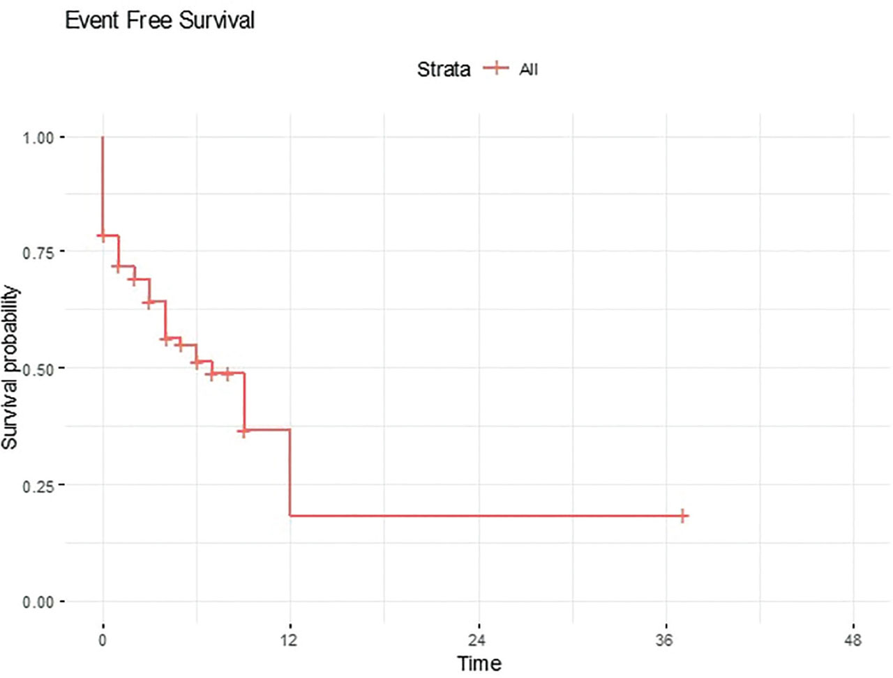
Figure 4.
Kaplan–Meier Curve of Event-Free Survival in Patients with B-cell Non-Hodgkin Lymphoma.
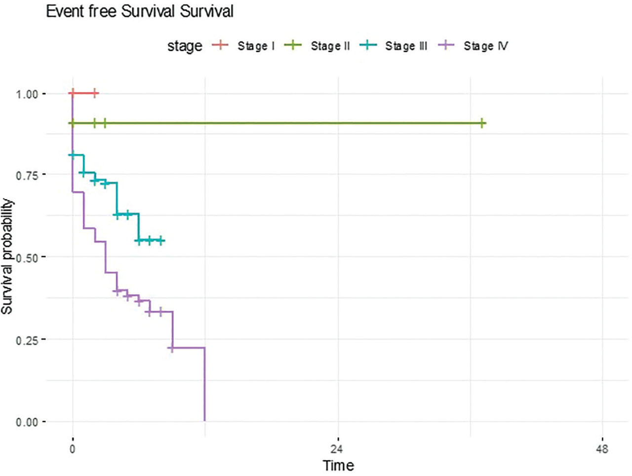
Figure 5.
Kaplan–Meier Curve of Event Free Survival Stratified by Clinical Stage in Patients with B-cell Non-Hodgkin Lymphoma
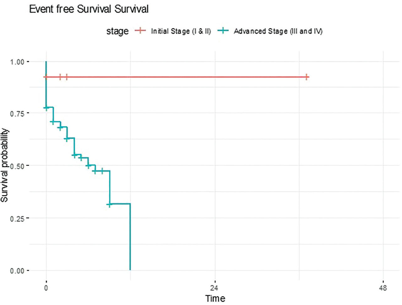
Figure 6.
Kaplan–Meier Curve of Event-Free Survival Stratified by Disease Burden in Patients with B-cell Non-Hodgkin Lymphoma.
Discussion
B-cell non-Hodgkin lymphoma is a highly curable disease in high-income countries with an intensive chemotherapy regimen based on LMB89 and BFM backbone (7). Survival outcome has been improved with the incorporation of Rituximab in the intensive chemotherapy regimen. Radhakrishnan et al. study showed that in Low- middle countries, survival outcome of B-cell non-Hodgkin lymphoma has been improved to 92% by reduction of 25% chemotherapy dose along with incorporation of Rituximab in advanced stage disease (3). However, our study showed that the long-term survival outcome is still less than 65%, and advanced stage IV disease has an OS of 38%. Due to inferior long-term survival in advanced-stage disease, institutional guidelines were made, and Rituximab was added as a frontline therapy in stage IV disease. However, only seven patients received rituximab, and the overall cohort was small; therefore, larger future studies are needed to evaluate its impact.
Our study showed that baseline LDH levels more than twice the normal value were an independent risk factor for poor prognosis. When combined with other risk factors such as tumor lysis syndrome, advanced stage, and specific disease sites, prognosis was even worse—findings consistent with numerous other studies (8). Patients who achieved a complete response on reassessment imaging after intensive induction chemotherapy (after the first CYM cycle in Group B and the second CYVE cycle in Group C) had an overall survival rate exceeding 90%. In contrast, nearly 80% of patients with progressive disease on reassessment imaging subsequently died. A study from University College London by Green K et al. suggested that scans at this point cause clinical uncertainty, as they do not always indicate residual disease or predict relapse (9).
A study by Burkhardt et al. from Germany examined the impact of age and gender on the biology, clinical features, and treatment outcomes of B-cell non-Hodgkin lymphoma. They found that more boys than girls were diagnosed, but there was no difference in long-term prognosis between the groups. In our study, over 75% of the patients were boys, and only 23% were girls; however, girls had a 4.0% higher hazard ratio compared to boys. The hazard ratio indicated that patients who underwent biopsy as a diagnostic procedure had a worse prognosis, possibly due to a higher disease burden. This finding was contrary to the study conducted by Vural et al. (11). This disparity highlights the importance of early monitoring of tumor lysis profiles and timely multidisciplinary intervention, especially in LMICs with a high incidence of B-cell non-Hodgkin lymphoma. Management of tumor lysis syndrome should include intensive care support, with pediatric intensivist involvement and CRRT when indicated, as also recommended in the study by Reiger et al. (12).
In LMICs, critical challenges include delays in diagnosis and referral to pediatric oncology centers, poor nutritional status, and advanced stage at the time of diagnosis. Due to these factors, our patients were at high risk for acute complications, serious infections, and sepsis (13). Sepsis was the leading cause of death in our study. Contributing factors included late presentation to the emergency department by families and the serious problem of resistant microorganisms.
One likely reason is poor tolerance to chemotherapy due to severe malnutrition, which results in prolonged and profound neutropenia and an inability to clear infections. Based on these results, there is a need to modify chemotherapy protocols with appropriate dose reductions to help prevent acute mortality and deaths from serious, life-threatening infections.
The European Intergroup and Children’s Oncology Group study (Inter-B-NHL Ritux 2010) demonstrated that adding rituximab to the LMB chemotherapy backbone for patients with high-risk B-cell non-Hodgkin lymphoma (stage III with elevated LDH or stage IV) improved event-free survival (EFS) from 82.3% to 93.9% (14).
The primary limitations of this study are its single-center, retrospective, descriptive design, which evaluates 5-year long-term survival outcomes in relation to various risk factors. Our center is a charitable tertiary cancer care hospital that treated a limited number of patients due to resource constraints. Although multiple risk factors influencing survival were identified, many of these could potentially be modified to improve outcomes in patients presenting with advanced-stage disease.
Our study emphasizes that prognosis for B-cell non-Hodgkin lymphoma in LMICs is significantly inferior to that in developed countries, primarily due to high disease burden at initial presentation. The development of tumor lysis syndrome further contributed to severe complications, with sepsis identified as the predominant cause of mortality in our patient population.