
Introduction
Endometrial carcinoma (EC) is one of the most common gynecologic malignancies worldwide, and its incidence continues to rise across both high-income and low- and middle-income countries (LMICs). According to GLOBOCAN 2022, cancers of the corpus uteri contribute substantially to the global cancer burden, reflecting demographic aging, increasing obesity, and metabolic risk factors [1]. In Pakistan, GLOBOCAN 2022 reported 185,748 new cancer cases and 118,631 cancer-related deaths, with uterine cancer ranking among the leading malignancies among women [2].
Although EC is frequently diagnosed at an early stage due to abnormal uterine bleeding, substantial geographic variation exists in disease biology, access to care, and outcomes [3,4,5]. In South and Southeast Asia, institutional data increasingly reflect an epidemiologic transition driven by rising obesity, diabetes, and increased postmenopausal longevity, all of which are established risk factors for EC [6,7,8].
Long-term Asian institutional experiences also suggest evolving histopathological patterns. A Japanese population-based series documented an increasing proportion of non-endometrioid histologies, including serous and mixed subtypes, which are associated with poorer outcomes [9]. Similarly, studies from Southeast Asia report a high prevalence of metabolic comorbidities and a significant proportion of intermediate- and high-risk disease among patients with early-stage EC [10].
Published data from Pakistan remain limited but consistently indicate a younger age at diagnosis compared with Western populations, under-recognition of precursor lesions, and wide variability in access to specialized oncologic care [11,12,13,14]. Reported mean ages at diagnosis range from 50 to 55 years, nearly a decade younger than those of Western counterparts. Although most patients present with FIGO stage I disease, a clinically relevant proportion demonstrate high-risk features such as deep myometrial invasion or high tumor grade [12,13,14,15]. Survival outcomes vary widely and are influenced by socioeconomic barriers, treatment completion, and availability of radiotherapy and brachytherapy services [13,14,15,16].
Randomized trials including GOG-99 and the PORTEC series have identified key prognostic factors—age, tumor grade, depth of myometrial invasion, lymphovascular space invasion (LVSI), and stage—that form the basis of contemporary risk-adapted adjuvant treatment strategies [17,18,19,20]. However, implementing these recommendations in LMICs such as Pakistan is challenging due to constraints in diagnostic capacity, surgical staging, and radiotherapy infrastructure.
Given the rising burden of EC in Pakistan and the paucity of large institutional datasets, there is a need for updated local evidence. This study presents a 10-year experience from a tertiary oncology center in Pakistan, describing patient demographics, tumor characteristics, treatment patterns, and survival outcomes to inform regional practice and align care with international standards.
Materials and Methods
This retrospective cohort study was conducted at Shaukat Khanum Memorial Cancer Hospital and Research Centre, Pakistan. Women aged ≥18 years with histologically confirmed endometrial carcinoma treated between January 2010 and December 2020 were included, while patients with incomplete records were excluded; however, this may introduce selection bias, which is an inherent limitation of retrospective studies. The study was approved by the Hospital’s Institutional Review Board (IRB exemption number: EX-17-12-25-01). Informed consent was waived due to the retrospective design, and patient data were anonymized.
Clinical, pathological, and treatment data were extracted from electronic medical records and the institutional tumor registry. Collected variables included age, body mass index, ECOG performance status, menopausal status, comorbidities, histologic subtype, tumor grade, depth of myometrial invasion, cervical stromal involvement, lymphovascular space invasion, lymph node status, and FIGO 2009 stage [21]. Treatment details included surgical approach, extent of staging, and use of adjuvant chemotherapy, external-beam radiotherapy, or vaginal brachytherapy. Chemotherapy was predominantly administered as platinum-based doublet regimens (carboplatin and paclitaxel) for 4–6 cycles in the adjuvant or concurrent setting, according to institutional protocols and risk stratification.
Radiotherapy was delivered either as external beam radiotherapy (EBRT) to the pelvis using conventional or intensity-modulated radiotherapy (IMRT) techniques with standard fractionation (typically 45–50.4 Gy in 25–28 fractions), or as vaginal brachytherapy using high-dose-rate (HDR) technique delivered in 3–5 fractions, depending on pathological risk factors and multidisciplinary tumor board recommendations. Treatment selection was individualized based on FIGO stage, histopathological risk factors, and multidisciplinary tumor board discussion. Non-endometrioid histologies and Uterine sarcomas were excluded from the primary survival analysis. Outcomes assessed were disease recurrence, disease-free survival, and overall survival.
All histopathology was centrally reviewed, and staging investigations included contrast-enhanced CT and pelvic MRI, with PET-CT performed when clinically indicated. Tumors were staged according to FIGO 2009 criteria [21], and management decisions were made through multidisciplinary tumor board review. Primary treatment consisted predominantly of total abdominal hysterectomy with bilateral salpingo-oophorectomy, with adjuvant therapy administered according to institutional protocols.
Patients were followed every 3 months during the first year, every 6 months during years 2–3, and annually thereafter. Recurrence was confirmed by clinical, radiological, or histopathological evidence.
Statistical Analysis
Survival outcomes were defined as follows: overall survival (OS) was calculated from the date of diagnosis to death from any cause or last follow-up, while disease-free survival (DFS) was defined as the time from completion of primary treatment to the first documented recurrence or death. Survival curves were estimated using the Kaplan–Meier method. Cox proportional hazards regression was used to evaluate factors associated with overall survival (OS). Variables with p < 0.10 on univariate analysis were considered for inclusion in the multivariate model. Clinically relevant variables, including age, were included a priori irrespective of univariate significance.
Continuous variables, including age and body mass index (BMI), were analyzed as continuous predictors to preserve statistical power and avoid arbitrary categorization. Treatment-related variables were not included in multivariate analysis to minimize confounding by indication, as patients with higher-risk disease were more likely to receive adjuvant therapy.
A complete-case analysis approach was used, and variables with substantial missing data were excluded from regression models. The proportional hazards assumption was assessed using Schoenfeld residuals. A two-sided p-value <0.05 was considered statistically significant. Statistical analyses were performed using IBM SPSS Statistics version 27.0.
Results
Patient Characteristics
A total of 622 patients with endometrioid endometrial carcinoma were included after exclusion of non-endometrioid histologies and incomplete records. The median age was 56 years (range 24–87), with 36.3% aged ≥60 years. The mean BMI was 32.8 kg/m2, reflecting a high prevalence of obesity.
Most patients presented with early-stage disease, with FIGO stage I accounting for 71.4% of cases. Stage II, III, and IV disease were observed in 10.9%, 14.2%, and 3.5% of patients, respectively. Deep myometrial invasion (≥50%) was present in 47.7% of cases, and nodal involvement was identified in 5.8%.
The majority of patients underwent primary surgical management (94.4% total abdominal hysterectomy with bilateral salpingo-oophorectomy). Adjuvant therapies were commonly administered, including external beam radiotherapy (57.2%), brachytherapy (60.6%), and chemotherapy (83.6%) (Table 1).
Table 1.
Baseline Clinicopathological Characteristics of Patients with Endometrioid Endometrial Carcinoma (n = 622).
| Characteristic | n (%) |
|---|---|
| Age | |
| <60 years | 396 (63.7) |
| ≥60 years | 226 (36.3) |
| Median age (years) | 56 (range 24–87) |
| Body Mass Index (BMI) | |
| Mean BMI (kg/m2) | 32.8 ± 7.3 |
| Underweight | 7 (1.1) |
| Normal weight | 64 (10.3) |
| Overweight | 143 (23.0) |
| Obesity class I | 184 (29.6) |
| Obesity class II | 135 (21.7) |
| Obesity class III | 89 (14.3) |
| Menopausal status | |
| Pre-menopausal | 169 (27.2) |
| Post-menopausal | 453 (72.8) |
| FIGO Stage (2009) | |
| IA | 275 (44.2) |
| IB | 169 (27.2) |
| II | 68 (10.9) |
| IIIA | 38 (6.1) |
| IIIB | 14 (2.3) |
| IIIC | 36 (5.8) |
| IV | 22 (3.5) |
| Myometrial invasion | |
| <50% | 325 (52.3) |
| ≥50% | 297 (47.7) |
| LVSI | |
| Negative | 316 (50.8) |
| Focal | 25 (4.0) |
| Diffuse | 27 (4.3) |
| Unknown/Missing | 253 (40.7) |
| ER status | |
| Positive | 135 (92.5) |
| Negative | 11 (7.5) |
| Unknown/Missing | 476 (76.5 of total cohort) |
Adjuvant therapies were frequently administered, including external beam radiotherapy, brachytherapy, and chemotherapy, in accordance with institutional protocols. Treatment patterns and additional clinical characteristics are summarized in Table 2.
Table 2.
Treatment Characteristics.
| Characteristic | n (%) |
|---|---|
| Surgical Procedure | |
| TAH + BSO | 587 (94.4) |
| Subtotal hysterectomy | 9 (1.4) |
| No surgery | 26 (4.2) |
| Lymph Node Dissection | |
| Yes | 113 (18.2) |
| No | 509 (81.8) |
| Nodal Status | |
| Negative | 586 (94.2) |
| Positive | 36 (5.8) |
| Adjuvant Radiotherapy (EBRT) | |
| Yes | 356 (57.2) |
| No | 266 (42.8) |
| Brachytherapy | |
| Yes | 377 (60.6) |
| No | 245 (39.4) |
| Concurrent Chemotherapy | |
| Yes | 595 (95.7) |
| No | 27 (4.3) |
| Induction/Adjuvant Chemotherapy | |
| Yes | 520 (83.6) |
| No | 102 (16.4) |
| Outcome | |
| Alive | 561 (90.2) |
| Dead | 61 (9.8) |
| Relapse Status | |
| No | 510 (82.0) |
| Yes | 112 (18.0) |
| Treatment at Relapse | |
| Chemotherapy | 60 (9.6) |
| Radiotherapy | 18 (2.9) |
| Surgery | 11 (1.8) |
| Supportive care | 17 (2.7) |
| Not applicable | 514 (82.6) |
Survival Outcomes
The estimated 5-year overall survival (OS) was 88.6% (95% CI: 85.6–91.7%), while the 5-year progression-free survival (PFS) was 80.1% (95% CI: 76.5–83.9%).
Five-year OS varied by FIGO stage:
Stage IA 93.1% (95% CI: 89.7–96.7)
Stage IB 92.4% (95% CI: 87.7–97.3)
Stage II 80.5% (95% CI: 68.8–94.1)
Stage IIIA 92.9% (95% CI: 83.8–100.0)
Stage IIIB 81.5% (95% CI: 61.1–100.0)
Stage IIIC 70.3% (95% CI: 55.4–89.3)
Stage IV 53.1% (95% CI: 32.1–87.7).
Survival varied significantly by FIGO stage (Figure 1). Stage IA demonstrated the highest 5-year OS (93.1%), while stage IV had the poorest survival (53.1%). Stage-specific survival curves are shown in Figure 1, while the stage III subgroup analysis is shown in Figure 2. Survival according to nodal status is presented in Figure 3. Estimates for stage III substages and stage IV should be interpreted with caution due to small sample sizes and wide confidence intervals. Kaplan–Meier survival curves are shown in Figures 1–3.
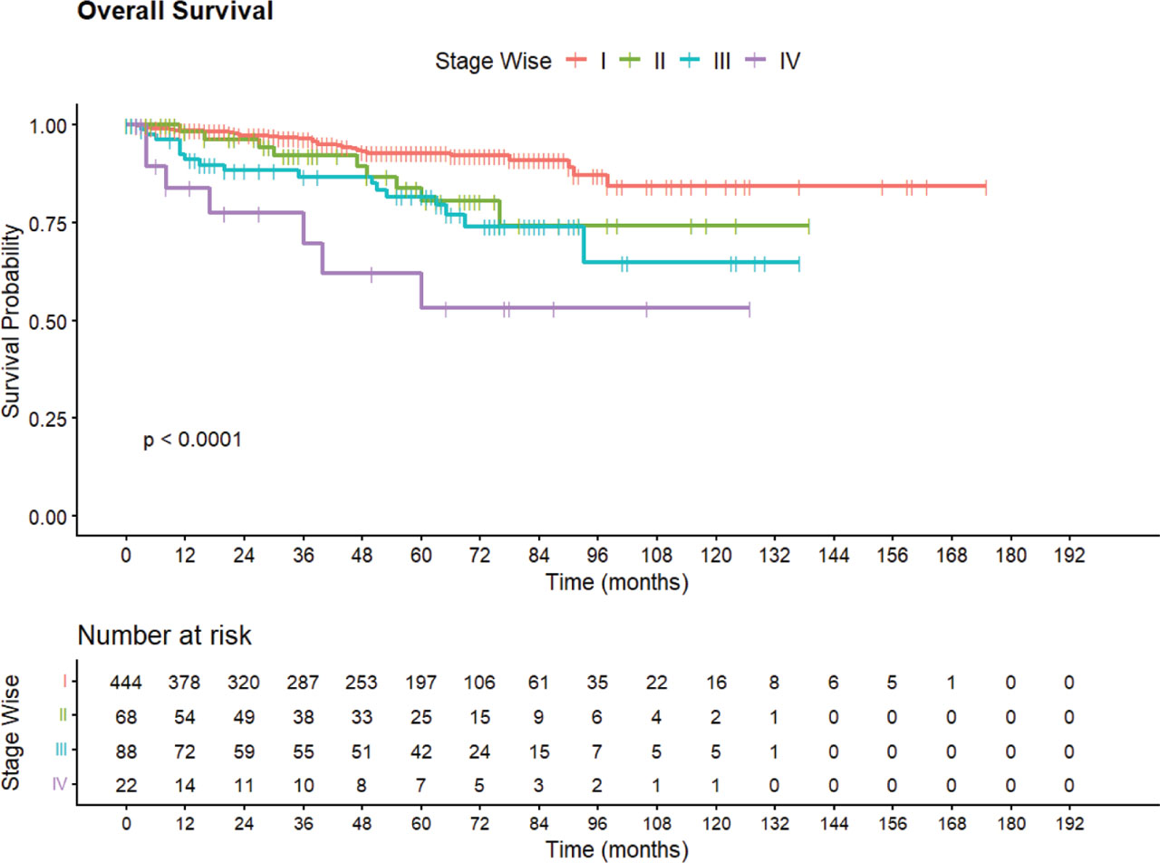
Figure 1.
Overall survival stratified by FIGO stage.
Kaplan–Meier curves illustrating overall survival (OS) according to FIGO stage (I–IV). Survival outcomes differed significantly across stages (log-rank p < 0.0001), with progressively worse survival observed with advancing stage. Patients with stage I disease demonstrated the most favorable survival, whereas stage IV disease was associated with the poorest outcomes. Tick marks represent censored observations, and numbers at risk at prespecified time intervals are displayed below the x-axis.
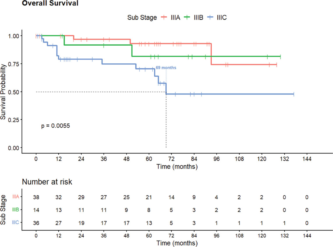
Figure 2.
Overall survival stratified by FIGO stage III subgroups (IIIA, IIIB, IIIC).
Kaplan–Meier curves demonstrating overall survival among patients with stage III endometrioid endometrial carcinoma, subclassified into stage IIIA, IIIB, and IIIC. Stage IIIC shows comparatively poorer survival outcomes, with an estimated median overall survival of approximately 69 months. Survival differences between subgroups were statistically significant (log-rank p = 0.0055). Tick marks indicate censored observations. The number of patients at risk at specified time intervals is displayed below the plot.
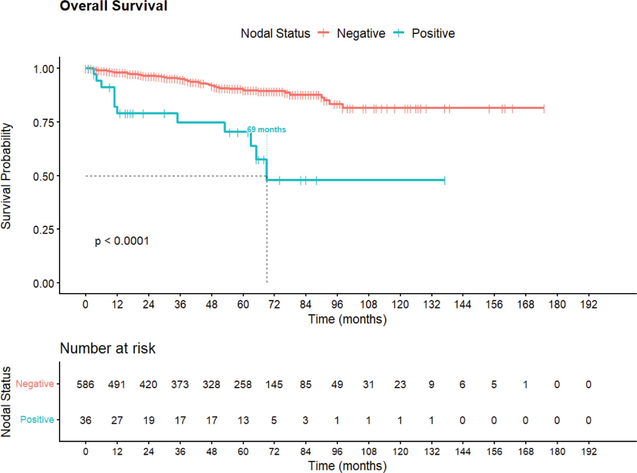
Figure 3.
Overall survival stratified by nodal status.
Kaplan–Meier curves demonstrating overall survival (OS) according to nodal status in patients with endometrioid endometrial carcinoma. Patients with node-positive disease exhibited significantly inferior survival compared with node-negative patients (log-rank p < 0.0001). The median OS for the node-positive cohort was approximately 69 months, while median OS was not reached in the node-negative group. Tick marks indicate censored observations. Numbers at risk at prespecified time intervals are provided below the x-axis.
Univariate Analysis
On univariate Cox regression analysis, advanced FIGO stage, nodal involvement, and higher tumor grade were significantly associated with poorer overall survival (Table 3). Compared with stage I disease, stage IV demonstrated the highest risk of mortality (HR 6.69, 95% CI 2.92–15.3, p < 0.001).
Table 3.
Univariate Cox Regression Analysis for Overall Survival.
| Variable | HR (95% CI) | p-value |
|---|---|---|
| FIGO Stage | <0.001 | |
| Stage I | Reference | |
| Stage II | 2.26 (1.07–4.79) | |
| Stage III | 3.08 (1.68–5.63) | |
| Stage IV | 6.69 (2.92–15.3) | |
| Nodal Status | <0.001 | |
| Negative | Reference | |
| Positive | 4.98 (2.64–9.40) | |
| Tumor Grade | 0.072 | |
| Grade I | Reference | |
| Grade II | 1.09 (0.62–1.90) | |
| Grade III | 2.40 (1.17–4.93) | |
| Myometrial Invasion | 0.101 | |
| <50% | Reference | |
| ≥50% | 1.53 (0.92–2.54) | |
| Age (per year) | 1.02 (0.99–1.04) | 0.140 |
| BMI (per unit) | 0.94 (0.90–0.97) | <0.001 |
| Menopausal Status | 0.124 | |
| Pre-menopausal | Reference | |
| Post-menopausal | 1.56 (0.87–2.79) |
Positive nodal status was strongly associated with worse survival (HR 4.98, 95% CI 2.64–9.40, p < 0.001). Tumor grade III was associated with increased mortality compared with grade I (HR 2.40, 95% CI 1.17–4.93, p = 0.018).
BMI demonstrated a significant inverse association with mortality (HR 0.94 per unit increase, 95% CI 0.90–0.97, p < 0.001). Age showed a non-significant trend toward worse survival (HR 1.02 per year, 95% CI 0.99–1.04, p = 0.14).
Treatment-related variables, including radiotherapy and surgery, appeared associated with improved survival on univariate analysis; however, these findings likely reflect treatment-selection bias.
Multivariate Analysis
On multivariate Cox regression analysis, advanced stage, nodal involvement, and increasing age were independently associated with poorer overall survival (Table 4).
Table 4.
Multivariate Cox Regression Analysis for Overall Survival.
| Variable | HR (95% CI) | p-value |
|---|---|---|
| FIGO Stage | ||
| Stage I | Reference | |
| Stage II | 2.46 (1.14–5.31) | 0.022 |
| Stage III | 1.14 (0.42–3.06) | 0.800 |
| Stage IV | 5.82 (2.43–13.9) | <0.001 |
| Nodal Status | ||
| Negative | Reference | |
| Positive | 5.89 (2.00–17.3) | 0.001 |
| Tumor Grade | ||
| Grade I | Reference | |
| Grade II | 0.71 (0.39–1.31) | 0.300 |
| Grade III | 1.18 (0.52–2.68) | 0.700 |
| Age (per year) | 1.03 (1.00–1.06) | 0.023 |
| BMI (per unit) | 0.95 (0.91–0.99) | 0.009 |
| Myometrial Invasion | ||
| <50% | Reference | |
| ≥50% | 1.02 (0.56–1.84) | 0.940 |
Compared with stage I disease, stage II (HR 2.46, 95% CI 1.14–5.31, p = 0.022) and stage IV disease (HR 5.82, 95% CI 2.43–13.9, p < 0.001) were significantly associated with increased mortality. Stage III disease did not retain independent significance after adjustment (HR 1.14, 95% CI 0.42–3.06, p = 0.80). Positive nodal status remained a strong independent predictor of worse survival (HR 5.89, 95% CI 2.00–17.3, p = 0.001). Increasing age was independently associated with poorer survival (HR 1.03 per year, 95% CI 1.00–1.06, p = 0.023). BMI retained a statistically significant inverse association with mortality (HR 0.95 per unit increase, 95% CI 0.91–0.99, p = 0.009).
Tumor grade and depth of myometrial invasion were not independently associated with survival after adjustment, suggesting that their prognostic effects may be mediated through stage and nodal status.
Proportional Hazards Assumption
The proportional hazards assumption was assessed using Schoenfeld residuals. No significant violation of the proportional hazards assumption was observed for the overall model (global p = 0.21).
Among individual variables, BMI demonstrated evidence of a time-varying effect (p = 0.008), while all other variables satisfied the proportional hazards assumption.
Discussion
This large institutional study provides real world experience on endometrial carcinoma (EC) from a low- to middle-income country (LMIC) setting. Major prognostic determinants largely mirror those reported in international and Asian populations, but important regional differences were noted in age at presentation, obesity burden, and histologic distribution.
Patients in this cohort were younger than typically reported in Western series (median 60–65 years) [3,5], consistent with other South and East Asian studies [6,7]. Despite the younger demographic, advancing age remained an independent predictor of poorer overall survival (OS), aligning with global evidence [22,23,24]. Obesity was highly prevalent (mean BMI >30 kg/m2), resembling Western rather than East Asian populations, reflecting the epidemiologic transition in LMICs and the contribution of metabolic risk factors to EC burden [25,26].
Adenocarcinoma was the predominant histology (72%), but clear cell carcinoma and uterine sarcomas were relatively more common than reported in Western or East Asian series, potentially reflecting referral bias, biological variation, or population-specific risk factors. Sarcomas remained independently prognostic, consistent with their aggressive behavior across geographic regions [28]. Two-thirds of patients presented with stage I disease, reflecting early detection due to abnormal uterine bleeding [3,29]. Deep myometrial invasion was common, suggesting delayed diagnosis after detection or more aggressive tumor biology. FIGO stage and nodal status were strong predictors of survival, consistent with international and Asian literature [30,31,32].
The association of adjuvant therapy with poorer outcomes in univariate analysis likely reflects treatment-selection bias, as patients with high-risk disease are more likely to receive multimodality treatment. Randomized trials support the benefit of adjuvant therapy in appropriately selected patients [19,20]. Disease relapse was among the strongest independent predictors of mortality, emphasizing the importance of early detection, complete surgical staging, and adherence to risk-adapted treatment, particularly in resource-limited settings.
The long-term outcomes observed in this cohort demonstrate favorable survival for patients with endometrial carcinoma, with a median follow-up of 49 months and a 10-year overall survival (OS) of 76.5%, with median OS not yet reached. These results are consistent with published population-based and institutional series reporting 10-year survival rates ranging from 70–85% for endometrial cancer, particularly among patients with early-stage disease [33,16]. The prolonged median disease-free survival (DFS) of 132 months further highlights that the favorable outcomes may be associated with surgical management combined with risk-adapted adjuvant therapy [5]. Lack of molecular classification is a limitation within our institution.
Significant differences in OS according to histology and FIGO stage reinforce their established prognostic significance (Figures 1C,D). Patients with endometrioid adenocarcinoma and early-stage disease demonstrated the most favorable survival, in line with the well-documented indolent biology and hormone responsiveness of this subtype [27,3]. In contrast, patients with sarcomatous histology and stage IV disease had the poorest outcomes, reflecting the aggressive clinical behavior, early hematogenous dissemination, and relative resistance to conventional therapies associated with uterine sarcomas and advanced-stage tumors [28,29]. These findings underscore the importance of accurate histopathological classification and stage-appropriate treatment planning, while highlighting the unmet need for improved systemic and targeted therapies for high-risk histologies and advanced-stage endometrial cancer [34].
The key strengths of this study include a large sample size, comprehensive clinicopathological and treatment data, and long follow-up, which allow robust survival analyses and meaningful comparisons with both international and regional cohorts. Limitations include its retrospective design, potential referral and selection bias, and reliance on existing medical records, which may have led to missing or incomplete data. Additionally, molecular profiling was not routinely available, limiting the analysis of genomic risk factors increasingly recognized as prognostic in EC. Though established prognostic factors such as age, stage, nodal status, histology, and ECOG performance are applicable in LMIC contexts, regional differences in age at diagnosis, obesity prevalence, and histologic distribution underscore the importance of context-specific strategies to improve outcomes. Prospective studies incorporating molecular profiling and standardized treatment pathways may further optimize risk stratification and survival in Pakistani women with EC.