
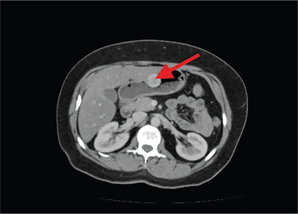
Figure 1.
Contrast-enhanced CT abdomen (arterial phase) showing a round, circumscribed, avidly enhancing mass along the greater curvature of the stomach at the junction of the gastric body and antrum, suspicious for a hypervascular submucosal neoplasm.
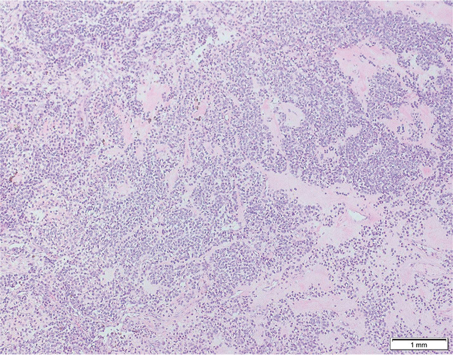
Figure 2.
Hematoxylin and eosin (H&E) stained section (10×) demonstrating neoplastic epithelioid cells with round nuclei and indistinct borders arranged around branching capillary-sized vessels lined by endothelial cells, consistent with glomangioma.
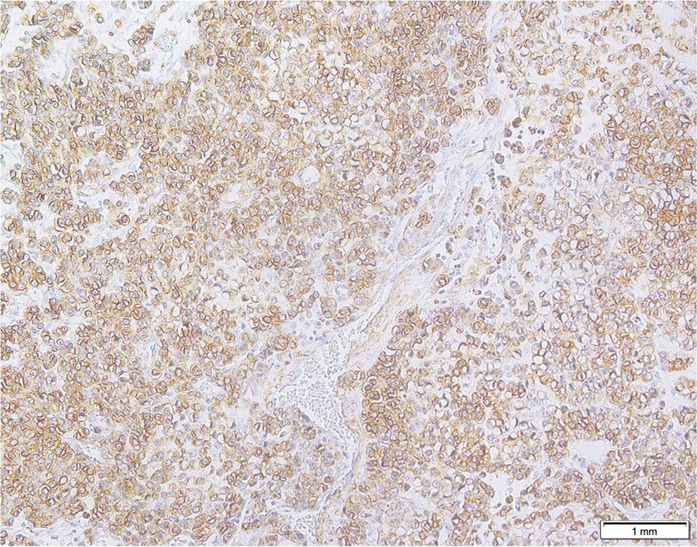
Figure 3.
Immunohistochemical staining showing diffuse cytoplasmic positivity for smooth muscle actin (SMA) in tumor cells.
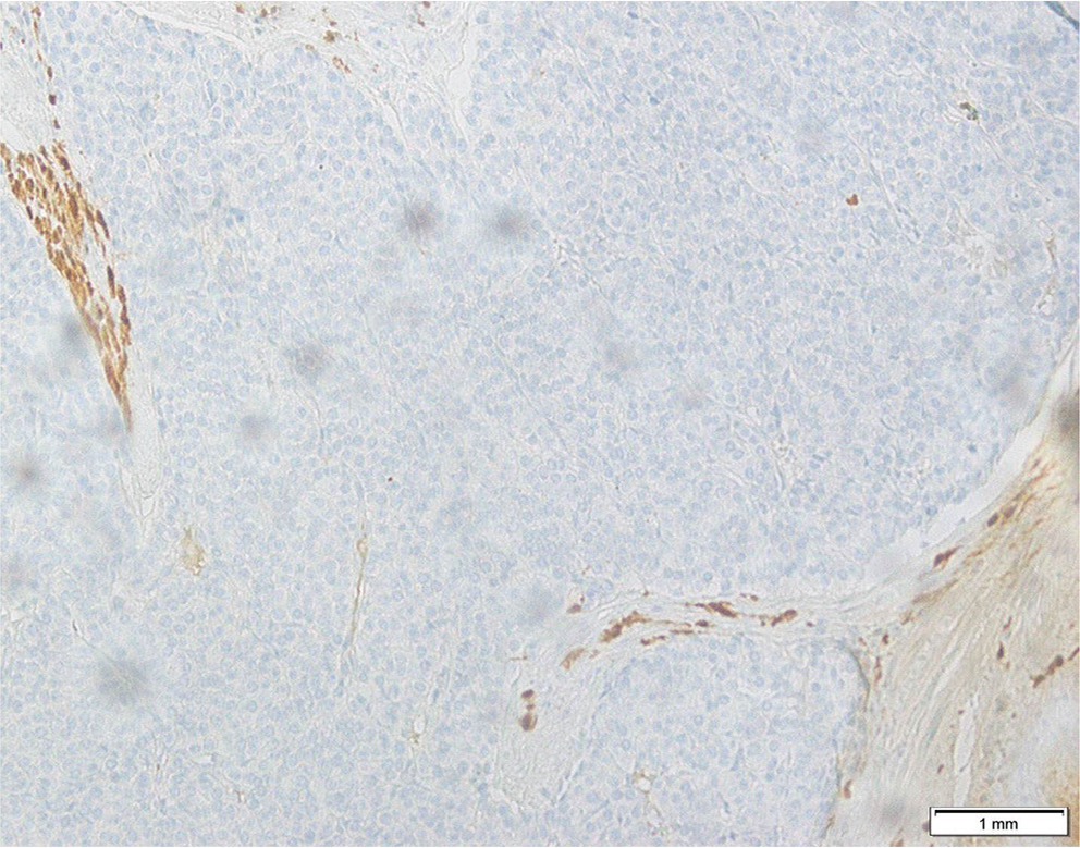
Figure 4.
S100 immunostaining negative in tumor cells, excluding neural differentiation (schwannoma).
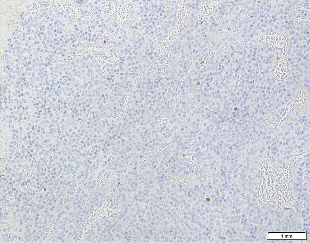
Figure 5.
Ki-67 immunostain demonstrating very low proliferative activity (~1%), consistent with benign behavior.
Table 1.
Comparative immunohistochemical profile of gastric submucosal neoplasms
| Marker | GGT | GIST | Gastric NET | Schwannoma | Leiomyoma | Hemangioma |
|---|---|---|---|---|---|---|
| CD117 (KIT) | Negative | Positive | Negative | Negative | Negative | Negative |
| DOG1 | Negative | Positive | Negative | Negative | Negative | Negative |
| SMA | Positive | Variable | Negative | Negative | Positive | Variable |
| Vimentin | Positive | Variable | Variable | Positive | Positive | Variable |
| S100 | Negative | Negative | Negative | Positive | Negative | Negative |
| Desmin | Negative | Negative | Negative | Negative | Positive | Negative |
| CD34 | Negative* | Variable | Negative | Negative | Negative | Positive |
| CD31 | Negative | Negative | Negative | Negative | Negative | Positive |
| Chromogranin | Negative | Negative | Positive | Negative | Negative | Negative |
| Synaptophysin | Variable** | Negative | Positive | Negative | Negative | Negative |
| Ki-67 | Very low (<2%) | Variable | Stratifies grade | Low | Low | Low |