
Table 1.
Summary of the blood analysis of the patient.
| Laboratory tests | Results | Reference range |
|---|---|---|
| Hemoglobin | 11.7 g/dl | 12–15 g/dl |
| Creatinine | 0.38 mg/dl | 0.50–0.90 mg/dl |
| eGFR | 212.49 mL/min/1.73 m2 | >60 mL/min/1.73 m2 |
| Calcium, corrected | 12.17 mg/dl | 8.5–10.5 mg/dl |
| 25 Hydroxy Vitamin D | 22.21 ng/ml | Insufficiency 20 - less than 30 ng/mL |
| PTH-Intact | 231.8 pg./mL | 18.5–88 pg./mL |
| Free T4 | 0.87 ng/dl | 0.89–1.76 ng/dl |
| TSH | 1.62 uIU/ml | 0.35–5.5 uIU/ml |
| GLUCOSE (FASTING) | 92 mg/dL | 70 – 99 mg/dl |
| Prolactin | 18 ng/ml | 1.90 – 25 ng/ml |
| Metanephrine, Normetanephrine | 54.71 pg/mL | Less than 90 pg/ml |
| 165.5 pg/mL | Less than 190 pg/ml | |
| CALCITONIN | 3.39 pg/mL | ND – 5.0 pg/ml |
| CEA | 0.643 ng/mL | 0.8–2.5 pg/ml |
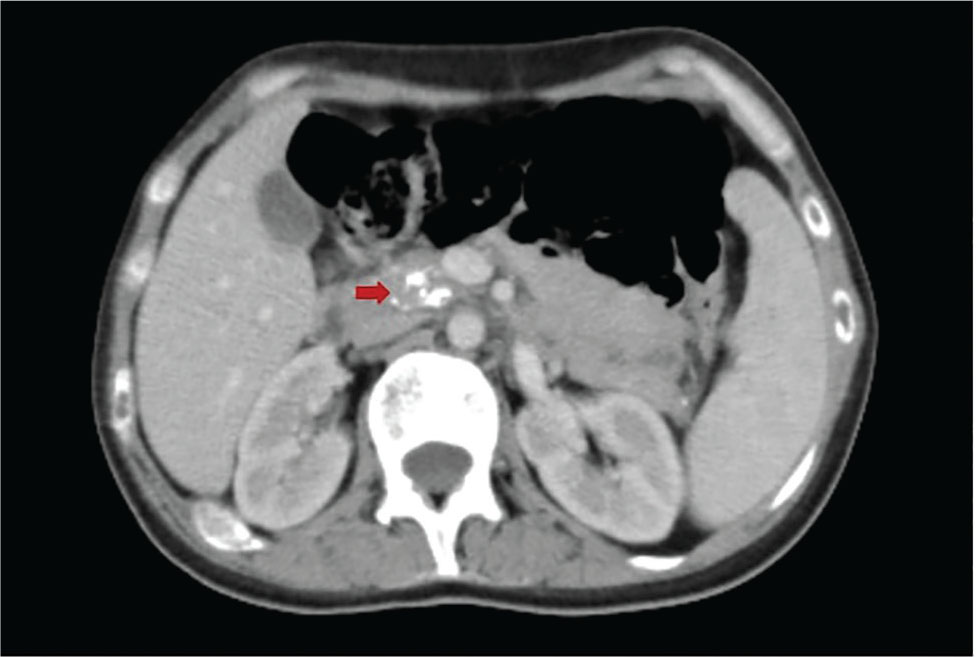
Figure 1.
Computed tomography scanning chest, abdomen, and pelvis showing pancreatic calcification (red arrow).
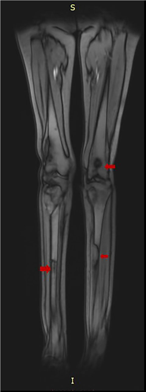
Figure 2.
Magnetic resonance imaging lower limbs showing bilateral lytic lesions (red arrows).
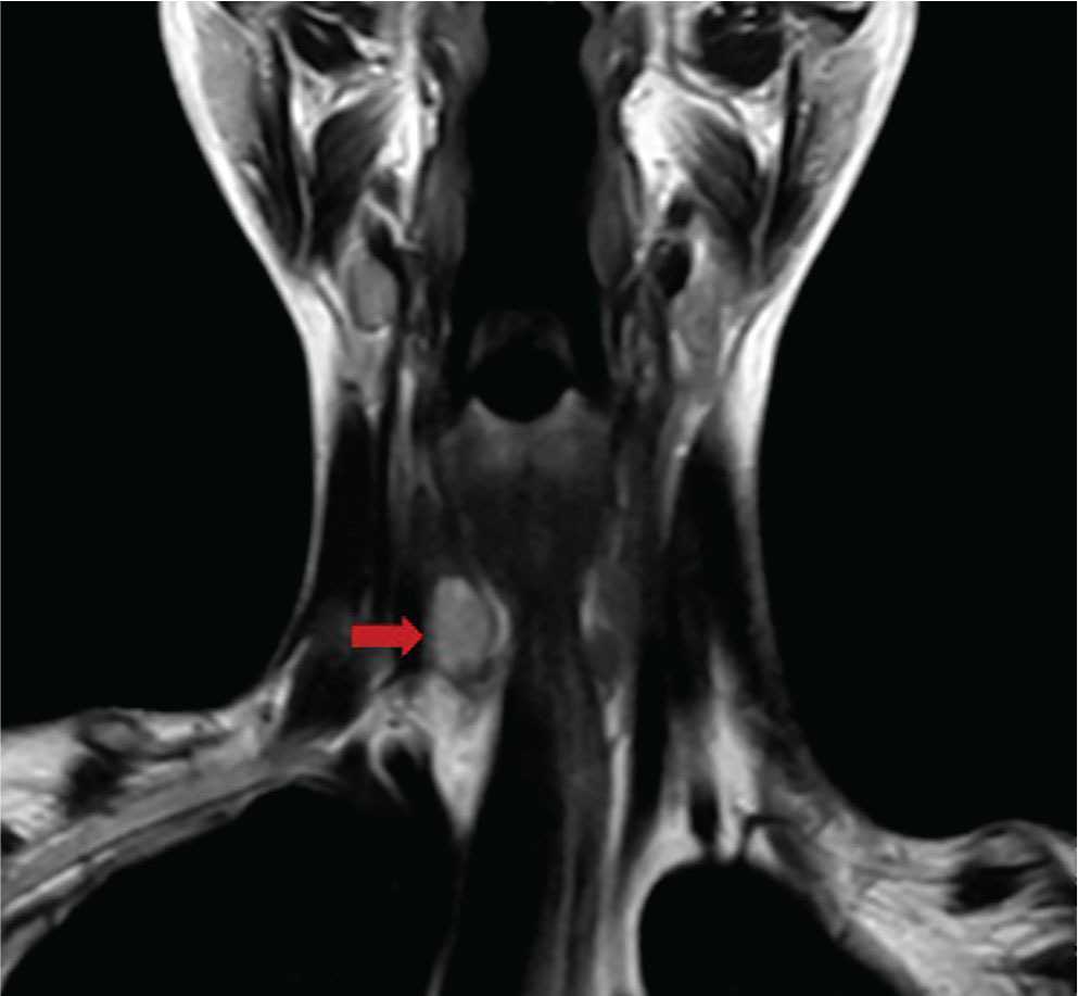
Figure 3.
Magnetic resonance imaging neck showing a mass adjacent to the right lobe of the thyroid at its inferior pole (red arrow).