
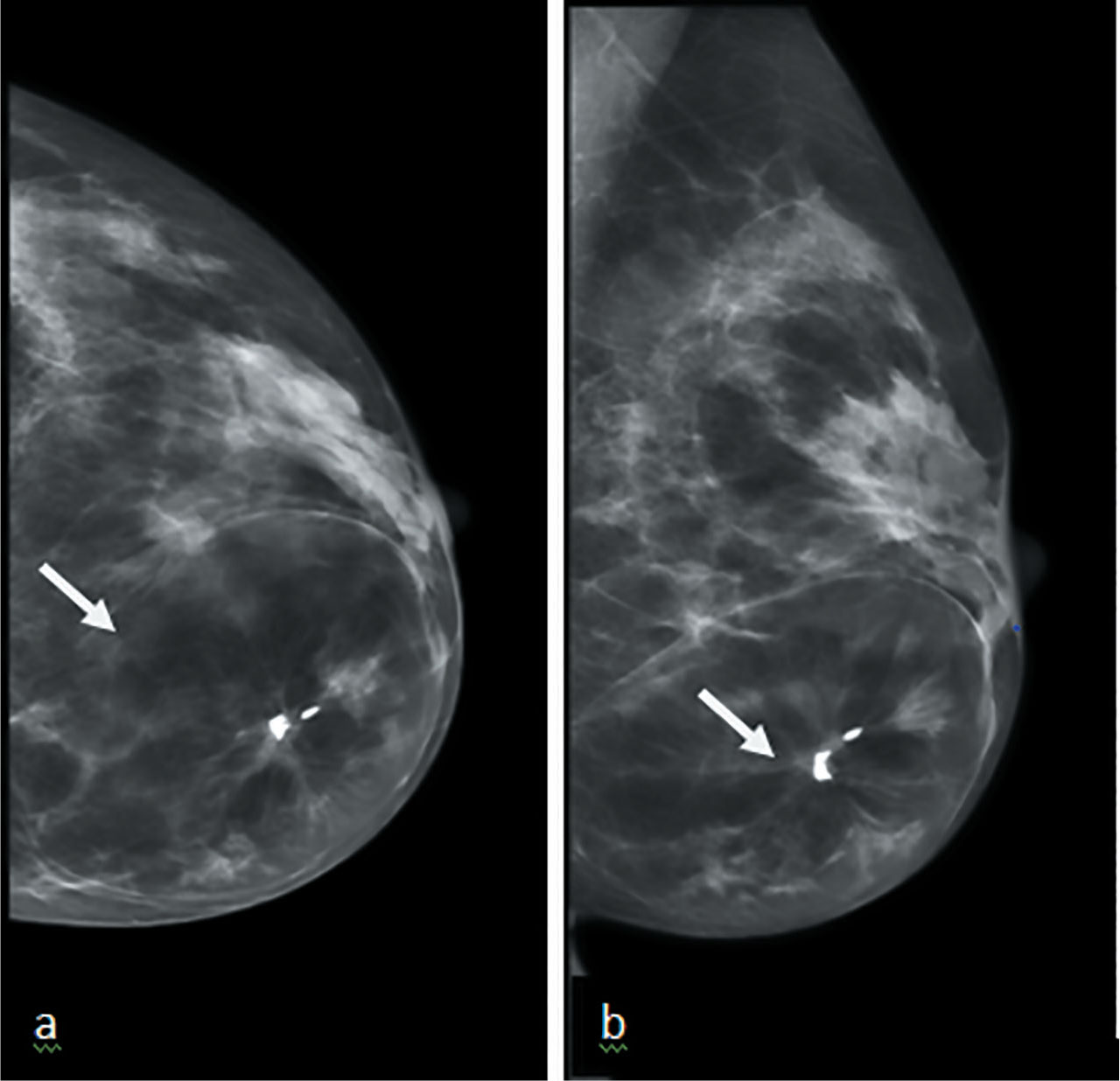
Figure 1.
(a) CC and (b) MLO views of hamartoma, seen as encapsulated heterogenous mass in the lower and inner breast with central macrocalifications and distortion (arrows).
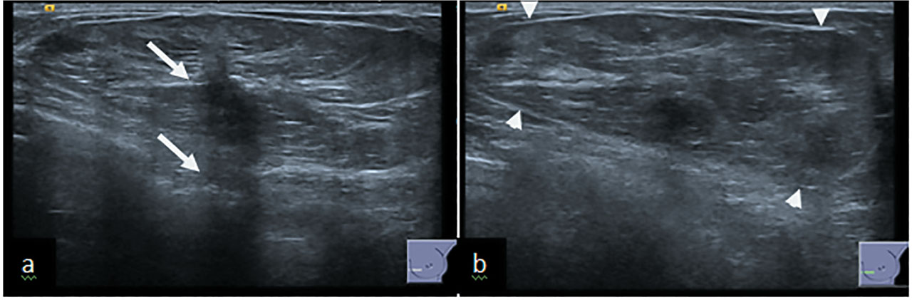
Figure 2.
(a) US of hamartoma showing a central small hypoechoic area with associated acoustic shadowing (arrow). (b) The thin echogenic pseudocapsule can also be appreciated (arrowheads).
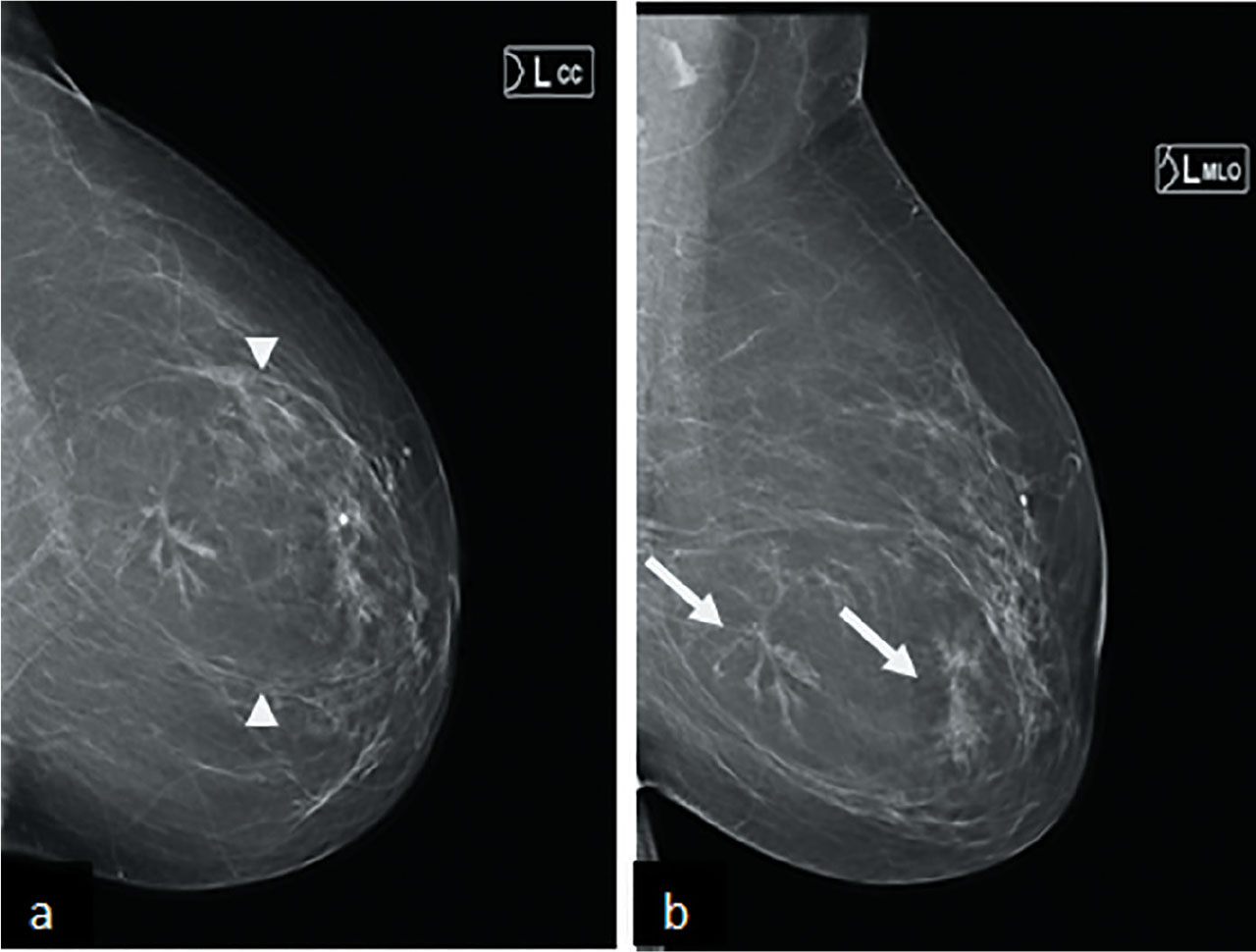
Figure 3.
(a) CC and (b) MLO views of mammogram, showing circumscribed mass with pseudocapsule (arrow heads) and central area of distorsion (arrow) in the left lower central breast.
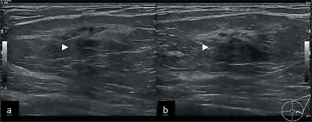
Figure 4a & b.
US shows well defined oval mass with central hypoechoic shadowing area (arrowheads) corresponding to mammographic distortion.
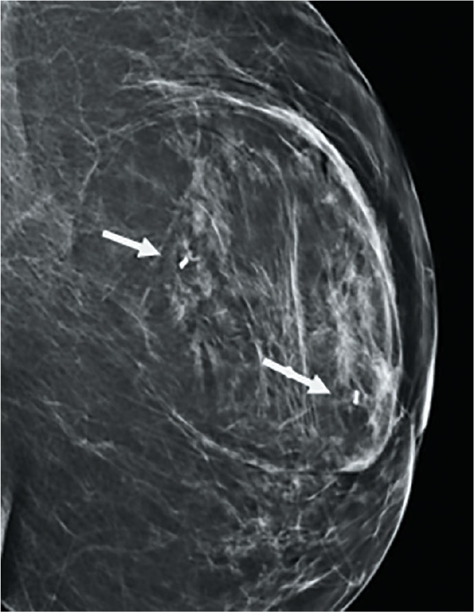
Figure 5.
Mammogram showing post VAB marker insertion (arrow).