
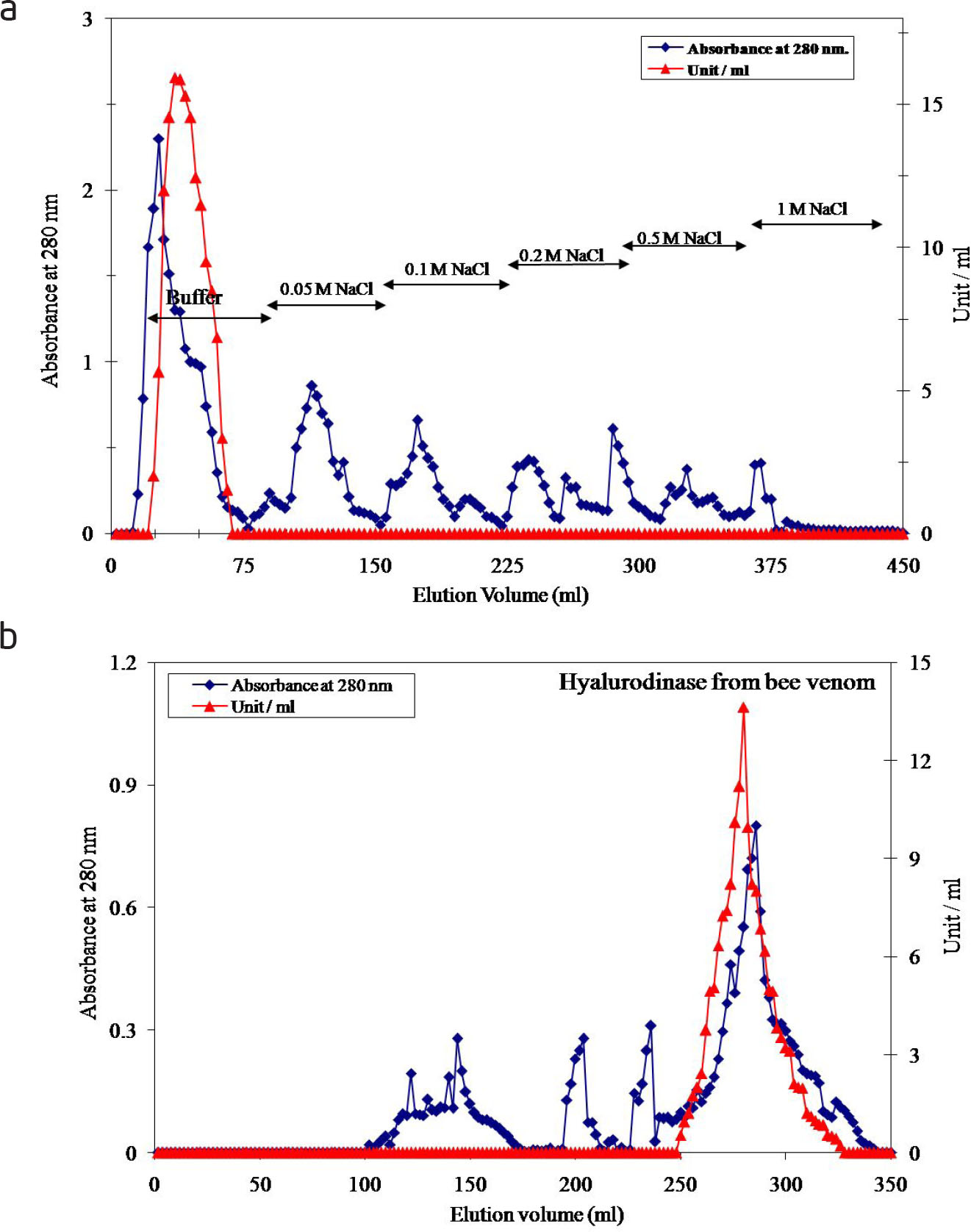
Fig. 1
(a): A typical elution profile for the chromatography of the crude honey bee venom on DEAE-cellulose column (12 cm × 2.4 cm i.d.) previously equilibrated with 0.02 M potassium phosphate buffer, pH 7.4. (b): A typical elution profile for the chromatography of the concentrated pooled DEAE-cellulose fractions containing hyaluronidase enzyme activity on Sephacryl S-300 column (142 cm × 1.75 cm i.d.) previously equilibrated with 0.02 M potassium phosphate buffer pH 7.4. The blue line indicates the protein b (Absorbance at 280 nm) present on the left vertical axis, while the red line indicates the hyaluronidase activity (Unit / ml) present on the right vertical axis.
Table 1
A typical purification scheme of honey bee venom hyaluronidase
| Purification steps | Total protein (mg) | Total Activity (unit) | Specific Activity | Yield (%) | Fold Purification |
|---|---|---|---|---|---|
| Crude honey bee venom | 27.2 | 3466.2 | 127.43 | 100 | 1.0 |
| DEAE-Cellulose | |||||
| Hyalurodinase (0 M NaCI) | 12.5 | 2932.8 | 234.62 | 84.6 | 1.84 |
| Sephacryl S-300 | |||||
| Hyalurodinase | 4.2 | 1729.1 | 411.7 | 49.9 | 3.23 |
[i] *One unit of hyaluronidase enzyme activity is identified as the amount of enzyme required to cause 50% turbidity reduction as one unit of international standard preparation. Turbidity reducing units (TRU) are expressed as the amount of enzyme required to hydrolyze 50% of the hyaluronic acid.
[ii] *Specific activity is expressed as units / mg protein.
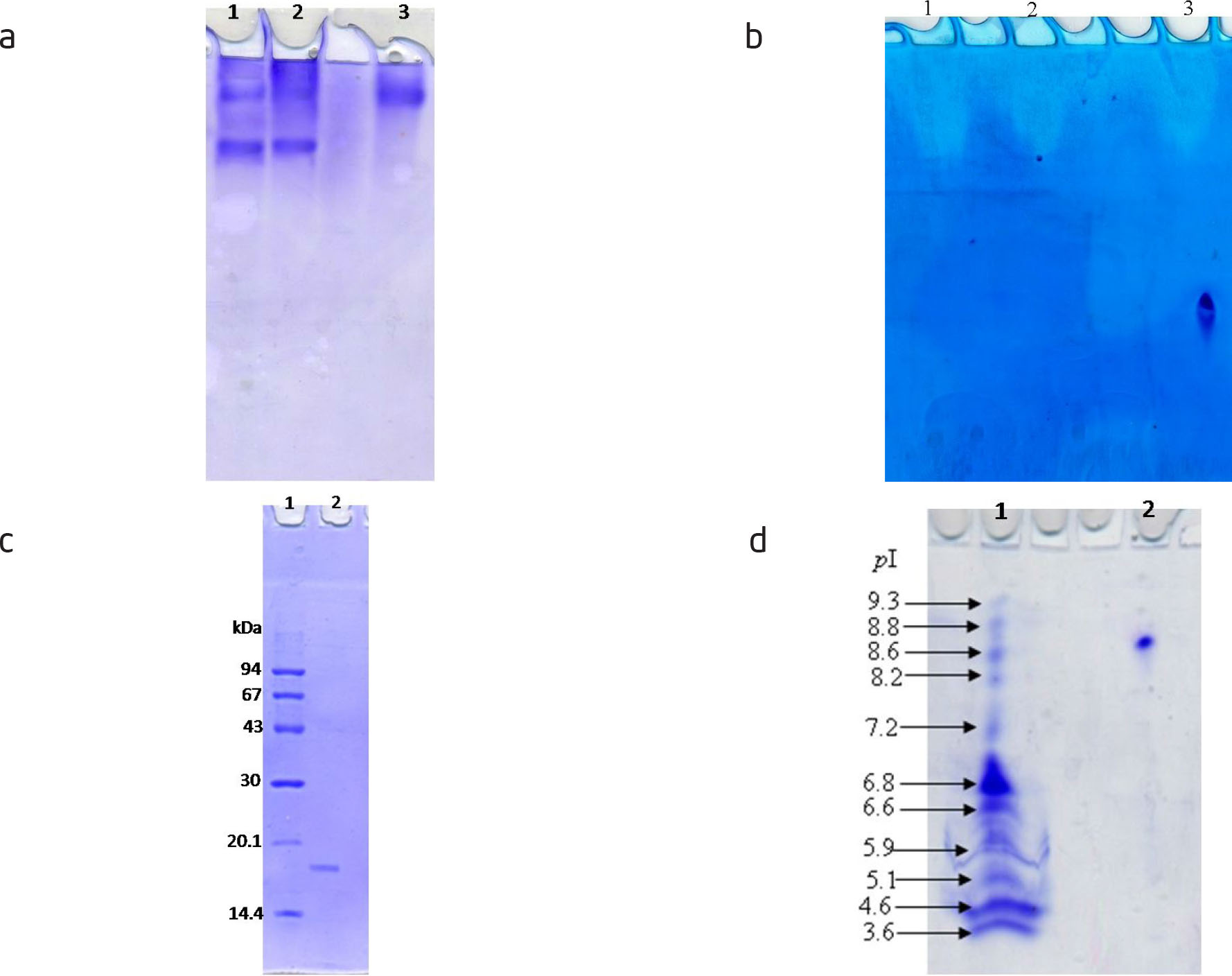
Fig. 2
(a) Electrophoretic analysis of hyaluronidase protein pattern of the different purification steps on 7% native polyacrylamide gel: (1) crude venom, (2) DEAE-cellulose fraction and (3) Sephacryl S-300 purified fraction of honey bee venom hyaluronidase. (b) Electrophoretic analysis of hyaluronidase isoenzyme pattern of the different purification steps on 7% native polyacrylamide gel: (1) crude venom, (2) DEAE-cellulose fraction and (3) Sephacryl S-300 purified fraction of honey bee venom hyaluronidase. (c): Subunit molecular weight determination by electrophoretic analysis of purified hyaluronidase on 12% SDS-polyacrylamide gel: (1) molecular weight marker proteins and (2) purified hyaluronidase. (d): Isoelectrofocusing; (1) Isoelectric point (pI) marker proteins and (2) the purified hyaluronidase.
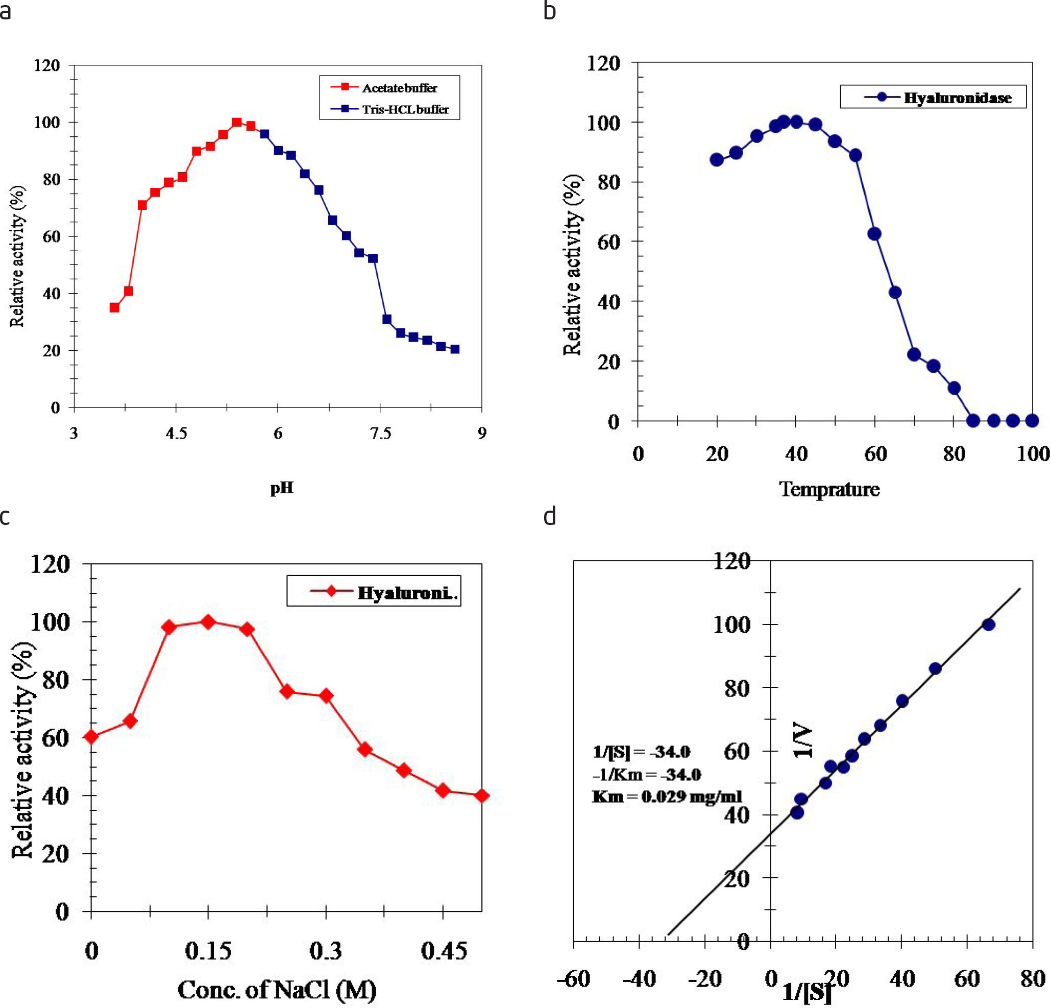
Fig. 3
(a) Effect of pH on the purified honey bee venom hyaluronidase using 0.02 M sodium acetate buffer, pH (3.2 – 5.6) and Tris-HCl buffer, pH (5.8 – 8.6). (b) Effect of temperature on the purified honey bee venom hyaluronidase. (c) Effect of NaCl concentration on the purified honey bee venom hyaluronidase. (d) Lineweaver-Burk plot relating the reciprocal of the reaction velocity of the purified hyaluronidase to hyaluronic acid concentration in mg/ml.
Table 2
Effect of divalent cations on honey bee venom hyaluronidase
| Reagent | Final Concentration (mM) | Residual activity (%) |
|---|---|---|
| Control | ----- | 100.0 |
| CaCl2 | 2.0 | 96.4 |
| 5.0 | 89.9 | |
| CoCl2 | 2.0 | 49.7 |
| 5.0 | 28.1 | |
| CuCl2 | 2.0 | 98.0 |
| 5.0 | 93.1 | |
| FeCl2 | 2.0 | 18.8 |
| 5.0 | 11.2 | |
| MgCl2 | 2.0 | 109.0 |
| 5.0 | 115.0 | |
| MnCl2 | 2.0 | 44.5 |
| 5.0 | 20.7 | |
| NiCl2 | 2.0 | 52.4 |
| 5.0 | 27.5 | |
| ZnCl2 | 2.0 | 112.0 |
| 5.0 | 118.0 |
Table 3
Effect of various inhibitors on honey bee venom hyaluronidase
| Reagent | Final Concentration (mM) | Inhibition (%) |
|---|---|---|
| Control | ----- | 0.0 |
| 1,10 Phenanthroline | 2.0 | 72.8 |
| 5.0 | 85.1 | |
| β-Mercaptoethanol | 2.0 | 37.6 |
| 5.0 | 48.4 | |
| Citric acid | 2.0 | 50.9 |
| 5.0 | 69.3 | |
| Dithiothreitol (DTT) | 2.0 | 15.6 |
| 5.0 | 23.3 | |
| EDTA | 2.0 | 52.2 |
| 5.0 | 71.8 | |
| Heparin | 5 U | 47.85 |
| 10 U | 85.73 | |
| Hydrogen peroxide (H2O2) | 2.0 | 30.2 |
| 5.0 | 43.6 | |
| Iodoacetamide | 2.0 | 21.3 |
| 5.0 | 29.1 | |
| Potassium cyanide (KCN) | 2.0 | 12.4 |
| 5.0 | 15.8 | |
| Sodium azide (NaN3) | 2.0 | 16.5 |
| 5.0 | 22.1 | |
| Phenyl methyl sulfonyl fluoride (PMSF) | 2.0 | 26.7 |
| 5.0 | 31.8 | |
| Reduced glutathione | 2.0 | 8.20 |
| 5.0 | 12.4 | |
| Sodium dodecyl sulphate | 2.0 | 6.20 |
| 5.0 | 9.60 | |
| Urea | 2.0 | 78.3 |
| 5.0 | 89.9 |
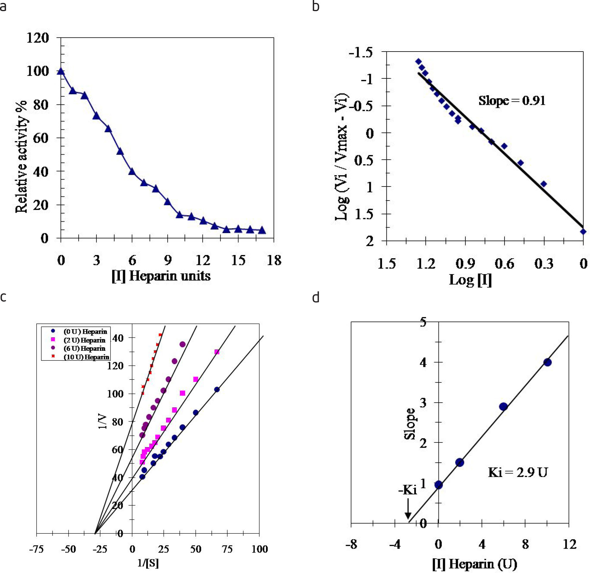
Fig. 4
(a) Inhibition of the purified honey bee venom hyaluronidase by varying concentrations of Heparin. (b) Hill plot for inhibition of the purified hyaluronidase by varying concentrations of Heparin where Vmax is the enzyme activity in absence of inhibitor, Vi is the enzyme activity in presence of inhibitor and [I] is inhibitor concentration in units of Heparin. (c) Lineweaver-Burk plots showing the type of inhibition of the purified honey bee venom hyaluronidase by Heparin. The activity of the purified hyaluronidase was measured with varying concentrations of the substrate hyaluronic acid in absence and presence of three various concentrations of Heparin. (d) Determination of the inhibition constant (Ki) value for the inhibition of the purified hyaluronic acid by Heparin. The plotted slope values were determined from the lines of the reciprocal plots of the different inhibitor concentrations.