
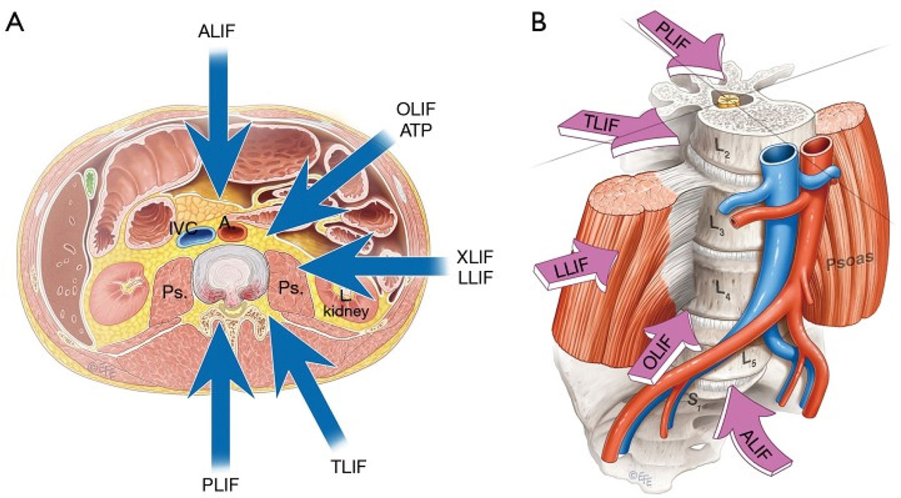
Figure 1:
A. Cross-sectional view, B. Normal view. LIF surgery types and their relevant access points [1]. LIF, lumbar interbody fusion.
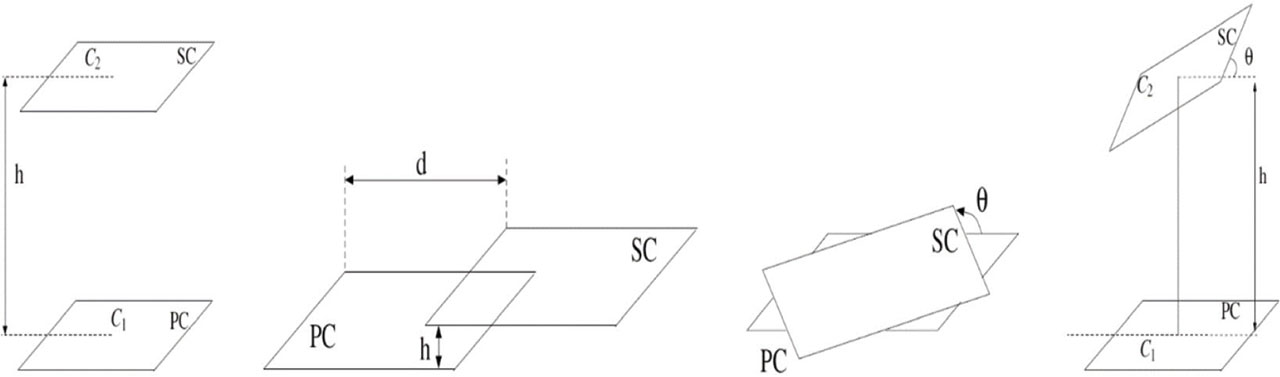
Figure 2:
Vertical, horizontal, planar, and angular misalignment [113].
Table 1:
EFM8BB52 microcontroller component current requirements [94].
| Parameter | Symbol | Typ | Unit |
|---|---|---|---|
| Normal mode at 80 kHz using low-frequency oscillator | INORMAL | 612 | µA |
| ADC supply current at full speed with 12-bit conversions | IADC | 250 | µA |
| External voltage reference input current | IEXTVREF | 5 | µA |
| Current for VDD divider when VDD used as VREF | IVDDRED | 16 | µA |
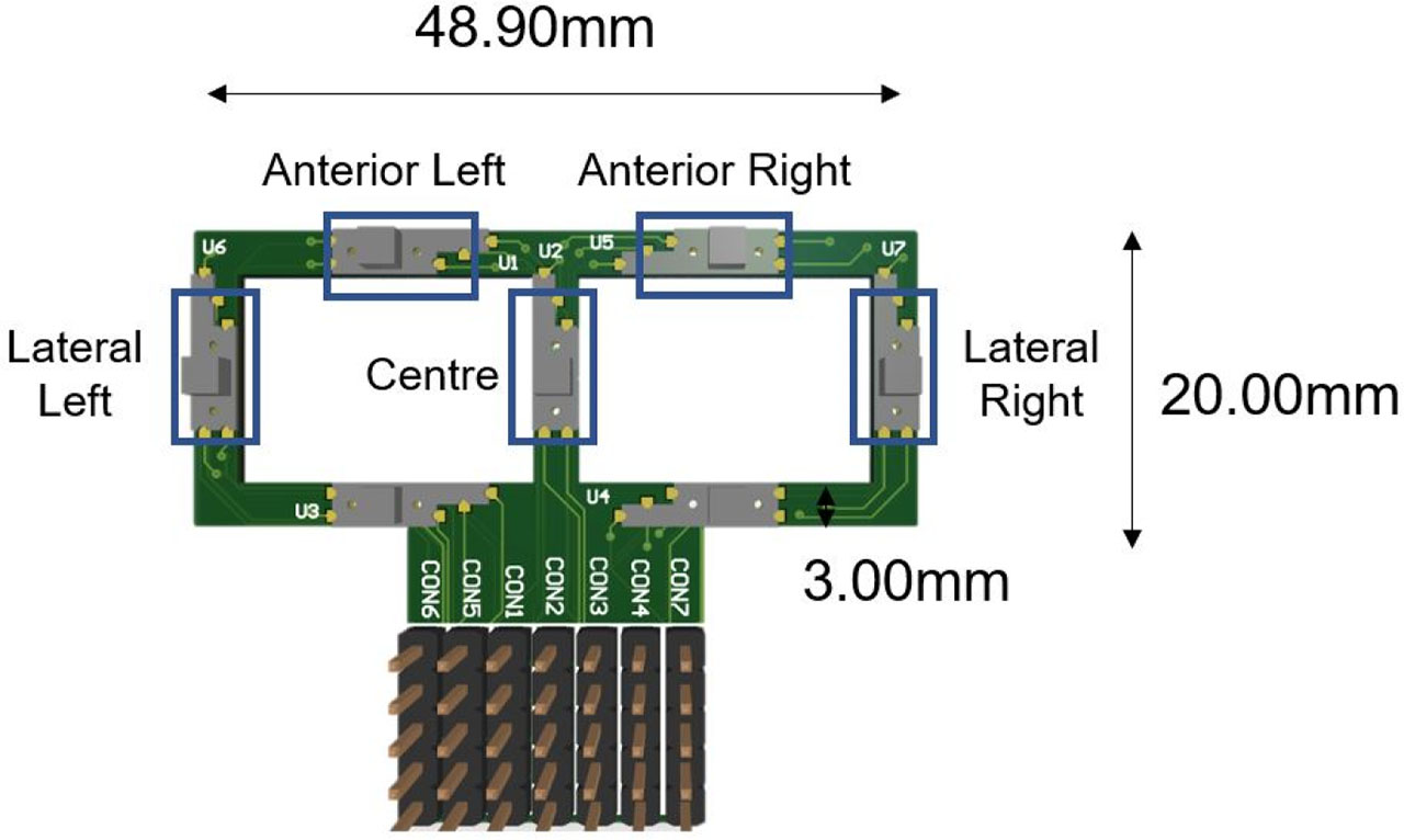
Figure 3:
Piezoresistive sensor array.
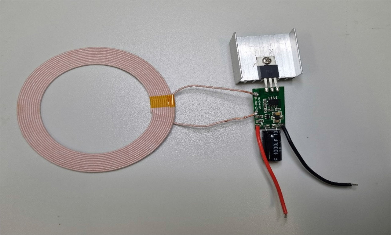
Figure 4:
High power transmitting coil and circuitry.
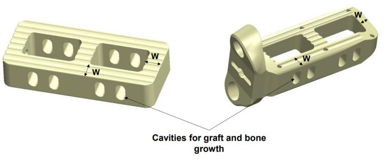
Figure 5:
Implant standalone version (left) and screwed version (right) [96].
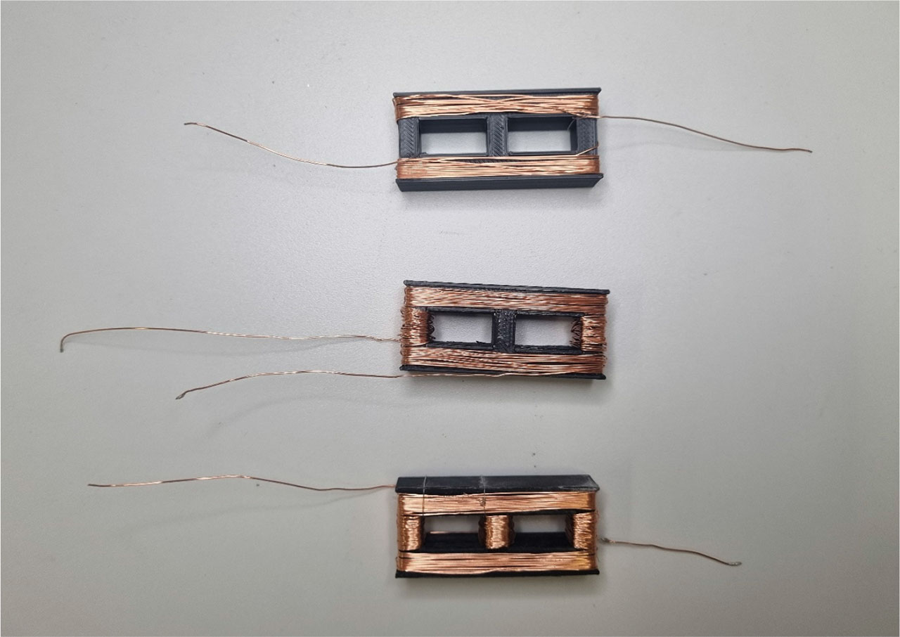
Figure 6:
Coil V1, V2, and V3 designs using standalone implant geometry.
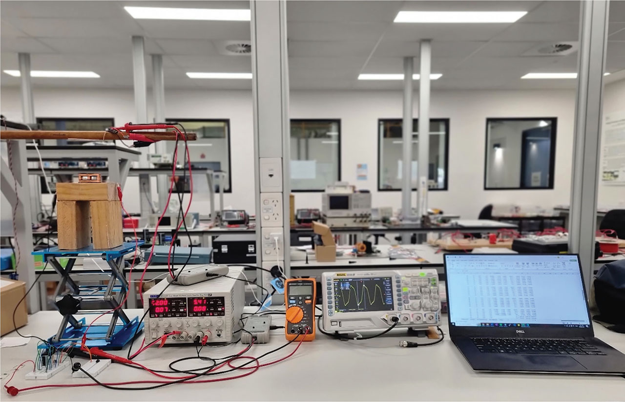
Figure 7:
Vertical misalignment testing setup.
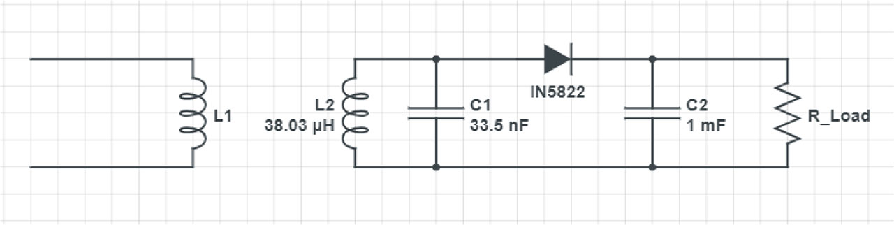
Figure 8:
Half-bridge receiver perfectly tuned for 0.4 mm standalone v3 coil.
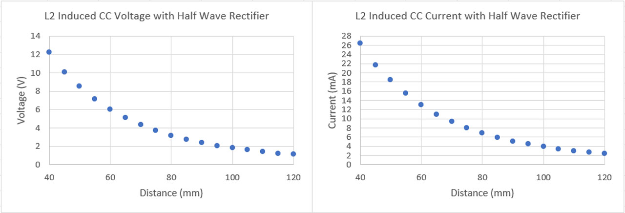
Figure 9:
Receiver half-bridge distant dependent CC results through air.
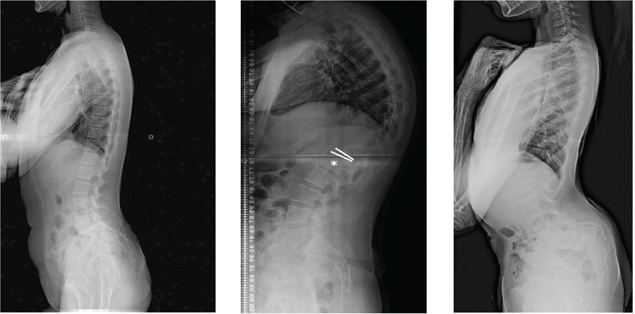
Figure 10:
X-ray of a natural spine [119], kyphosis [118], and lordosis [117].
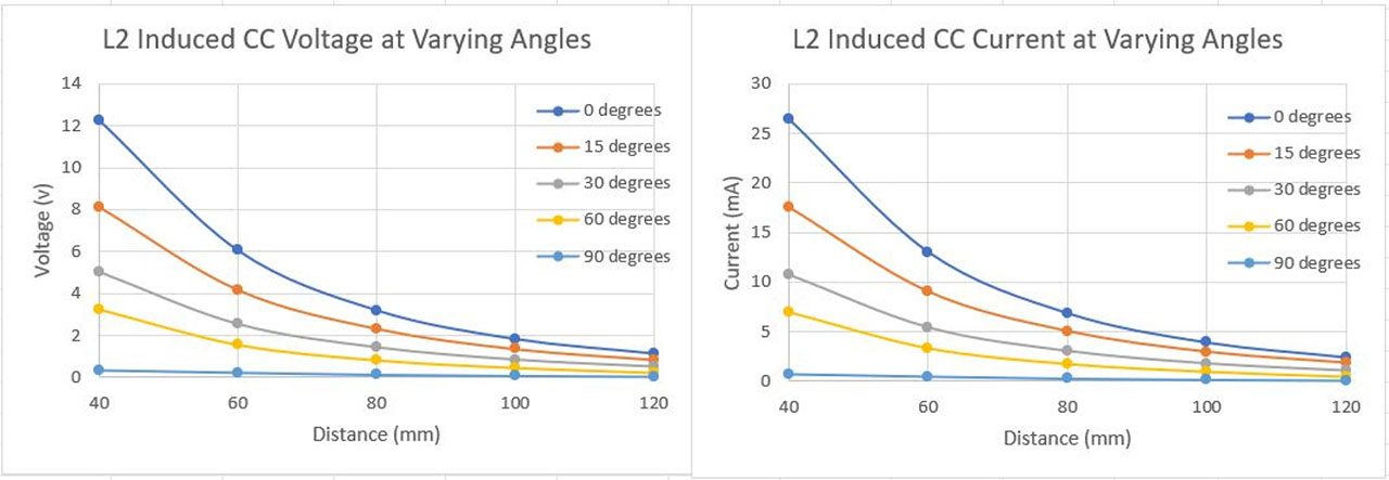
Figure 11:
Receiver CC results at different distances and angles.
Table 2:
PTE and PDL losses due to angular misalignment.
| Misalignment (°) | Loss in PTE (%) | Loss in PDL (%) |
|---|---|---|
| 0 | 0 | 0 |
| 15 | 24–34 | 24–34 |
| 30 | 56–59 | 56–59 |
| 60 | 74–81 | 74–81 |
| 90 | 95–97 | 95–97 |
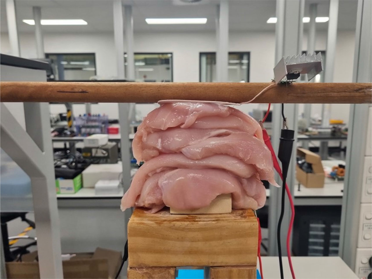
Figure 12:
Vertical misalignment testing setup using tissue substitutes.
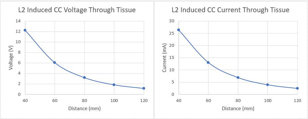
Figure 13:
Receiver CC results at different distances through tissue.
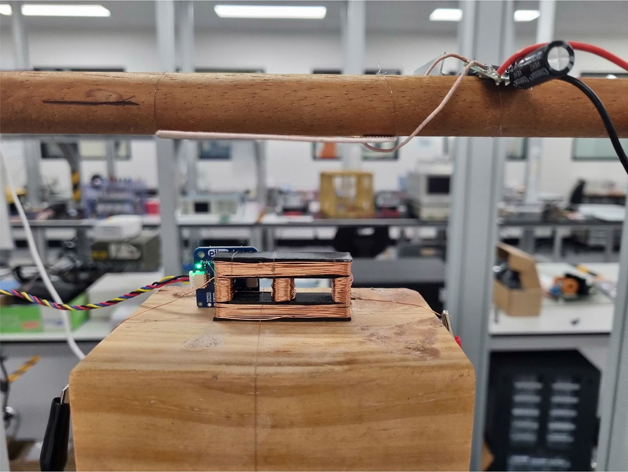
Figure 14:
Temperature sensor setup.
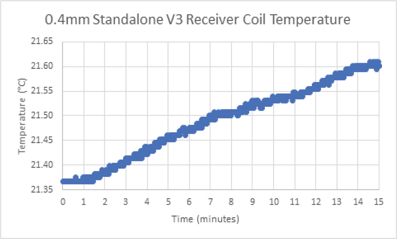
Figure 15:
Receiver coil temperature as a function of time.