
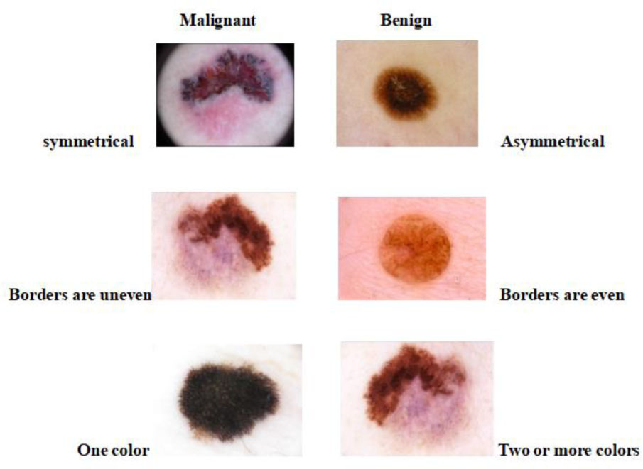
Figure 1.
Image showing comparison between bengin and maligant skin lesion
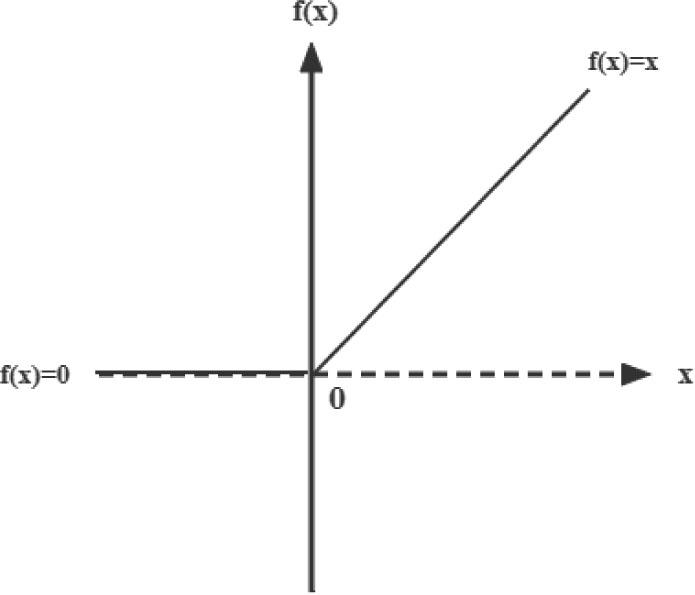
Figure 2.
The Relu function in two dimensions
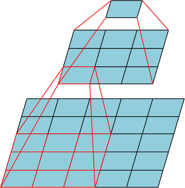
Figure 3.
Mapping relationship between 3*3 convolution kernel and 5*5 convolution kernel
TABLE I.
Feasibility of 3*3 Convolution Kernels Replace 5*5 Convolution Kernels
| Assuming: feature_map = 28*28 | Convolution step = 1 | Padding = 0 |
|---|---|---|
| 1-Layer 5×5 convolutional kernel | 2-Layer 3×3 convolutional kernel | |
| Layer1: (28-5) / 1 + 1 = 24 | Layer1:(28-3) / 1 + 1 = 26 | |
| Output: Feature map = 24×24 | Layer2:(26-3) / 1 + 1 = 24 | |
| Output:Feature map = 24×24 |
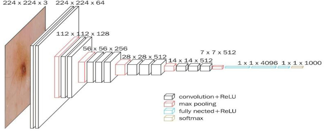
Figure 4.
Overall network structure of VGG16
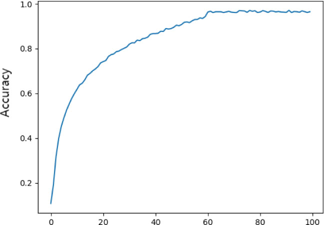
Figure 5.
Training Accuracy Curves of the proposed method on the ISIC-2016 datasets
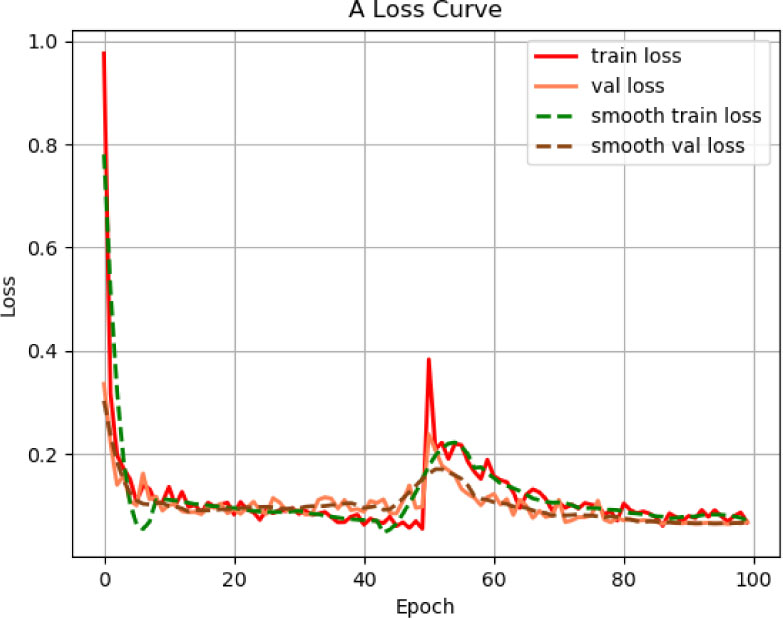
Figure 6.
Training Loss Curves of the proposed method on the ISIC-2016 datasets
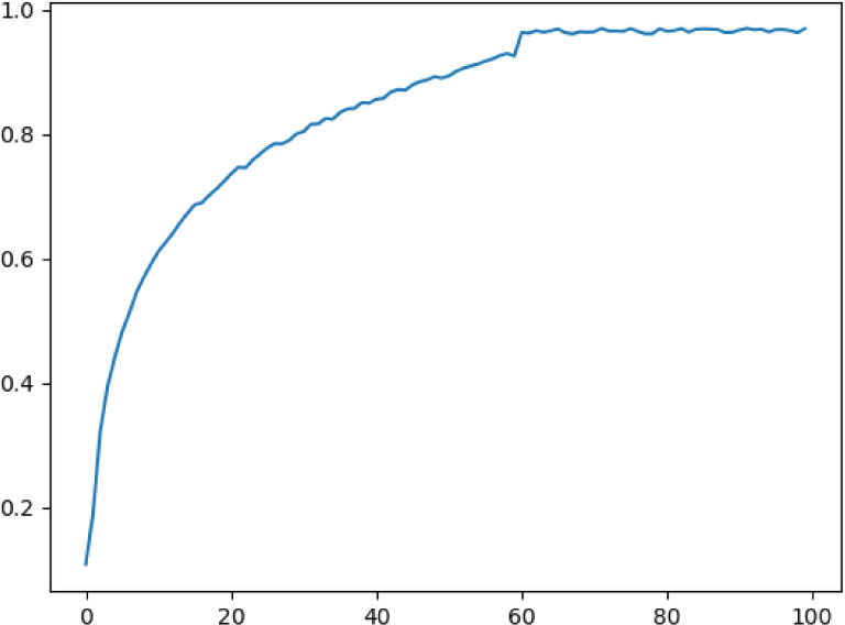
Figure 7.
Training Eval_Top1 Curves of the proposed method on the ISIC-2016 datasets
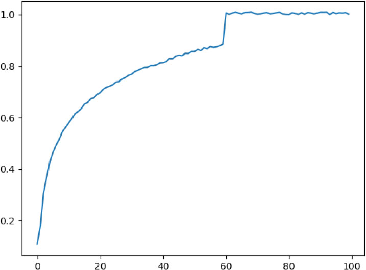
Figure 8.
Training Eval_Top5 Curves of the proposed method on the ISIC-2016 datasets
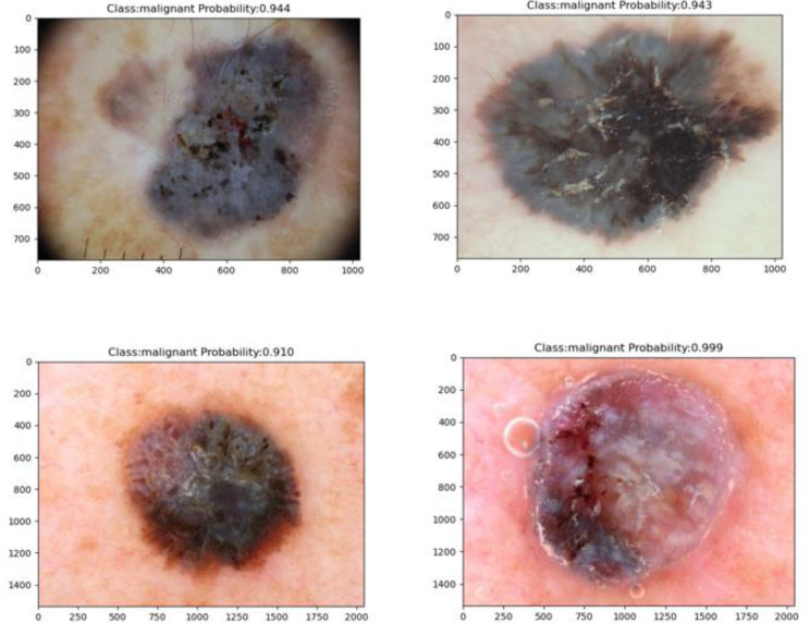
Figure 9.
Prediction results of the proposed method for malignant melanoma in this study
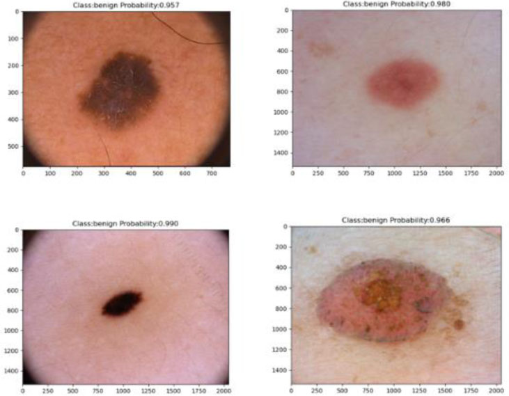
Figure 10.
Prediction results of the proposed method for benign melanoma in this study