
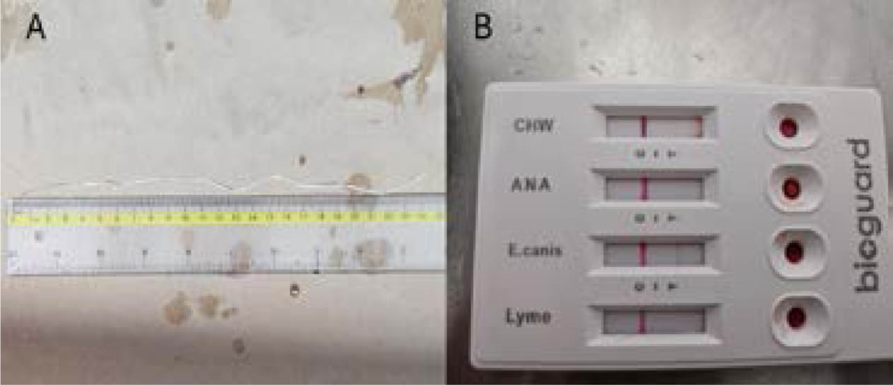
Fig. 1.
A. Adult Dirofilaria immitis nematodes expelled from a canine patient. An adult specimen of Dirofilaria immitis, approximately 24.5 centimeters in length, collected from the sputum expectorate by the patient. B. Bioguard Vlabs 4Dx® rapid immunochromatographic test demonstrating co-infection. The positive line for Ehrlichia canis antibodies indicates previous exposure or active infection with E. canis. In contrast, the faint positive line for Dirofilaria immitis antigens (CHW) confirms the presence of the parasite.
Table 1.
Complete blood count (CBC) analysis of the affected canine patient. This table summarizes the whole blood hemogram data generated by the Mindray Auto Hematology Analyzer BC-5150. Key hematological parameters, including red blood cell indices, white blood cell differentials, and platelet counts, are provided with their respective units. Alterations from established canine reference intervals are highlighted, indicating, e.g., anemia, leukocytosis, and eosinophilia consistent with parasitic infection.
| Parameter (Reference range) | Patient value |
|---|---|
| WBC (6 – 17) | 16.76 |
| Lym# (0.83 – 4.91) | 11.65 |
| Mon# (0.14 – 1.97) | 1.29 |
| Gran# (3.62 – 12.3) | 3.82 |
| Lym% (12 – 33) | 69.5 |
| Mon% (2 – 13) | 7.7 |
| Gran% (52 – 81) | 22.8 |
| RBC (5.1 – 8.5) | 2.59 |
| HGB (11 – 19) | 8.9 |
| HCT (33 – 56) | 17.9 |
| MCV (60 – 76) | 69.1 |
| MCH (20 – 27) | 34.2 |
| RDW (12.5 – 17.2) | 25.8 |
| PLT (200 – 490) | 77 |
| MPV (8 – 17.1) | 12.6 |
| PDW (12 – 17.5) | 9.4 |
| PCT% (0.09 – 0.58) | 0.1 |
| Eos% (0.5 – 10) | 0.2 |
| Eos# (0.04 – 1.62) | 0.03 |
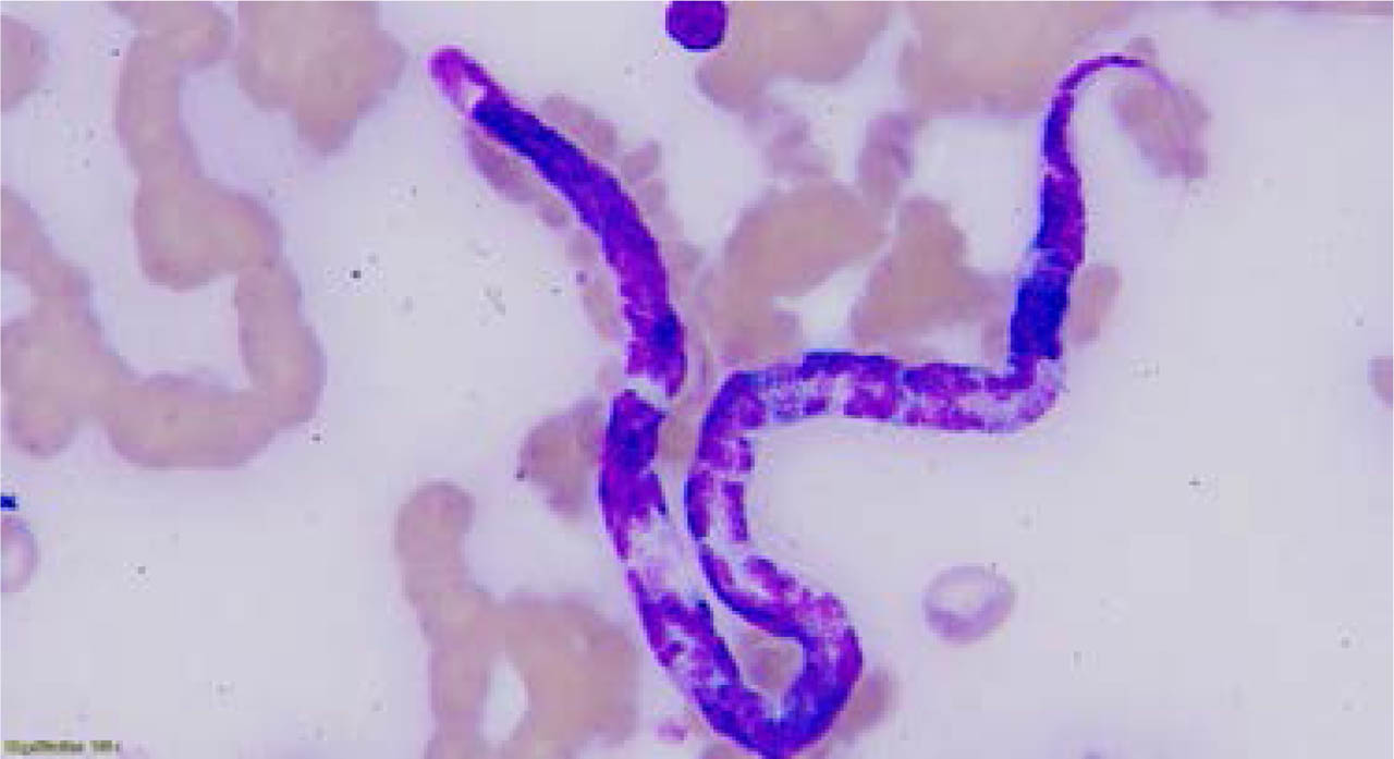
Fig. 2.
Dirofilaria immitis microfilaria in a peripheral blood smear (Hematoxylin-Eosin stain). This photomicrograph, taken at 1000X oil-immersion, shows a single Dirofilaria immitis microfilaria in a peripheral blood smear. The microfilaria exhibits a fusiform body, a distinct diffuse nuclear distribution, and a notable tapered caudal extremity, consistent with the morphology of D. immitis.
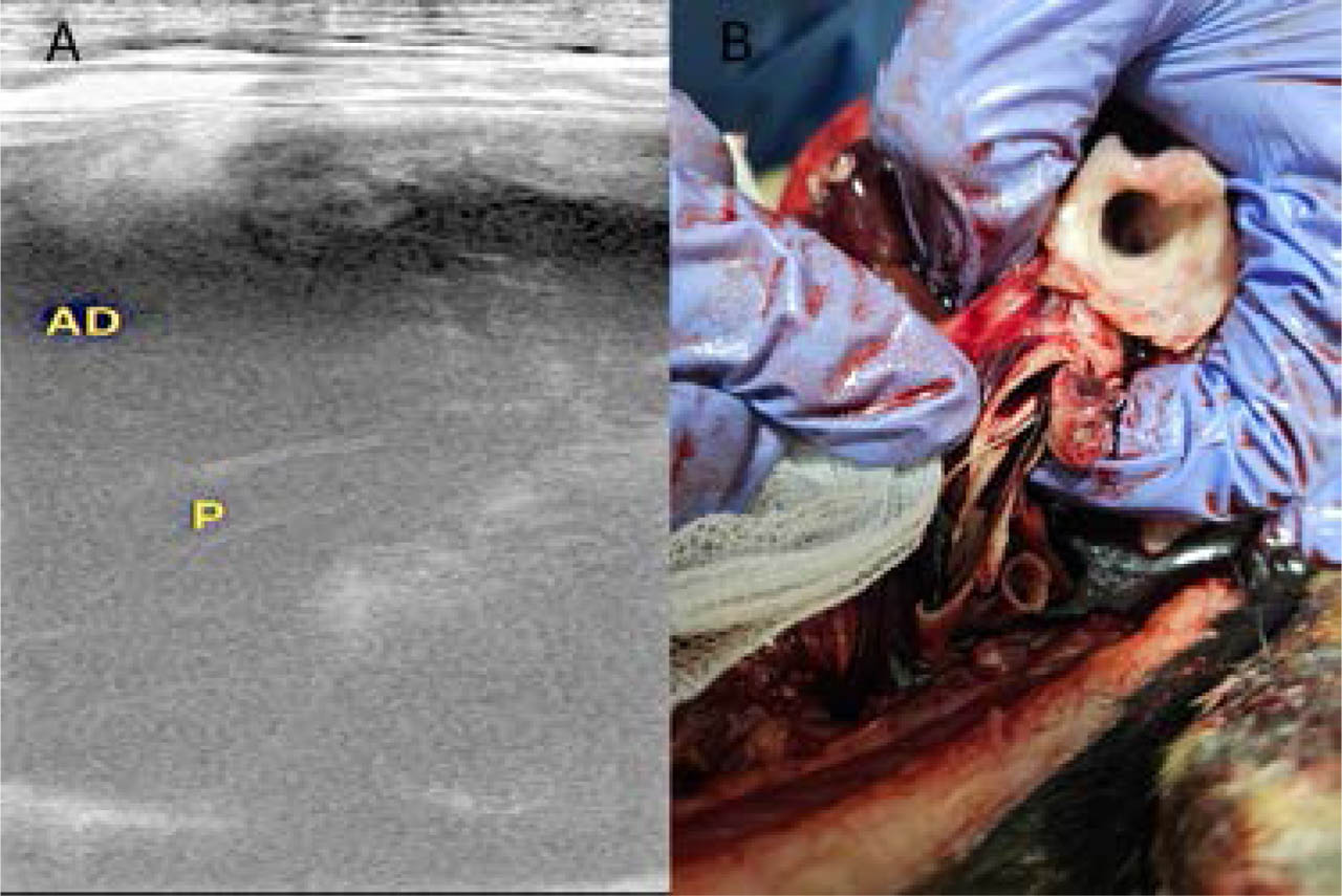
Fig. 3.
A. Echocardiographic visualization of adult Dirofilaria immitis in the right atrium. This ultrasound image captures the characteristic hyperechoic, parallel lines of adult D. immitis (P) within the lumen of the patient's right atrium (AD). B. Macroscopic view of adult Dirofilaria immitis nematodes within the pulmonary artery.
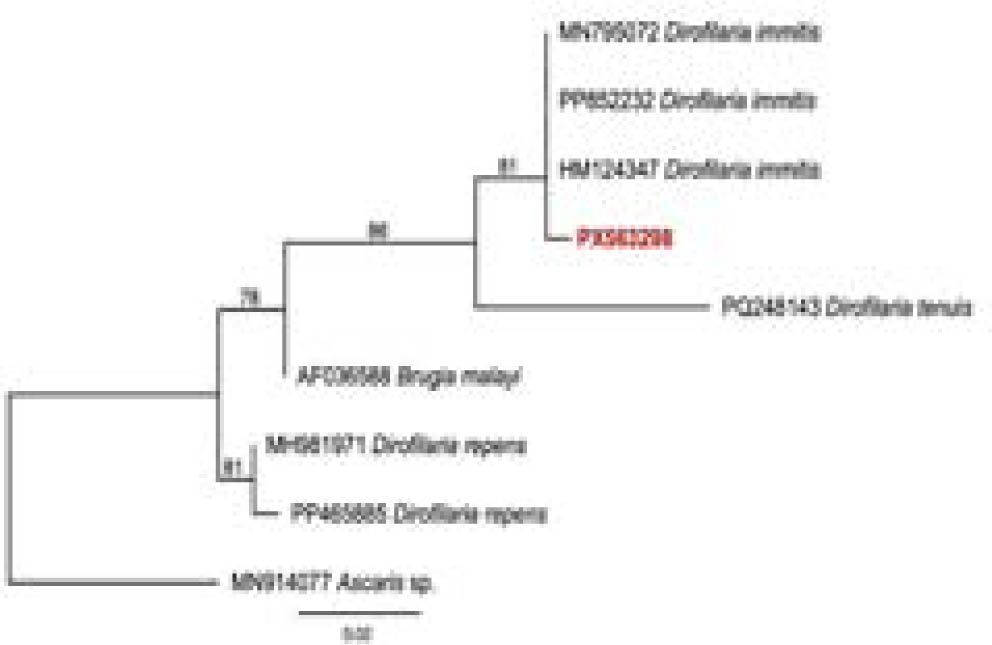
Fig. 4.
Phylogenetic tree inferred by the Maximum Likelihood method and the HKY+F+I evolutionary model based on an alignment of 249 bp of the 16S rRNA gene. Sequence from the present study is highlighted in bold red. Ascaris sp. was used as an outgroup, and node numbers indicate bootstrap values.