
Introduction
Dirofilaria immitis, commonly known as heartworm, is an obligate parasitic nematode belonging to the family Onchocercidae (Canestri Trotti et al., 1997). It's notable for its global distribution and the wide range of definitive hosts it infects, including domestic animals like canines (Anvari et al., 2020), felines (Smith et al., 2022), and various orders of wild mammals (Artiodactyla, Carnivora, Edentata, Lagomorpha, Perissodactyla, Primates, and Rodentia) (Dantas-Torres & Otranto, 2013). These parasites are transmitted by mosquito vectors, with at least 77 species identified as competent, including the genera Culex, Aedes, Anopheles, Mansonia, Coquillettidia, Psorophora, and Culiseta (Riahi et al., 2021). This highlights the complexity and persistence of its transmission cycle.
In Latin America, vector-borne diseases are increasing and evolving as public health concerns. Dirofilariasis fits into this picture as an emerging zoonosis, worsened by a complicated interplay of the region's unique socio-environmental factors (Cardona-Ospina et al., 2021). High population density, combined with the close co-habitation of domestic animals in both urban and rural settings, and a tropical climate that supports sustained proliferation of mosquito vectors for much of the year (Maggi & Krämer, 2019), creates an ideal epidemiological scenario for D. immitis persistence, trans-mission, and spread. This complex situation is further aggravated by large populations of stray and abandoned animals, particularly dogs. These animals not only act as key reservoirs but also facilitate parasite dispersal to new areas and significantly increase the risk of infection for other definitive hosts, including humans (Garde et al., 2013; Mota-Rojas et al., 2021). Therefore, effective control of these zoonoses in the region is a crucial strategic priority to lessen their impact on public and animal health and to prevent new endemic areas from consolidating (Rodriguez-Morales et al., 2021)
In Panama, dirofilariasis has garnered increasing attention, with initial reports in 2021 based on immunochromatographic tests (Candanedo Chacón & Candanedo, 2021). Subsequent research explored pulmonary and cardiac histopathological damage associated with D. immitis infection (Candanedo Chacón & Candanedo, 2023) and even confirmed the presence of the parasite in dogs with Ehrlichia spp. co-infections (Candanedo Chacón et al., 2024) and, recently, a population genomics study of D. immitis that included sequences obtained from Panama (Power et al., 2026). It is important to note that all these previous contributions focused solely on the western part of Panama. Despite Panama's critical epidemiological role as a “biological bridge” and biodiversity corridor, the lack of molecular diagnostic confirmation of D. immitis in pet and wild animal populations across various regions of the country has limited a precise characterization of the parasite and a comprehensive understanding of its true national distribution (Aththanayaka et al., 2024). This deficiency not only hindered the implementation of an effective control strategy but also diminished Panama's ability to contribute robust data to regional and global epidemiological models. This report details a severe clinical case of an approximately 8-year-old rescued stray dog that presented with expectoration of an adult nematode. This led to a diagnosis of D. immitis, and post-mortem findings showed severe cardiac insufficiency. This case highlights the importance of comprehensive diagnosis and the severe pathological sequelae of advanced canine dirofilariasis.
Material and Methods
Case presentation: An approximately 8-year-old rescued stray female dog, of medium stature and 20 kg, presented with anorexia, lethargy, a temperature of 39.7°C, and palpable lymphadenopathy. The primary reason for consultation was the expectation of an adult nematode in sputum (Fig. 1A).
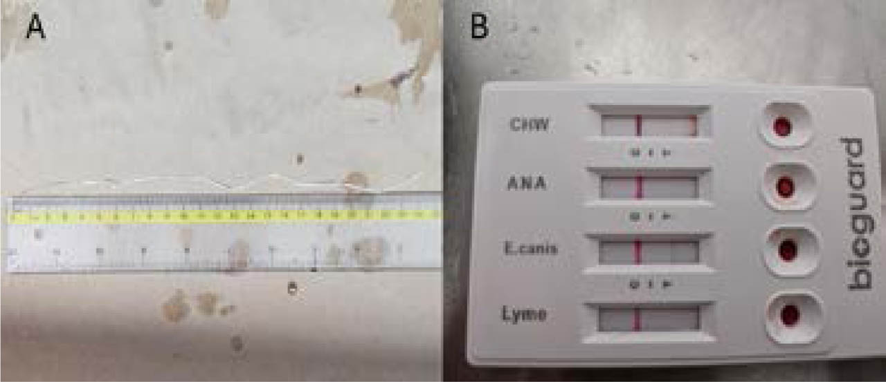
Fig. 1.
A. Adult Dirofilaria immitis nematodes expelled from a canine patient. An adult specimen of Dirofilaria immitis, approximately 24.5 centimeters in length, collected from the sputum expectorate by the patient. B. Bioguard Vlabs 4Dx® rapid immunochromatographic test demonstrating co-infection. The positive line for Ehrlichia canis antibodies indicates previous exposure or active infection with E. canis. In contrast, the faint positive line for Dirofilaria immitis antigens (CHW) confirms the presence of the parasite.
Blood examinations: A blood sample was collected by venipuncture from the V. cephalica antebrachia into tubes containing K2ED-TA and submitted for routine hematological examination using an automatic cell counter, the Mindray Auto Hematology Analyzer BC-5150 (Mindray Bio-Medical Electronics, Shenzhen, China). Whole blood was used in a rapid immunochromatographic test, Bioguard VLabs 4x® (Bioguard Corporation, New Taipei City, Taiwan), for antibodies against Ehrlichia canis, Anaplasma sp., and Leishmania sp., and for Dirofilaria immitis antigens. Finally, a thick blood smear was stained with the Diff-Quik procedure (Medion Diagnostics AG, Switzerland) for morphological identification of the microfilariae, using the dichotomous keys of Liotta et al. (2013).
Echocardiographic Evaluation: An echocardiographic examination was performed using a Samsung HS-40 ultrasound machine (Samsung Electronics, South Korea) with a 2 – 5 MHz convex transducer.
Necropsy: The necropsy was conducted following the guide established by McDonough and Southard (2017).
Molecular identification
To confirm the identity of the adult nematode, molecular characterization was performed. Genomic DNA was isolated using the MagMAX™ Viral/Pathogen II (MVP II) Nucleic Acid Isolation Kit (Applied Biosystems, Thermo Fisher Scientific, Waltham, MA, USA), following the manufacturer's instructions. Polymerase Chain Reaction (PCR) was executed to amplify a partial segment of the mitochondrial 16S rRNA gene, utilizing the Dirofilaria immitis-specific primers ROR 91 (5′-GCATCTTAGAACTTGGTC-CATCC-3′) and ROR 92 (5′-CAAAGGCGTATTTACCGCCAC-3′) originally designed by Watts et al. (1999). The amplification profile followed the optimized thermal cycling conditions described by Vezzani et al. (2011). Assays were conducted using the HotStart Taq DNA Polymerase (BIORON GmbH, Römerberg, Germany). The reaction was initiated with a 10-minute denaturation step at 95°C to ensure complete enzyme activation. This was followed by 35 cycles of 95°C for 60 s, 55°C for 45 s, and 72°C for 60 s, with a final extension hold.
During the assembly pipeline, reads were assembled into a consensus contig using the CAP3 assembly algorithm integrated into UGENE software version 51.0 (Unipro LLC, Novosibirsk, Russia), followed by quality-based end-trimming to remove low-confidence noise from both ends. This bioinformatic procedure yielded a high-fidelity 249-bp consensus fragment, which was subsequently used for definitive taxonomic identification via the Basic Local Alignment Search Tool (BLASTn).
Phylogenetic analysis
The sample sequence was assembled, visually checked, and trimmed to the partial 16S rRNA region using Geneious Prime 2026.0.01 (Kearse et al., 2012). The resulting sequence was aligned with previously published 16S rRNA data from Dirofilaria specimens, as well as other closely related species, and with Ascaris sp. (MN914077) as the outgroup, using MAFFT's (Katoh et al., 2019) L-INS-i translational alignment plugin of Geneious. The phylogenetic tree was estimated using the maximum-likelihood criterion in IQ-TREE (Minh et al., 2020). The best-fitting nucleotide-evolution model was selected using the corrected Akaike information criterion in IQ-TREE, and nodal supports were estimated by running 1,000 standard nonparametric bootstrap replicates.
Ethical Approval and/or Informed Consent
The authors declare that all examinations performed were within the scope of a veterinary examination.
Results
A complete blood count (CBC) revealed lymphocytosis and eosinopenia in the white cell line, anemia in the red cell line, and thrombocytopenia (Table 1). These hematological findings suggested a chronic inflammatory response, bone marrow suppression, and potential platelet consumption, all of which are common in co-infections with other pathogens.
Table 1.
Complete blood count (CBC) analysis of the affected canine patient. This table summarizes the whole blood hemogram data generated by the Mindray Auto Hematology Analyzer BC-5150. Key hematological parameters, including red blood cell indices, white blood cell differentials, and platelet counts, are provided with their respective units. Alterations from established canine reference intervals are highlighted, indicating, e.g., anemia, leukocytosis, and eosinophilia consistent with parasitic infection.
| Parameter (Reference range) | Patient value |
|---|---|
| WBC (6 – 17) | 16.76 |
| Lym# (0.83 – 4.91) | 11.65 |
| Mon# (0.14 – 1.97) | 1.29 |
| Gran# (3.62 – 12.3) | 3.82 |
| Lym% (12 – 33) | 69.5 |
| Mon% (2 – 13) | 7.7 |
| Gran% (52 – 81) | 22.8 |
| RBC (5.1 – 8.5) | 2.59 |
| HGB (11 – 19) | 8.9 |
| HCT (33 – 56) | 17.9 |
| MCV (60 – 76) | 69.1 |
| MCH (20 – 27) | 34.2 |
| RDW (12.5 – 17.2) | 25.8 |
| PLT (200 – 490) | 77 |
| MPV (8 – 17.1) | 12.6 |
| PDW (12 – 17.5) | 9.4 |
| PCT% (0.09 – 0.58) | 0.1 |
| Eos% (0.5 – 10) | 0.2 |
| Eos# (0.04 – 1.62) | 0.03 |
The rapid immunochromatographic test was positive for antibodies against Ehrlichia canis and for Dirofilaria immitis antigens, thereby confirming co-infection (Fig. 1B). Subsequently, the thick blood smear revealed abundant peripheral blood microfilariae (+++), corroborating active D. immitis infection (Fig. 2).
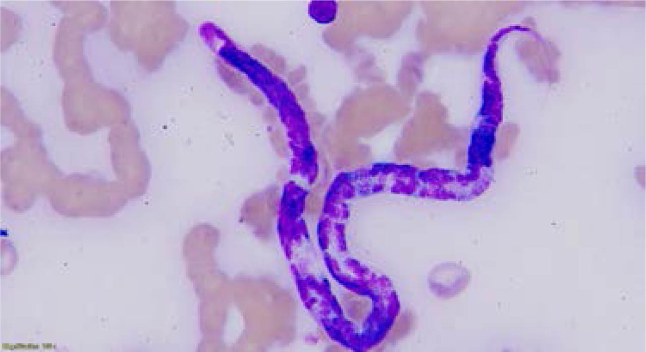
Fig. 2.
Dirofilaria immitis microfilaria in a peripheral blood smear (Hematoxylin-Eosin stain). This photomicrograph, taken at 1000X oil-immersion, shows a single Dirofilaria immitis microfilaria in a peripheral blood smear. The microfilaria exhibits a fusiform body, a distinct diffuse nuclear distribution, and a notable tapered caudal extremity, consistent with the morphology of D. immitis.
On ultrasonography, mobile, hyperechoic, linear structures consistent with adult D. immitis parasites were visualized within the right atrium and pulmonary artery (Fig. 3A). Additionally, severe right ventricular dilation was observed, along with moderate tricuspid regurgitation and findings consistent with severe pulmonary hypertension. These findings indicated significant hemodynamic compromise and severe cardiac dysfunction secondary to the parasitic burden.
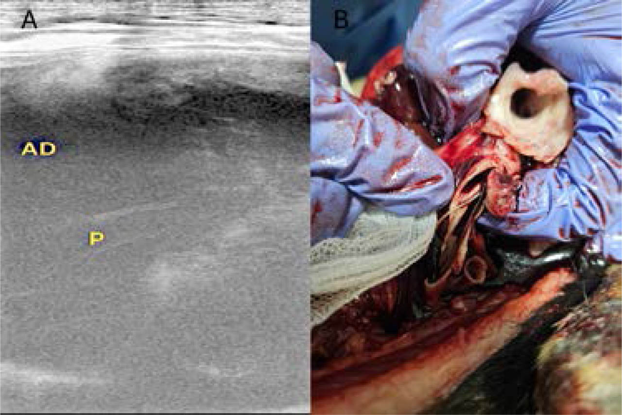
Fig. 3.
A. Echocardiographic visualization of adult Dirofilaria immitis in the right atrium. This ultrasound image captures the characteristic hyperechoic, parallel lines of adult D. immitis (P) within the lumen of the patient's right atrium (AD). B. Macroscopic view of adult Dirofilaria immitis nematodes within the pulmonary artery.
Necropsy revealed the extensive and severe nature of the disease. A severe dilation of the right atrium was found, with rupture of the atrial wall and presumed extravasation of blood into the pericardial cavity (hemopericardium). This finding is consistent with atrial rupture secondary to chronic pressure overload, a mortal complication of advanced dirofilariasis. Numerous adult nematodes were observed within the right cardiac chambers (atrium and ventricle) as well as in the pulmonary artery (Fig. 3B). Obstruction of blood flow and hemodynamic overload from this massive parasitic infestation were the primary factors contributing to the dilation and eventual rupture of the right atrium.
At the molecular level, a 453-base-pair PCR product was successfully obtained, and the sequence was deposited in GenBank under accession number PX563298.
Maximum-likelihood phylogenetic analysis confirmed the species identification as D. immitis, placing the specimen at the base of a well-defined clade composed exclusively of D. immitis representatives (Fig. 4).
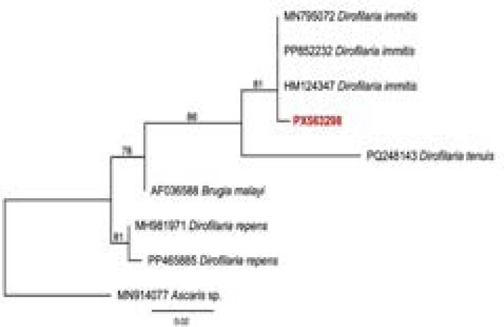
Fig. 4.
Phylogenetic tree inferred by the Maximum Likelihood method and the HKY+F+I evolutionary model based on an alignment of 249 bp of the 16S rRNA gene. Sequence from the present study is highlighted in bold red. Ascaris sp. was used as an outgroup, and node numbers indicate bootstrap values.
Discussion
The clinical signs observed in this case, including lethargy, anorexia, and the expectoration of an adult nematode, are highly consistent with the course of canine filariasis caused by D. immitis (Atwell, 2018). The severity of the condition, culminating in severe cardiac insufficiency and fatal atrial rupture, is compatible with Stage III dirofilariasis, which is associated with a reserved to unfavorable prognosis (Polizopoulou et al., 2000). In addition to this clinical complexity, a co-infection with E. canis was identified, an association previously reported in Panama's Chiriquí province (Candanedo Chacón et al., 2024). The presence of multiple vector-borne pathogens can significantly worsen the patient's condition, adversely affecting clinical presentation, disease progression, and hematological findings through a potential synergistic effect (Candanedo Chacón et al., 2024; Dantas-Torres & Otranto, 2013). The patient's history of street rescue is a crucial factor and a significant limitation in this case. The absence of prior medical history and prophylaxis, coupled with constant exposure to an uncontrolled environment, likely contributed to the acquisition of these infections. This situation underscores the vulnerability of homeless animals, which not only suffer the consequences of neglected diseases but also act as reservoirs and sources of infection for other animals and, potentially, humans, within a One Health perspective (Nametov et al., 2025). The lack of long-term follow-up and the impossibility of specific treatment for dirofilariasis due to the patient's acute deterioration further highlight the therapeutic challenges in the region. Critically, a significant limitation in Panama is the absence of government-approved, specific adulticidal medications for canine dirofilariasis on the local market. This severely hinders the complete and effective therapeutic management of the disease in the country, especially in areas with socioeconomic difficulties, where the owners cannot afford the government-approved prophylactic compounds on the market with proven efficacy. This barrier not only impacted the prognosis of this particular animal but also represents a constant and significant challenge for animal health in the region, underscoring the difficulties in accessing essential treatments for such a dangerous disease.
In Panama, mosquito vectors pose an epidemiological risk factor for multiple diseases. While the vector competence of local species for D. immitis has not been demonstrated, there is evidence that some species can serve as carriers of filarial parasites, including Dirofilaria repens, in Darien province, Panama (Rivera et al., 2025). Genera such as Culex and Aedes are known as confirmed dirofilariasis vectors in other regions (Ledesma & Harrington, 2011). In Panama's urban environment, highly anthropophilic species such as Culex quinquefasciatus and Aedes aegypti are widely circulating, alongside other Culex and Aedes species (Whiteman et al., 2019; Lopez-Solis et al., 2023). Although their primary role in D. immitis transmission to humans in the region is not fully elucidated, the ability of these mosquito species to colonize new ecological niches and invade human dwellings (Loaiza et al., 2017) poses a potential risk not only for zoonotic transmission but also for maintaining the disease cycle within the country.
Conclusion
This case underscores the relevance of targeted molecular tools for the definitive diagnosis of canine dirofilariasis and for providing reliable epidemiological data. The selection of the mitochondrial 16S rRNA gene as a molecular marker offered distinct analytical advantages over other conventional loci; unlike the highly conserved nuclear 18S rRNA gene or the cox1 gene, which is frequently hindered by the co-amplification of nuclear pseudogenes (Numts) in nematoda, the 16S rRNA locus provided clean, high-fidelity reads with ideal species-level resolution. Confirming the presence of D. immitis by capturing this specific hypervariable region significantly strengthens the region's diagnostic precision and collaborative epidemiological surveillance capacity.
Collaboration between clinics and reference centers, such as the Gorgas Memorial Institute (ICGES), is crucial for supporting the surveillance and control of canine dirofilariasis and other zoonotic diseases, as well as addressing their public health implications in Panama. More studies are needed to understand the distribution of this parasite and the extent of the threat it poses to public health in Panama.
This work represents the first molecular identification of Dirofilaria immitis in Panama City.
Acknowledgments
The authors wish to express their sincere gratitude to the team at La Fe Veterinary Clinic. We extend our thanks to DMV Ana Velasco, DMV Bredio Velasco, and staff members Alexander Andreve and Carlos Herrera for their crucial clinical assistance and professionalism in managing this case. We are also grateful to Brigitte Henriquez and Angelica Castro for their help with the molecular laboratory work. Finally, we would like to thank NutriServi Panama and Boehringer Ingelheim for their financial support of the publication process.