
Introduction
The nematode phylum is a highly diverse group of animals. Estimates range from 100.000 to 100 million species (Bouamer et al., 2006; Hammond, 1992; Hugot et al., 2001; Lambshead, 1993). Many species are free-living; others live as parasites in plants, arthropods and vertebrates, including humans (Anderson, 2000; Yamaguti, 1961; Yeates et al., 2009). Among parasitic nematodes, a wide range of species are found in birds as final hosts (Cram, 1927; Rebelo et al., 2024; Leung & Koprivnikar, 2016; Malik et al., 2022; Movsesyan et al., 2020; Okulewicz, 2013; Saparov et al., 2013; Santoro et al., 2016; Zhang et al., 2012). There, they can cause air sac inflammation, pneumonia, bronchopneumonia, and other diseases. Symptoms include deterioration in general condition, lethargy, and reduced flight performance (Abdu et al., 2023; Freeman et al., 2025; Leung & Koprivnikar, 2016; Sterner & Cole, 2008).
Here we report the first record of the nematode Diplotriaena obtusa in a Eurasian nuthatch (Sitta europaea) from southwestern Germany.
Material and Methods
Sampling and necropsy of the bird
The nuthatch was caught in a net on 5 September 2024 in Wangen im Allgäu, Baden-Württemberg, Germany (47°41′ N, 9°48′ E; Fig. 1) and ringed with the ring number Hiddensee C4E9091 (Fig. 1).
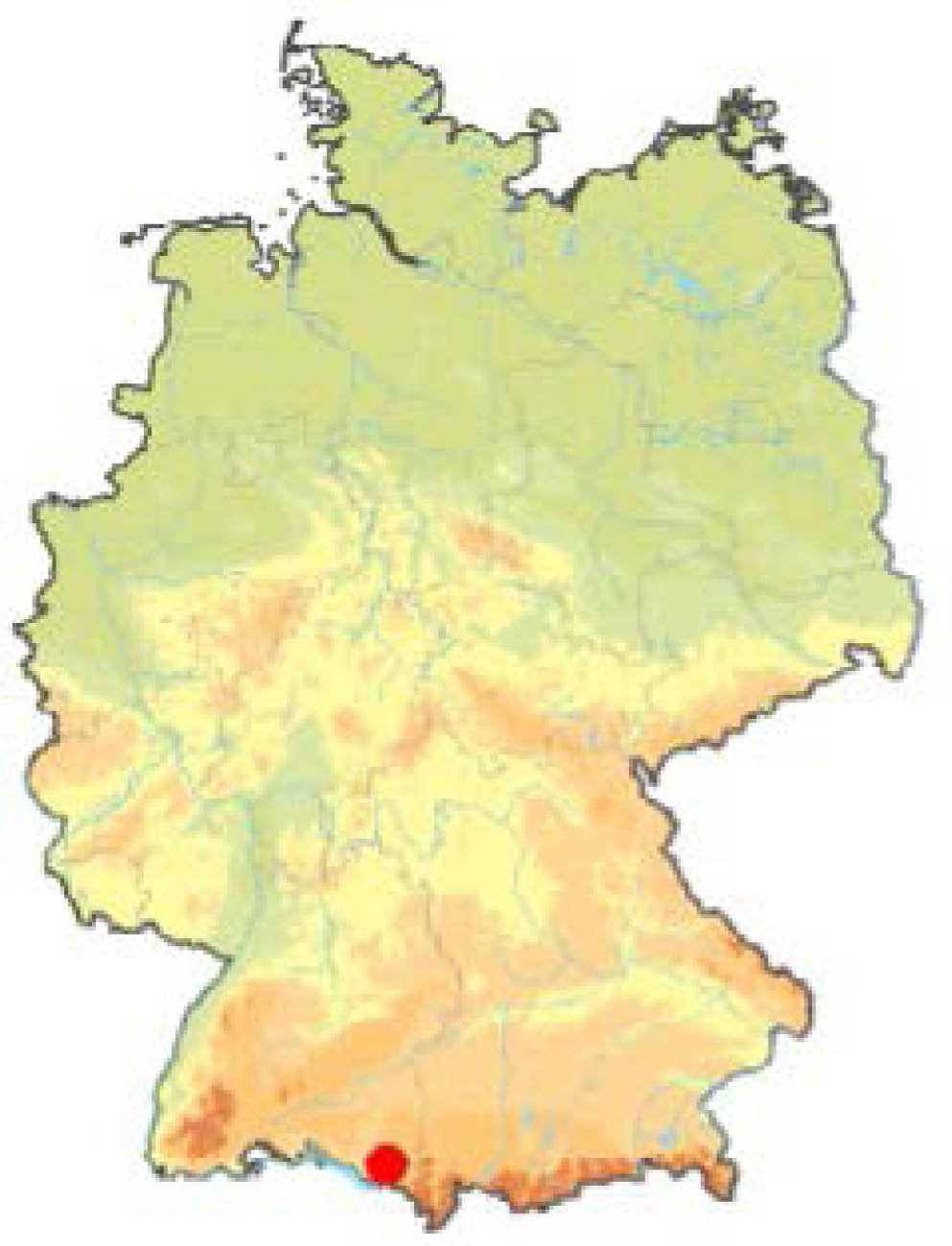
Fig. 1.
Collection locality (Wangen im Allgäu, Baden-Württemberg, Germany, 47°41′ N, 9°48′ E, red dot).
The bird was recognized as a juvenile male and weighed 23.2 g. It was noticed that the bird was rattling. Two days later, the nuthatch was found dead nearby and then dissected to determine the cause of death and confirm its age and sex. There were no signs of injury, for example, from being hit by a windscreen or a predator, but two nematodes were found in the area of the esophagus.
Altogether, three nematodes were fixed in ethanol (70 %) and sent to the first author as an incidental finding as part of a research program funded by members of the Saxon State Parliament to record louse flies (Freick et al., 2025). The morphological and molecular examinations and species determination were conducted at the Institute of Parasitology, University of Leipzig.
Morphological identification of parasites
Two fixed specimens were examined via light microscopy using a Leica DMLB clinical microscope. For clearing, one nematode was placed in lactophenol. Unfortunately, specific morphologic features of the helminth were unrecognizable. Therefore, a single female nematode was dissected, and eggs were obtained for morphological characterization.
DNA extraction, amplification and sequencing
One nematode was used for molecular analysis, and DNA was extracted using the NucleoSpin Tissue Kit (Macherey-Nagel, Düren, Germany) according to the manufacturer's instructions. Afterward, a PCR was conducted to amplify an approximately 900 bp fragment of the 18S small subunit rRNA gene using the primer pair Nem_18SF (5′-cgcgaatrgctcattacaacagc-3′) and Nem_18SR (5′-gggcggtatctgatcgcc-3′) according to Floyd et al. (2005). Each reaction mixture contained: 2.5 μl 10X DreamTaq Buffer (Thermo Scientific TM), 0.8 μl dNTP-Mix (0.2 μM of each deoxynucleoside triphosphate; Thermo Scientific TM), 0.5 μl of Primer each (25 μM), 0.1 μl DreamTaq Green DNA Polymerase (Thermo ScientificTM), 3 μl of the DNA sample and DEPC water to a total volume of 25 μl. The initial denaturation at 94°C for 5 min was followed by 35 cycles at 94°C (30s), 54°C (30s), and 72°C (1 min). A positive and a negative control were included to confirm the PCR result. Final extension was performed at 72°C for 7 min. After gel electrophoresis, the PCR products were visualized on a 1.5 % agarose gel. The purified PCR product was bidirectionally sequenced by a commercial service (Microsynth Seqlab, Göttingen, Germany).
Data analysis
The consensus sequence was compared with sequences in Gen-Bank using the Basic Local Alignment Search Tool (Altschul et al., 1990), and exemplary sequences (E-value < 0.0; percent identity > 96 %) were used for phylogenetic comparison. All sequences (inclusive references) used for this analysis are given as supplementary data (Supplementary data 1). A Toxocara cati sequence (EF180059) was used as the outgroup. Evolutionary analysis was performed twice on the Phylogeny. fr platform (Dereeper et al., 2010; Dereeper et al., 2008), with MUSCLE (v3.8.31) for alignment and Gblocks (v0.91b) for refinement (Edgar, 2004; Castresana, 2000). Subsequently, the maximum-likelihood method (100 bootstrap replicates) implemented in PhyML (v3.1/3.0 aLRT) was used, and the default substitution model was selected to construct the phylogenetic tree, which was edited in TreeDyn (v198.3) (Anisimov & Gascuel, 2006; Chevenet et al., 2006; Guindon & Gascuel, 2003). Two analyses were conducted, one with a final length of 791 bp (Fig. 3A) and another with a length of 492 bp (Fig. 3B), including exemplary sequences of D. bargusinica and D. anthreptis.
Ethical Approval and/or Informed Consent
For this study, formal consent was not required.
Results
Since no specific morphological features were recognizable via light microscopy in the adult nematodes, as might be expected in the case of Diplotriaena spp. Eggs, such as those with size, specific trident structures at the anterior end, and lateral pores at the oral opening (Freeman et al., 2025; Rentería-Solís et al., 2021, Anderson et al., 2009), were removed from a female specimen for morphological characterization. These nematodes' eggs were identified as Spirurid eggs according to Schmäschke (2025) and Beck & Pantchev (2013), with a thick, smooth wall and a larval stage inside (Fig. 2). Measurements averaged 46 μm x 30 μm.
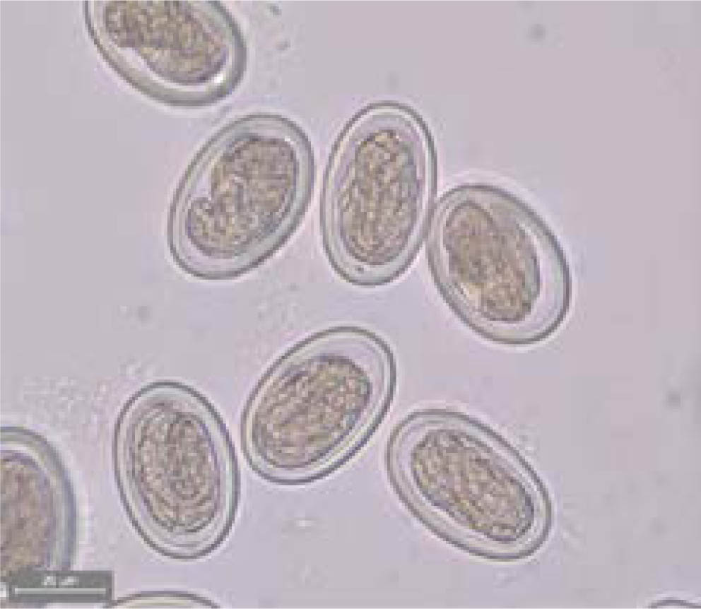
Fig. 2.
Spirurid eggs removed from a female specimen. Shells were thick and smooth with a larval stage inside.
Phylogenetic analysis of the herein obtained sequence (Isolate_Eurasian_nuthatch) for 791 bp (Fig. 3A) and 492bp (Fig. 3B) with those available from GenBank (e.g., MT129516, MT129509) employed the maximum likelihood method. The T. cati sequence (EF180059) was used as the outgroup. Only branches with support values greater than 70 % are shown (bootstrapping method; 100 bootstrap replicates). The sequence obtained was submitted to GenBank (accession number PV942266, 861 bp) and showed the highest percent identity (99.75 %) with isolates of Diplotriaena obtusa (e.g., accession number MT129516, query cover 95 %; accession number MT129509, query cover 94 %). Evolutionary analysis showed that the herein obtained sequence clustered together with other D. obtusa sequences (Fig. 3A). The specimen branched between a specimen of D. obtusa found in a blue tit from Germany (accession number: MT705333) (Fig. 3A) and the other specimens of D. obtusa (accession numbers: MT129516, MT129509, MT129524, MT129521, MT129507) which showed very high percent identities (99.75 %) and were detected in barn swallows and cliff swallows from the USA hinting on some potential genetic variations due to geographical conditions, which should be confirmed with higher-resolution markers and broader sampling. Including sequences from other Diplotriaena species in the phylogenetic analysis further supported the consensus between the isolate herein found and other isolates of D. obtusa (Fig. 3B).
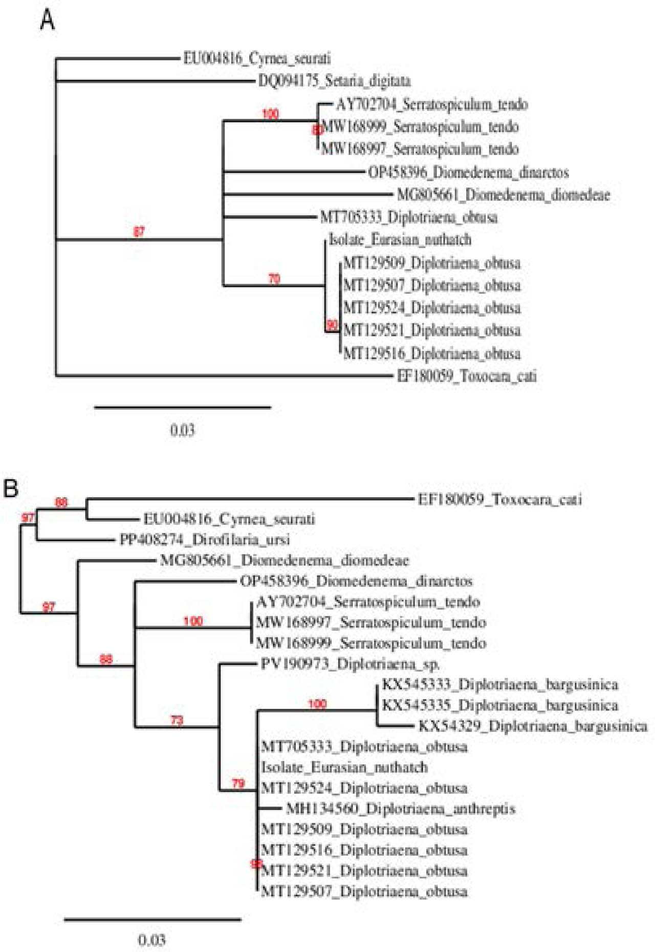
Fig. 3.
Phylogenetic analysis of herein obtained sequence for 791 bp (A) and 492bp (B) with those available from GenBank (e.g., MT129516, MT129509) using the maximum likelihood method. The sequence of T. cati (EF180059) was used as an outgroup. Only branches with support values greater than 70% are shown (bootstrapping method; 100 bootstrap replicates).
Discussion
Among the nematodes affecting birds, particularly passerines, there were approximately 77 described species of the genus Diplotriaena sp. (Hong et al., 2019), to which D. obtusa found in the nuthatch was attributed. D. obtusa was recorded in different passerines of the northern hemisphere and from Madagascar, whereby, according to the frequency of publication, swallows (Hirundinidae Rafinesque, 1815) were particularly frequently observed as hosts (Tab. 1). The assignment of D. obtusa to the hosts Junco hyemalis (Linnaeus, 1758) and Quiscalus quiscula (Linnaeus, 1758) by Leidy (1885, 1886) and Walton (1927) was incorrect and based on a misidentification of the parasites (Anderson, 1959).
Table 1.
Bird species with records of Diplotriaena obtusa (names of subspecies only if given in the cited references).
| Family | Species | Location | References |
|---|---|---|---|
| Apodidae Hartert, 1897 | Apus apus (Linnaeus, 1758) | Czech Republic | Sitko & Heneberg (2023), |
| Hirundinidae Rafinesque, 1815 | Delichon urbicum (Linnaeus, 1758) | No information | Schreibers (1811) |
| Poland | Jaroń (1969) | ||
| Czech Republic | Sitko & Heneberg (2023), Sitko & Okulewitcz (2010) | ||
| No information | Leidy (1890) | ||
| Hirundo rustica Linnaeus, 1758 | Poland | Jaroń (1969) | |
| Slovenia | Brglez (1982) | ||
| Slovakia | Birová (1990) | ||
| Czech Republic | Sitko & Heneberg (2023), Sitko & Okulewitcz (2010) | ||
| Upper Midwest, United States | Michalski et al. (2021) | ||
| No information | Rudolphi (1802) | ||
| Hirundo rustica gutturalis Scopoli, 1786 | Yunnan, China | Wu (1973) | |
| Hirundo rustica saviginii Stephens, 1817 | Egypt | Ahmed (1997) | |
| Petrochelidon pyrrhonota (Vieillot, 1817) | Upper Midwest, United States | Michalski et al. (2021) | |
| Progne chalybea (Gmelin, JF, 1789) | No information | Diesing (1851) | |
| Progne subis (Linnaeus, 1758) | No information | Diesing (1851) | |
| Ontario, Canada | Webster (1974) | ||
| Riparia riparia (Linnaeus, 1758) | No information | Schreibers (1811) | |
| Czech Republic | Sitko & Heneberg (2023) | ||
| Tachycineta bicolor (Vieillot, 1808) | Colorado, United States | Sherwin & Schmidt (1988) | |
| Muscicapidae Fleming, 1822 | Ficedula albicollis (Temminck, 1815) | Czech Republic | Sitko & Heneberg (2023), |
| Paridae Vigors, 1825 | Cyanistes caeruleus (Linnaeus, 1758) | Germany | Rentería-Solís et al. (2021) |
| Parus major Linnaeus, 1758 | Russia | Sonin (1958) | |
| Poland | Rząd et al. (2023) | ||
| Parulidae Wetmore et al., 1947 | Setophaga occidentalis (Townsend, 1837) | Wyoming, United States | Orrell T, Informatics and Data Science Center - Digital Stewardship (2025) |
| Ploceidae Sundevall, 1836 | Foudia madagascariensis (Linnaeus, 1766) | Madagascar | Henry & Ozoux (1909) |
| Sittidae Lesson, 1828 | Sitta europaea Linnaeus, 1758 | Germany | This paper |
| Sturnidae Rafinesque, 1815 | Sturnus vulgaris Linnaeus, 1758 | Armenia | Akhumian (1966), Movsesyan et al. (1985) |
| Sylviidae Leach, 1820 | Sylvia atricapilla (Linnaeus, 1758) | Poland | Stanicka et al. (2021) |
Until now, there has been no scientific evidence that D. obtusa has been found in the European nuthatch, of which only the species Acuaria subula (Dujardin, 1845) and D. henryi (Blanc, 1919) have been known as helminth parasites (Sitko & Okulewicz, 2010; Okulewicz, 2013).
The only available study of D. obtusa from Germany identified the blue tit as the host (Rentería-Solís et al., 2021). A report of D. obtusa parasitizing the great tit was from Poland (Rząd et al., 2023). European nuthatches live in loose associations with tits during autumn and winter, and also in similar breeding habitats (Blotzheim, 1993). It is therefore possible that the birds were infected with nematodes in habitats similar to those in which they were found. Parasitic helminths of the genus Diplotriaena sp. can cause significant pathology in their hosts (Abdu et al., 2023; Michalski et al., 2021; Stanicka et al., 2021). Many are (Leung & Koprivnikar, 2016). Sitko & Heneberg (2023) showed that the prevalence, and thus the infection intensity, of this nematode in Europe has increased in recent years.
The trivial name “air sac worm“ refers to the leading site of infection, which can also affect the body cavity (Sonin, 1975). In our case, the nematodes were found in the tracheal region, but they likely originated from the neighboring air sacs. The dissected nut-hatch showed no external injuries. However, a rattle was through-out its life, likely due to the nematode infection. This symptom may be associated with the dead bird, given the nuthatch's ringing. Although Dipoltriaena infections are usually subclinical, a high parasite load can lead to morbidity and mortality (Freeman et al., 2025; Stanicka et al., 2021). Whether the nematode infection itself was the cause of death of the bird cannot be said with certainty, as no histological or pathological evidence was available. However, it is at least possible that it contributed to the bird's death.
A limitation of this study is the small number of specimens, or, rather, the limited potential for morphometric analysis. However, this matter was mitigated by the clear identification of the parasites' eggs and subsequent molecular analysis to identify the species.
D. obtusa was found for the first time in a European nuthatch. This finding broadens the spectrum of potential hosts for this nematode species and, consequently, its distribution, as this bird species is (Blotzheim, 1993). The larvae are transmitted when they are ingested by their definitive hosts (Anderson, 2000). This occurs via invertebrates as intermediate hosts (Abdu et al., 2023; Rentería-Solís et al., 2021; Sitko & Heneberg, 2023), which are part of the European nuthatch's and many other passerines' diet (Brehm, 1867) as part of the natural chain of infection.
Acknowledgements
We want to thank Lidija Nemec Golobič, University of Ljubljana, and Anja Großmann, University of Applied Sciences Dresden, for providing us with literature that was difficult to obtain.
The publication of this article is funded by the Open Access Publication Fund of Hochschule für Technik und Wirtschaft Dresden – University of Applied Sciences.
We are indebted to PhD Anna Stanicka (Nicolaus Copernicus University, Torun) and Prof. Dr. Markus Freick (Martin Luther University Halle-Wittenberg) for their valuable comments on the manuscript.