
Introduction
Fasciolosis is one of the most neglected food- and water-borne zoonotic diseases of cattle, buffaloes, sheep, goats, horses, and humans of all ages. It is the most important helminth infectious disease worldwide (Alkahtani et al., 2024). Fasciolosis is caused by the ingestion of encysted metacercariae of digenean fasciolid trematodes, Fasciola hepatica and Fasciola gigantica (Najib et al., 2020; Nkurunziza et al., 2024). Fasciolid trematodes are commonly called flatworms or liver flukes, which belong to the family Fasciolidae of the class Trematoda of the phylum Platyhelminthes. The presence of cephalic cones and shoulders in the body of Fasciola hepatica and their absence in Fasciola gigantica are the two most common morphological characteristics that distinguish these two species (Ahmed et al., 2005; Valino et al., 2017). According to geographical distribution, there are some differences between the occurrence of F. hepatica and F. gigantica because F. hepatica is distributed across nearly all continents of the world, while F. gigantica is mostly specific to the parts of Asia and Africa, including Pakistan (Abdullah, 2023; Caravedo & Cabada, 2020; Tang et al., 2021; Afshan et al., 2014a). To complete their life cycle, fasciolids use two types of hosts: an intermediate host (specific host) and a definitive host (Ibrahim, 2017). Two species of snails, Lymnaea natalensis and Lymnaea truncatula, are the most common intermediate hosts of fasciolosis in rainy muddy areas. While cattle, buffalo, goats, sheep, and horses are the definitive hosts (Ulhaq et al., 2022). During the transmission of the disease, buffaloes, sheep, goats, horses, and humans ingest the encysted metacercarial form of Fasciola, and the worm then migrates to the liver, where it causes liver damage; the mature worm then resides in the bile duct (Seldemir, 2000). After the parasite enters the liver, it causes an acute infection, and the incubation period lasts 2 – 4 weeks. When metacercariae migrate to the bile ducts and mature into adult flukes, chronic disease occurs, with an incubation period of 3 – 11 months (Phalee et al., 2015). The acute form of fasciolosis includes fever, skin rashes, hair coat browning, liver cirrhosis, and reduced reproductive performance. In contrast, the chronic form is commonly associated with anemia, jaundice, and persistent hair discoloration (Mas-Coma et al., 2009). Fasciolosis leads to significant weight loss and reduced meat and milk production in livestock, causing severe economic losses to the agricultural sector, particularly in developing countries.
According to a survey conducted by the World Health Organization (WHO, 2021), fasciolosis affects at least 17 million individuals, and 180 million are at risk of fasciolosis in more than 70 countries worldwide. Developing and underdeveloped countries are at higher risk of fasciolosis-related consequences than developed countries (Sarkari et al., 2018). In different areas of Pakistan, fasciolosis is one of the major diseases of small ruminants, including sheep and goats (Akhtar et al., 2012). In the development of livestock production, fasciolosis is one of the major problems (Bilal et al., 2009). The prevalence of fasciolosis was recorded as 1.48 times higher in cattle as compared to sheep and goats (Kruchynenko et al., 2022). The overall prevalence of fasciolosis in cattle was 6.41 %, while in small ruminants, it was 2.03 % (Afshan et al., 2014b; Afshan et al., 2022). Different studies conducted on the epidemiology of fasciolosis among different animals show that risk factors like age, sex, breed, livestock management, and health conditions have a significant influence on the prevalence of fasciolosis (Nkurunziza et al., 2024; Bulla-Castaneda et al., 2023; Kurnianto et al., 2022; Sarwar et al., 2025; Khan et al., 2025b). Fasciolosis remains a major parasitic disease of small ruminants, particularly in endemic regions where its burden is high. Although comprehensive data on prevalence, associated risk factors, and phylogenetic analyses are well documented globally, such data remain limited in Pakistan, particularly in the Malakand Division. Therefore, this study aims to address this information gap by investigating the prevalence, phylogenetic analysis, and risk factors associated with fasciolosis in sheep and goats in Malakand Division, Pakistan. The findings of the current study would help inform the government's parasitic control authorities to improve diagnostic efforts and practices.
Materials and Methods
Study area
The study was conducted in the Malakand Division of Khyber Pakhtunkhwa (KP), Pakistan. Malakand Division is famous for live-stock rearing and agricultural activities. Geographically, Malakand Division is situated in the northern part of the country, approximately 86.30 km (53.51 miles) by road from Peshawar, the capital of Khyber Pakhtunkhwa province of Pakistan. Its north latitude lies 35°29 – 59′99″ N and its east longitude lies 72°00 – 00′0″ E with 8.4 inches of annual rainfall. The area of Malakand Division is about 12,358 square miles (32,007 km2), including both plains and mountains, with an estimated population of 8.7 million. Maximum temperatures range from 16 – 32°C in June and July in the summer, and the minimum temperatures range from 11 – 2°C in January and February in the winter season (Fig. 1).
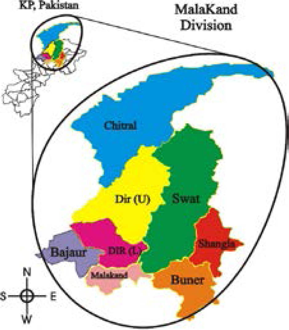
Fig. 1.
The figure shows different locations of study area.
Coprological examination and assessment of risk factors
Collection and Processing of Fecal Samples
In the current prevalence study, fresh feces from small ruminants (1307 sheep and 1274 goats) were collected across eight district-wise localities in the study area over a full year, from January 2024 to December 2024, including all four seasons. Fresh fecal materials (10 – 15 g) were collected directly from the rectum of randomly selected small ruminants using gloved fingers. Then, feces were placed in collecting bottles containing 70 % ethanol. The collected bottles were labeled with host sex, age, location, and date (Raza et al., 2014). To identify risk factors, the owners of small ruminants were asked about the host's gender, geographic location, grazing pattern, age, feeding type, health condition, prior anthelmintic treatment, and drinking water source. The collected fecal samples were brought to the parasitology laboratory in the Department of Zoology at the University of Malakand, Pakistan, for the diagnosis of Fasciolid trematodes eggs.
Sedimentation technique and egg diagnosis
The sedimentation technique was used to detect Fasciolid trematodes eggs because their eggs are larger than those of other parasites; therefore, they settle quickly in the test tube when centrifuged in a NaCl solution (Soulsby, 1982; Zajac & Conboy, 2012). In this technique, the first 3 grams of the fecal sample were dissolved in 20 ml of water, then filtered through a sieve several times. The filtrate was centrifuged for 3 – 5 min. Afterward, the supernatant was discarded, and the pellet was resuspended in normal saline. A drop of the mixture was placed on a glass slide, covered with a coverslip, and examined under the microscope at 4×, 10× and 40× for the presence of parasite eggs (Mumtaz et al., 2024; Ruhoollah et al., 2021; Soulsby, 1982).
Fasciolid eggs identification
The identification of Fasciolid eggs was based on observations of egg shape, coloration, contents, and operculum morphology, as described by Aghayan et al. (2019). The Fasciola egg is the largest helminth egg, oval in shape, with yellowish-brown to brown or golden-yellowish color, a thin shell, and an operculum at one end, as described by Hussein et al. (2010).
DNA extraction
DNA was extracted from individual Fasciola gigantica samples, which were obtained from selected slaughterhouses of the study area using a commercial DNA extraction kit (Qiagen, Hilden, Germany) following the manufacturer's instructions.
PCR amplification
For molecular characterization, DNA was extracted from adult Fasciola specimens, and the internal transcribed spacer region (ITS), a nuclear ribosomal DNA marker, was amplified using polymerase chain reaction (PCR) following the protocol of Mehmood et al. (2024) and Ruhoollah et al. (2025). ITS was selected for molecular characterization because it is a well-established nuclear ribosomal DNA marker widely used for species-level identification, phylogenetic analysis, and taxonomy of fasciolid trematodes such as Fasciola hepatica and Fasciola gigantica. It is highly conserved within species and its use is supported by the availability of reference data in public databases (Mas-Coma et al., 2009). The reaction mixture contained template DNA, forward primer (5′ GCGACCTGAAAATCTACTCTTACACAAGCG 3′) and reverse primer (5′ GACGTACGTATGGTCAAAGACCAGGTT 3′), dNTPs, Taq polymerase, and buffer in a total volume of 25 μL. Thermocycling conditions included an initial denaturation at 96 °C for 8 minutes, followed by 30 cycles of 96 °C for 60 seconds, annealing at 54°C for 60 seconds, extension at 72°C for 60 seconds, and a final extension at 72°C for 8 minutes. The amplified ITS gene PCR products were verified by agarose gel electrophoresis and visualized under UV illumination, as described by (Amor et al., 2011; Ndosi et al., 2023; Khan et al., 2025a). The amplified ITS fragment was approximately 420 bp in length for both isolates (Fig. 2).
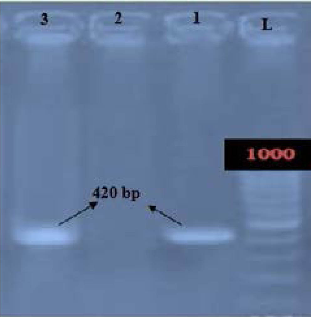
Fig. 2.
Agarose gel electrophoresis showing PCR amplification of ITS region of F. gigantica.
Phylogenetic tree construction and sequencing
The amplicons were confirmed through gel electrophoresis. Two amplified DNA products (one each from goat and sheep samples) were randomly selected, purified, and sequenced at Macrogen Inc. in Seoul, South Korea. The sequences were assembled and aligned using ClustalW multiple alignment implemented in MEGA11. Sequence analysis was performed using BLASTn and the National Center for Biotechnology Information (NCBI) databases. A phylogenetic tree was constructed using a neighbor-joining method implemented in MEGA 11.
Statistical analysis
The Statistical Package for the Social Sciences (SPSS) version 22 was used for data analysis. Data were submitted, appropriate tests were performed for each parameter, and P-values < 0.05 were considered significant. Infection rate was calculated as the percentage of animals positive for Fasciolid trematodes within specific categories (age, sex, season, or management factors), while prevalence represents the overall proportion of infected animals in the total sampled population and for species-wise distribution of Fasciola hepatica and Fasciola gigantica. Both measurements were calculated as the number of positive samples divided by the total number of samples examined × 100, and associations with risk factors were assessed using the Chi-square test at P < 0.05, as described by Irshad et al. (2023) and Hussain Shah et al. (2015).
Ethical Approval and/or Informed Consent
The study protocol (UOM/Admin/2024/501) was ethically approved by the Advanced Studies and Research Board, Malakand University, Lower Dir, Pakistan. All applicable national and institutional guidelines for the care and use of animals were followed during this research. Informed consent was obtained from the owners prior to sample collection.
Results and Discussion
Overall prevalence
A total of 77 (2.98 %) samples were positive for Fasciolid trematodes out of 2,581 fecal samples examined. The infection rate was higher in sheep, 3.9 % (51/1307), than in goats, 2.04 % (26/1274), indicating a comparatively lower occurrence of fasciolosis in goats (Table 1).
Table 1.
Overall prevalence of Fascioliasis in sheep and goats.
| S/No | Type of Host | Total Sample Examine | Total positive | Overall prevalence % |
|---|---|---|---|---|
| 1 | Sheep | 1307 | 51 | 3.9 % |
| 2 | Goat | 1274 | 26 | 2.04 % |
The overall prevalence of F. hepatica was higher than that of F. gigantica. In the case of sheep, 42 samples were positive for F. hepatica (3.21 %) and 9 for F. gigantica (0.68 %), while in goats, 19 were positive for Fasciola hepatica (1.49 %) and 7 for Fasciola gigantica (0.54 %) (Table 2).
Table 2.
Overall prevalence of Fasciolides trematodes species in sheep and goats.
| Parasite species | Total samples of Sheep (1307) | Total samples of Goats (1274) | ||
|---|---|---|---|---|
| Positive samples | Prevalence % | Positive samples | Prevalence % | |
| F. hepatica | 42 | 3.21 | 19 | 1.49 |
| F. gigantica | 9 | 0.68 | 7 | 0.54 |
The results of the present study are in line with the study conducted by Hussain Shah et al. (2015) in Peshawar, Khyber Pakhtunkhwa, who reported the prevalence of trematodes (4.6 %) and (3.4 %) in goats and sheep, respectively. Similar findings were also reported by Haleem et al. (2016) in Mardan, Khyber Pakhtunkhwa, who reported 5.89 % F. hepatica and 3.83 % F. gigantica in sheep. In contrast to the current study, Ruhoollah et al. (2021) reported that the prevalence of F. hepatica was 13.58 % and 8.39 % in sheep and goats, respectively. This difference may be due to differences in sample size, study area, and diagnostic procedures. The study conducted by Rizwan et al. (2022) in Quetta, Pakistan, which is in contrast to the present study, reported that the prevalence of F. hepatica and F. gigantica in goats was 0.0698 % and 0.209 %, respectively. This difference may be due to the arid environment of the study area, where the intermediate hosts are negligible.
Risk factors Analysis influencing fasciolosis in small ruminants
Risk factors such as host gender, age, area, season, grazing system, health condition, source of drinking water, and treatment status of small ruminants were analyzed as variables (Table 3). Chi-square analysis showed that the prevalence of fasciolosis was statistically associated with the host gender, age, area, season, grazing system, health condition, source of drinking water, and treatment status of the host animal.
Table 3.
Shows the risk factors associated with the prevalence of fascioliasis.
| Risk Factors | Variables | Sheep (1307) | Goats (1274) | P-value | ||||
|---|---|---|---|---|---|---|---|---|
| Sample | Positive % | Prevalence % | Sample | Positive | Prevalence % | |||
| Sex | Male | 526 | 23 | 4.372 | 508 | 10 | 1.96 | 0.611 |
| Female | 781 | 28 | 3.58 | 766 | 16 | 2.088 | ||
| Age | <1.5 years | 268 | 19 | 7.08 | 312 | 9 | 2.88 | 0.003 |
| >1.5 years | 1039 | 32 | 3.07 | 962 | 17 | 1.76 | ||
| Area | Dir (U) | 200 | 23 | 11.5 | 250 | 10 | 4 | 0.97 |
| Dir (L) | 445 | 20 | 4.49 | 395 | 7 | 1.77 | ||
| Bajaur | 101 | 1 | 0.99 | 114 | 2 | 1.75 | ||
| Swat | 100 | 1 | 1 | 100 | 1 | 1 | ||
| Buner | 150 | 2 | 1.33 | 150 | 2 | 1.33 | ||
| Shangla | 83 | 1 | 1.20 | 65 | 1 | 0.60 | ||
| Malakand | 150 | 2 | 1.33 | 109 | 2 | 1.83 | ||
| Chitral | 78 | 1 | 1.28 | 91 | 1 | 1.11 | ||
| Grazing system | Grazing | 504 | 16 | 3.17 | 507 | 10 | 1.97 | 0.32 |
| Stall fed | 803 | 35 | 4.35 | 767 | 16 | 2.08 | ||
| Health condition | Healthy | 1018 | 34 | 3.33 | 1041 | 14 | 1.34 | 0.0001 |
| Emaciated | 289 | 17 | 5.88 | 233 | 12 | 5.15 | ||
| Drinking water source | Open | 427 | 19 | 4.44 | 412 | 10 | 2.42 | 0.039 |
| Bore | 225 | 4 | 1.77 | 167 | 2 | 1.19 | ||
| Stream | 388 | 21 | 5.41 | 358 | 9 | 2.51 | ||
| Tap water | 267 | 7 | 2.62 | 337 | 5 | 1.48 | ||
| Season | Autumn | 294 | 10 | 3.40 | 308 | 6 | 1.94 | 0.0004 |
| Winter | 216 | 4 | 1.85 | 199 | 2 | 1.005 | ||
| Spring | 319 | 6 | 1.88 | 331 | 5 | 1.51 | ||
| Summer | 478 | 31 | 6.48 | 436 | 13 | 2.98 | ||
| Treatment | Treated | 320 | 7 | 2.18 | 308 | 4 | 1.29 | 0.036 |
| Untreated | 987 | 44 | 4.45 | 966 | 22 | 2.277 | ||
Sex-wise prevalence
To determine sex-wise prevalence, sheep and goats were categorized into two groups: males and females. Of the total animals evaluated, 526 were male sheep, 508 were male goats, 781 were female sheep, and 766 were female goats. The infection rate in male sheep was 4.372 % (23/526), higher than the rate in female sheep, which was 3.58 % (28/781). In goats, the infection rate in females was 2.088 % (16/766), compared to 1.96 % (10/508) in males. The prevalence rate according to the sex of sheep and goats in the study area was statistically nonsignificant (P = 0.611) (Table 2).
The results of the current study align, to some extent, with those documented by Mumtaz et al. (2024); Ruhoollah et al. (2021); Durrani et al. (2017); Haleem et al. (2016); and Hussain Shah et al. (2015), who also reported variations in fasciolosis prevalence correlated with the sex of sheep and goats. These variations may be linked to management practices, as female sheep are often confined, whereas male goats are typically allowed to graze freely. Moreover, variables including breed, species, and regional distribution may affect infection rates. In Bangladesh, Mazid et al. (2006) documented a greater incidence in female sheep and goats, ascribed to pregnancy and hormonal variations.
Age-wise prevalence
To find out the age-wise prevalence of infection, animals were classified as young (<1.5) and adults (>1.5) because this age approximates sexual maturity and the transition from immature to fully developed immune responses in small ruminants (Soulsby, 1982; Radostits et al. 2007). A total of 1307 (<1.5=268,>1.5=1039) fecal samples of sheep, while 1274 (<1.5=312, >1.5=962) fecal samples of goats were investigated. It was observed that the prevalence of fasciolosis in young sheep was 7.08 % (19/268), and in goats was 2.88 % (9/312), which was higher than in adult sheep, 3.07 % (32/1039), and in goats, 1.76 % (17/962), respectively. Thus, age-wise prevalence showed that infection was higher in young animals than in older animals, with a statistically significant difference (P = 0.003). Our findings are similar to the results of Mumtaz et al. (2024) and Rashid et al. (2016), who reported that young animals were more infected than older ones. The results of Durrani et al. (2017) contradict those of the present study, which found that the prevalence of fasciolosis was higher in older animals as compared to young ones. The higher rate of fasciolosis in young small ruminants in the current study may be due to young animals often being allowed to graze freely, increasing their exposure to Fasciolid eggs. In contrast, adults are mostly kept captive, limiting their risk of infection. Secondly, it may be due to the weaker immune systems of young small ruminants compared with older animals.
Season-wise prevalence
In the current study, it was found that the prevalence of fasciolosis was higher in sheep, 6.48 % (31/478), and goats, 2.98 % (13/436), in summer, and the lowest prevalence in sheep, 1.005 % (2/199), and in goats, 1.85 % (4/216), was recorded in winter. In spring, the prevalence in sheep and goats was 1.88 % (6/316) and 1.51 % (5/331), respectively, while in autumn, the prevalence was 3.40 % (10/294) and 1.94 % (6/308), respectively. The results were statistically significant (P = 0.0004) (Table 3). The findings of this study are similar to those of Ruhoollah et al. (2021). They also observed the highest prevalence of fasciolosis in small ruminants during summer. The findings of Ayele et al. (2018) and Durrani et al. (2017) are in contrast to the current study, as they reported the highest prevalence in sheep and goats in autumn, whereas Ashoor & Wakid (2023) reported the highest prevalence in spring. In the current study, the highest prevalence of infection in summer may be due to high temperatures and moisture during the Monsoon season, thereby increasing fluke ova hatching and the encystation of Fasciolid cercariae, ultimately increasing the population of intermediate host snails. Consequently, increased cercarial production and encystation on vegetation enhance the transmission risk of fasciolosis in grazing small ruminants (Mas-Coma et al., 2005). In the winter season, the prevalence of fasciolosis was lowest due to cool, dry conditions that reduced the population of intermediate hosts.
Body condition-wise prevalence
The health status of the host animals in the current study was also considered an important parameter for the prevalence of fasciolosis. In the present study, sheep and goats were categorized into two groups, i.e., normal and emaciated. In the current study, the prevalence of fasciolosis was high in emaciated sheep, 5.88 % (17/289), and goats, 5.15 % (12/233), while the prevalence of fasciolosis in normal sheep and goats was 3.33 % (34/1018) and 1.34 % (14/1041), respectively. The results were statistically significant (P = 0.0001) (Table 3). These findings are similar to the findings of Ayele et al. (2018). They also reported a higher prevalence of fasciolosis in sheep and goats with poor body condition than in healthy animals. However, the results of Dabasa et al. (2017) are in contrast to those of the current study, which reported a high prevalence in normal sheep and goats. The high infection rate among emaciated animals in the current study may be due to a weakened immune system.
Area-wise prevalence
In the study area, which includes eight distinct locations, the highest prevalence of fasciolosis in sheep was observed in Dir Upper (11.5 %), followed by Dir Lower (4.49 %), Buner and Malakand (1.33 %), Chitral (1.28 %), Shangla (1.20 %), Swat (1 %), and the lowest prevalence in Bajaur (0.99 %). On the other hand, the highest prevalence in goats was also recorded in Dir Upper (4 %), followed by Malakand (1.83 %), Dir Lower (1.77 %), Bajaur (1.75 %), Buner (1.33 %), Chitral (1.11 %), Swat (1 %), and the lowest prevalence in Shangla (0.60 %). No statistically significant (P = 0.97) difference in prevalence was observed among various regions of the study area (Table 3). The findings of Tasleem et al. (2023) align with those of the present investigation, since they observed varying prevalence rates of fasciolosis infection in livestock across different sites within the study area. The variation in fasciolosis prevalence among the research locations was attributable to factors such as differing grazing systems, feeding environments, management practices, and owner care behaviors.
Grazing system-wise prevalence
The prevalence of fasciolosis was higher in stall-fed sheep (4.35 %) and goats (2.08 %) compared to grazing sheep (3.17 %) and goats (1.97 %), with a p-value of 0.32, indicating statistical in-significance (Table 3). The findings of the present study differ from those of Mohammed et al. (2023), who observed that goats kept on open grazing had higher infection rates than those in stalled feeders. The elevated occurrence of fasciolosis in stall-fed small ruminants may result from inadequate cleanliness in stalls or the harvesting of plants and grasses from regions inhabited by metacercariae, such as wetlands and marshes.
Treatment-wise prevalence
The prevalence of fasciolosis was significantly higher in untreated sheep (4.45 %) and goats (2.27 %) compared to treated sheep (2.18 %) and goats (1.29 %), with a p-value of 0.036 indicating statistical significance (Table 3). The findings of our study align with those of Mohammed et al. (2023), who similarly report a higher prevalence of fasciolosis in untreated sheep and goats than in treated animals, attributable to the administration of anthelmintic medications. The elevated infection rate in untreated animals within the study area may result from inadequate treatment procedures, diminished attention, and a negligent attitude among owners towards small ruminants.
Water source-wise prevalence
The prevalence of fasciolosis in drinking water sources was highest in sheep (5.41 %) and goats (2.51 %) using stream water, followed by sheep (4.44 %) and goats (2.42 %) drinking open water, and finally sheep (2.62 %) and goats (1.48 %) using tap water. The lowest infection rates were seen in sheep (1.77 %) and goats (1.19 %) consuming bore water, with a p-value of 0.036, indicating statistical significance (Table 3). The results of the current investigation parallel the findings of Ruhoollah et al. (2021), who reported a high incidence of Fasciolid in sheep and goats that drink from open water sources. The elevated incidence of fasciolosis in animals that consume stream and open water may be attributed to the higher levels of pollution and contamination in these water sources. However, bore and tap water sources are often safe, clean, and fit for consumption.
Phylogenetic tree
After DNA extraction and ITS gene amplification, two representative PCR products, one from sheep and one from goat isolates, were selected for sequencing analysis. Following sequencing, the phylogenetic tree was constructed using the Neighbor-Joining method, comparing the two isolates from the present study with 11 published Fasciola gigantica sequences from Bangladesh, Iran, Nigeria, Vietnam, India, Egypt, Saudi Arabia, and Thailand. The phylogenetic analysis demonstrated that the first isolate (Malakand-1) was closely related to an Iranian isolate (KM085323), supported by a bootstrap value of (93 %) indicating a strong genetic relationship. Malakand-1 also showed close association with isolates from Saudi Arabia and Egypt, suggesting genetic similarity among geographically related Asian and Middle Eastern isolates.
The second isolate (Malakand-2) was close to isolates from Nigeria and Saudi Arabia, but this node received weak bootstrap support (4 %), indicating an unclear relationship. The whole tree shows both genetic similarities and differences between F. gigantica isolates from different places.
The similarity of the Malakand-1 isolate to Iranian and Middle Eastern genomes indicates a genetic connection between these regions, possibly due to livestock movements or historical transmission pathways. The distinctive location of Malakand-2, however lacking robust proof, may indicate the presence of multiple F. gigantica lineages in the vicinity. The results also indicate that additional molecular investigations using additional genetic markers and larger sample sizes are recommended to better understand the population structure and evolutionary relationships of the fasciolid trematodes (Fig 3).
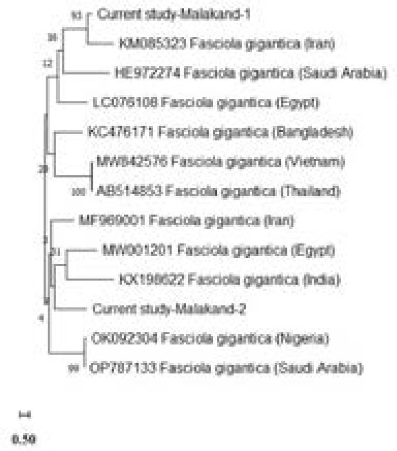
Fig. 3.
Phylogenetic tree of F. gigantica in goats and sheep utilising the ITS marker. The generated tree depicts the genetic relationships among the isolates of the present investigation and reference sequences from various regions. Bootstrap values are provided at branch points indicating the confidence levels of the groups.
Conclusion
The current study demonstrated a moderate prevalence of fasciolosis in sheep and goats in Malakand Division, Pakistan. It was significantly associated with prior anthelmintic treatment, host age, drinking water quality, feeding pattern, and health status. Molecular analysis additionally confirmed that the species was F. gigantica and showed that it was closely related to Egyptian, Iranian, Nigerian, and Saudi Arabian isolates. The high bootstrap support in phylogenetic analysis indicates that the local isolates are genetically similar, warranting continued molecular monitoring. Effective control measures, such as the periodic use of antihelminthics and fasciolicides, are recommended to maximize benefits in small ruminants and prevent economic losses.
Acknowledgment
The authors would like to acknowledge the Deanship of Graduate Studies and Scientific Research at Taif University for funding this work. We are grateful to the laboratory staff, slaughterhouse workers, and livestock farmers in Malakand Division for their co-operation and assistance during sample collection and fieldwork.