
Introduction
Dipylidium caninum (Cestoda: Dipylidiidae) is a globally distributed tapeworm that inhabits the small intestine of domestic dogs and cats and may occasionally infect humans, particularly children (Bowman et al., 2002; Jiang et al., 2017). Given the documented genetic heterogeneity and unresolved taxonomy within the Dipylidium caninum group, the lineage detected in the present study is hereafter referred to as Dipylidium caninum within the D. caninum species complex, unless otherwise specified. Transmission occurs through an indirect life cycle in which cysticercoids develop in flea intermediate hosts, mainly Ctenocephalides felis and Ctenocephalides canis, and definitive hosts become infected by ingesting infected fleas (Bowman et al., 2002).
Although D. caninum is among the most frequently reported cestodes in companion animals, human dipylidiasis has traditionally been considered rare. However, confirmed human cases reported in Asia and Eastern Europe indicate that zoonotic transmission continues in regions with high flea exposure (Ramana et al., 2011; Jiang et al., 2017; Bronstein et al., 2020). A recent global synthesis further supports the ongoing public health relevance of this parasite (Rousseau et al., 2022).
Recent molecular studies have challenged the long-standing view of D. caninum as a genetically uniform species. Substantial mitochondrial divergence has been documented, and at least two host-associated genotypes, adapted to dogs and cats, respectively, have been proposed (Labuschagne et al., 2018; Beugnet et al., 2018). In addition, morphologically similar taxa such as D. carracidoi may have been historically misidentified as D. caninum, highlighting unresolved taxonomic complexity within Dipylidiidae (Millán & Casanova, 2009; Enitez-Bolivar et al., 2022).
In Türkiye, numerous coprological surveys have reported D. caninum in both owned and shepherd dog populations across multiple provinces, with prevalence varying by geography, diagnostic approach and dog population structure (Yaman et al., 2006; Orhun & Ayaz, 2006; Kozan et al., 2007; Yıldırım et al., 2007; Öter et al., 2011; Işık et al., 2014; Nas & Biçek, 2018; Karakuş & Denizhan, 2019; Ceylan et al., 2024). These findings are consistent with earlier studies dating back to the late 1970s, indicating long-term endemic transmission in both rural and urban settings (Güralp et al., 1977; Taşan, 1983; Tınar et al., 1989; Doğanay & Öge, 1993; Şahin et al., 1993; Aydenizöz, 1997; Ayçiçek et al., 1998).
Despite this extensive parasitological background, molecular data on Dipylidium caninum in dogs from Türkiye remain scarce. Although recent work has demonstrated the presence of D. caninum DNA in flea vectors (Özdemir et al., 2025), molecular prevalence and sequence-based characterization studies in canine hosts are still limited, particularly at the regional level. In eastern Türkiye, previous studies have mainly focused on the molecular epidemiology of zoonotic and parasitic agents in animals and environmental samples, highlighting their prevalence and potential public health significance (Ertaş et al., 2022; Ertaş & Ayan, 2022; Kılınç et al., 2023; Koca et al., 2023; Akkuş & Ertaş Oğuz, 2024; Ertaş Oğuz et al., 2025). However, molecular information on Dipylidiidae cestodes in shepherd dog populations from this region remains limited.
Therefore, the present study aimed to investigate the molecular presence of Dipylidiidae cestodes in shepherd dogs from Iğdır Province, eastern Türkiye, using a COX1-based PCR approach and to characterize the detected lineage through partial COX1 gene sequencing. The resulting data provide new molecular evidence from an under-studied region and contribute to a better understanding of Dipylidium diversity and transmission dynamics in shepherd dog populations.
Materials and Methods
Sample Collection
In 2023, a total of 100 fecal samples were collected from shepherd dogs in Iğdır Province, eastern Türkiye. Before sampling, dog owners were informed about the study, and voluntary consent was obtained. During field visits, dogs were evaluated individually with assistance from their owners. Fresh fecal samples were collected non-invasively either directly from the ground when they could be attributed to the relevant dog or immediately after observed defecation. To prevent sample mixing, each sample was collected using disposable gloves, placed into a separate leak-proof container, and labeled with a unique sample code. Age, sex, and flock/farm information were also recorded. The samples were first transported to the laboratory of Iğdır University Animal Hospital and then transferred under cold-chain conditions to the Department of Genetics, Faculty of Veterinary Medicine, Van Yüzüncü Yıl University, for molecular analyses.
DNA extraction
Genomic DNA was extracted from approximately 200 mg of each fecal sample using the GeneMATRIX Stool DNA Purification Kit (Eurx, Poland), following the manufacturer’s protocol. DNA concentration and purity were assessed spectrophotometrically, then all extracts were stored at −20 °C until PCR analysis.
PCR amplification
A 446-bp fragment of the mitochondrial cytochrome c oxidase sub-unit I (COX1) gene was amplified using the primer pair JB3 and JB4.5 described by Bowles et al. (1992). The primer sequences were as follows:
JB3 (forward): 5′-TTTTTTGGGCATCCTGAGGTTTAT-3′
JB4.5 (reverse): 5′-TAAAGAAAGAACATAATGAAAATG-3′
PCR reactions were performed in a final volume of 25 μL containing 200 μM of each dNTP, 1.5 mM MgCl2, 10 pmol of each primer, 1.25 U Taq DNA polymerase, 1× PCR buffer, nuclease-free water, and 2 μL of template DNA. Amplification was carried out in a Veriti™ Thermal Cycler (Applied Biosystems) with the following cycling conditions: initial denaturation at 95 °C for 15 min, followed by 35 cycles of denaturation at 95 °C for 1 min, annealing at 45 °C for 1 min, and extension at 72 °C for 1 min, with a final extension step at 72 °C for 10 min. PCR products were resolved by electrophoresis on 1.5 % agarose gels stained with Safe-T-Stain, run at 90 V for 60 min, and visualized under UV light using a Syngene Bio-Imaging System.
Sequencing and sequence quality control
PCR amplicons that produced clear bands of the expected size were purified and bidirectionally sequenced by BM Labosis Laboratory (Ankara, Türkiye). Forward and reverse chromatograms (.ab1 files) were examined in MEGA11 (Tamura et al., 2021), and base calls were manually inspected. Only sequences with clear chromatogram peaks and agreement between forward and reverse reads at all variable positions were used for further analyses. A 393-bp consensus COX1 sequence was generated from the forward and reverse reads.
BLAST analysis
The consensus COX1 sequence was searched against the NCBI nucleotide database using BLASTn to assess molecular similarity with reference sequences. BLASTn hits were evaluated according to query coverage, percentage identity, E-value, accession number, host, country of origin, submission date, and reference/source information. The relevant BLASTn matches to Dipylidium caninum and related cestode sequences are presented in Supplementary Table 1.
Supplementary Table S1.
BLASTn comparison and phylogenetic dataset of Dipylidium caninum COX1 sequences
| No. | Accession | Isolate / description | Host | Country | Identity (%) | Query cover | Sequence length (bp) |
|---|---|---|---|---|---|---|---|
| 1 | PZ299077.1 | D. caninum isolate Igdir_Dog1 | Shepherd dog / Canis lupus familiaris | Türkiye | 100 | 100 % | 396 |
| 2 | PX765914.1 | D. caninum isolate Hp03 | Dog | Ghana | 92.12 | 93 % | 373 |
| 3 | OR251824.1 | D. caninum isolate Sv_PS_Amz_22 | Speothos venaticus / bush dog | Colombia | 91.92 | 100 % | 1602 |
| 4 | OR251823.1 | D. caninum isolate Sv_PS_Amz_21 | Speothos venaticus / bush dog | Colombia | 91.92 | 100 % | 1602 |
| 5 | OK523384.1 | D. caninum isolate Dcan_canine_FL1, complete mitogenome | Canis lupus familiaris | USA | 91.92 | 100 % | 14296 |
| 6 | PZ165824.1 | D. caninum isolate DICAA | Canis lupus familiaris | Croatia | 91.92 | 100 % | 396 |
| 7 | PX765913.1 | D. caninum isolate Hp02 | Dog | Ghana | 91.85 | 93 % | 373 |
| 8 | PX765912.1 | D. caninum isolate Hp01 | Dog | Ghana | 91.85 | 93 % | 373 |
| 10 | MN099047.1 | D. caninum complete mitogenome | Stray dog | China | 91.67 | 100 % | 14226 |
| 11 | PP842203.1 | D. caninum isolate DogSRTDc2 | Dog | Türkiye | 91.27 | 95 % | 383 |
| 12 | PP842202.1 | D. caninum isolate DogSRTDc1 | Dog | Türkiye | 91.27 | 95 % | 383 |
| 13 | MG587892.1 | D. caninum isolate R166, complete mitogenome | Feline | South Africa | 89.8 | 99 % | 13598 |
| 14 | OQ281679.1 | D. caninum isolate VCG6 | Felis catus | Mexico | 89.8 | 99 % | 416 |
| 15 | OK523385.1 | D. caninum isolate Dcan_feline_KS1, complete mitogenome | Felis catus | USA | 89.8 | 99 % | 13598 |
| 16 | MT806359.1 | D. caninum voucher 4245P18 | Vulpes vulpes / red fox | Italy | 89.8 | 99 % | 411 |
| 17 | PX765915.1 | D. caninum isolate Hp04 | Dog | Ghana | 89.67 | 93 % | 373 |
| 18 | OR511473.1 | D. caninum isolate Alex-2 | Stray cat | Egypt | 89.53 | 92 % | 363 |
| 19 | PZ165825.1 | D. caninum isolate DICAB.1 | Felis catus | Croatia | 89.39 | 90 % | 358 |
| 20 | ON506044.1 | D. caninum isolate Isb1 | Cat | Pakistan | 89.34 | 92 % | 384 |
| 21 | PP054312.1 | D. caninum haplotype A | Felis catus familiaris | Ethiopia | 89.27 | 96 % | 963 |
| 22 | OR511472.1 | D. caninum isolate Alex-1 | Stray cat | Egypt | 88.71 | 92 % | 363 |
| 23 | ON954760.1 | D. caninum isolate dog1 | Dog | Iraq | 88.01 | 92 % | 384 |
| 24 | ON954628.1 | D. caninum isolate cat1 | Cat | Iraq | 88.01 | 92 % | 384 |
| 25 | ON514129.1 | D. caninum isolate Isb2 | Cat | Pakistan | 88.01 | 92 % | 384 |
| 26 | ON954763.1 | D. caninum isolate cat1 | Cat | Iraq | 87.91 | 92 % | 383 |
| 27 | ON954761.1 | D. caninum isolate dog2 | Dog | Iraq | 86.58 | 92 % | 384 |
| 28 | PP054311.1 | Dipylidium sp. 01-SA haplotype A | Proteles cristata / aardwolf | South Africa | 85.86 | 100 % | 1647 |
| 29 | PZ165826.1 | D. caninum isolate DICAB.2 | Canis lupus familiaris | Croatia | — | — | 358 |
| 30 | ON954762.1 | D. caninum isolate dog3 | Dog | Iraq | — | — | 384 |
| 31 | ON954764.1 | D. caninum isolate cat2 | Cat | Iraq | — | — | 384 |
| 32 | ON954765.1 | D. caninum isolate cat3 | Cat | Iraq | — | — | 384 |
Multiple sequence alignment
Selected reference COX1 sequences of D. caninum and related dipylidiid taxa were retrieved from GenBank. All sequences were combined into a single FASTA dataset. The sequences were first checked, and multiple sequence alignment was then performed using MAFFT with the “adjust direction” option. Poorly aligned, ambiguous, and non-overlapping regions were removed to improve alignment quality, resulting in a final alignment of 358 bp. The accession numbers and metadata of these sequences are listed in Supplementary Table 1. ON981095 (Joyeuxiella pasqualei) was used as the outgroup.
Phylogenetic analysis
Phylogenetic analysis was performed in IQ-TREE v3 using the Maximum Likelihood method. The best-fit nucleotide substitution model was selected with ModelFinder Plus, and the GTR+I+G model was used for tree construction. Branch support was assessed with 1,000 ultrafast bootstrap replicates and 1,000 SH-aL-RT replicates. The resulting tree was rooted using Joyeuxiella pasqualei ON981095 as the outgroup and visualized in FigTree v1.4.5. Branch values are shown as SH-aLRT support (%) / ultra-fast bootstrap support (%).
Literature data extraction
To place the findings from Iğdır in a broader epidemiological context, published prevalence data for Dipylidium caninum infections in dogs were compiled from the literature. For each country, mean prevalence values were calculated across available studies irrespective of diagnostic method, and these country-level estimates were subsequently grouped to generate continent-level summaries. Although this approach does not constitute a formal meta-analysis, it provides a comparative framework for interpreting regional variation and situating the Iğdır data within global transmission patterns. A descriptive overview of the global prevalence of D. caninum in dogs is presented in Supplementary Figure S1. Data handling and visualization were performed using R statistical software (R Core Team, 2025).
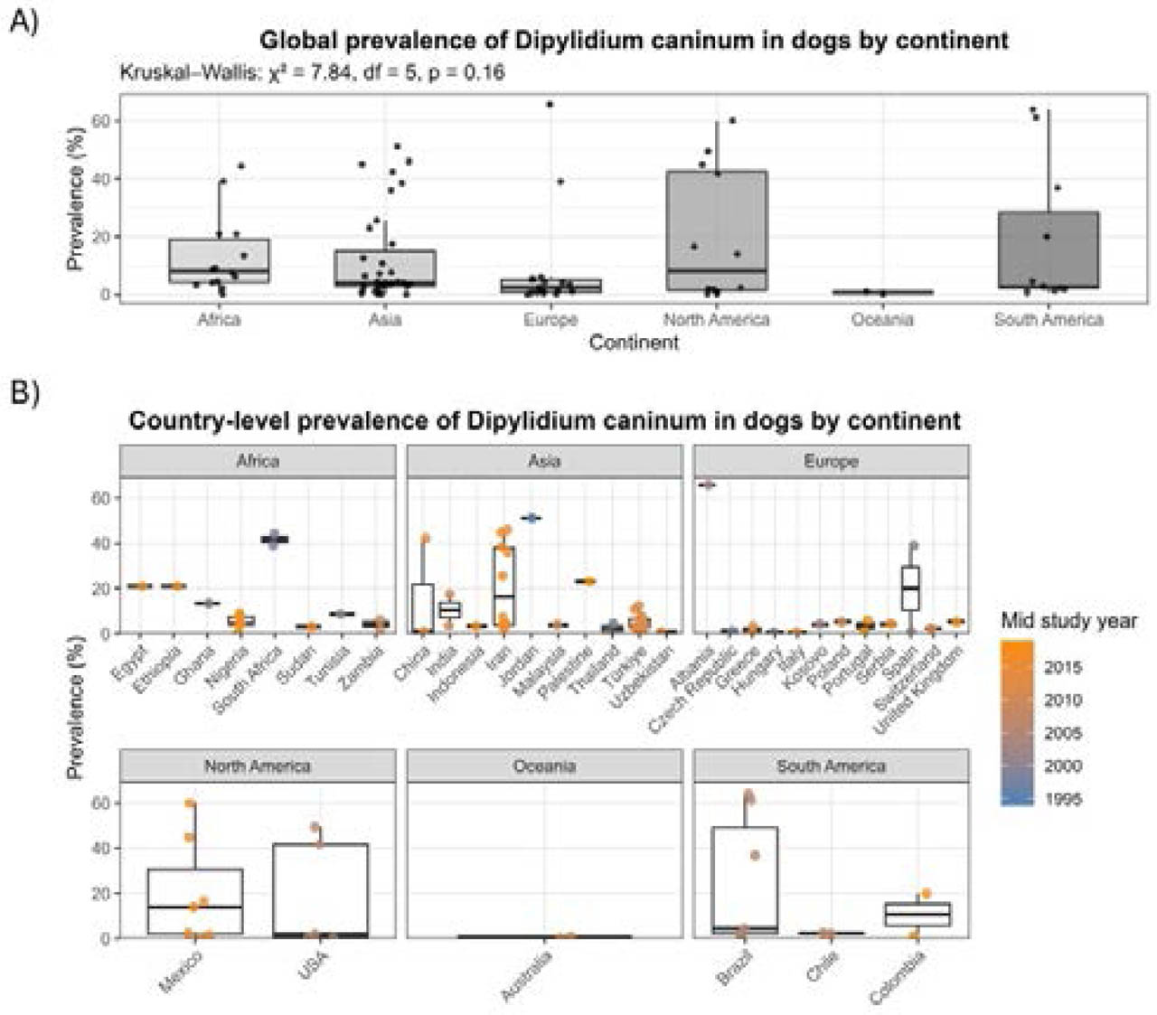
Supplementary Fig. S1.
Global prevalence of Dipylidium caninum in dogs.
Reported canine D. caninum prevalence data from previously published studies were descriptively summarized by continent and country to provide broad epidemiological context. (A) Prevalence values across continents. Box plots show the median, interquartile range, and individual study points. (B) Country-level prevalence grouped by continent. Colored points represent individual studies according to approximate mid-study year. No molecular sequence data were included in this overview. Because the included studies differed in sampling design, dog population, geographic setting, and diagnostic method, this figure should be interpreted only as descriptive context and not as a formal quantitative comparison among regions.
Ethical Approval and/or Informed Consent
Permission was obtained from the animal owners for this study. Additionally, fecal samples were collected from the ground without coming into contact with the animals. According to Turkish HADYEK (Local Ethics Committee for Animal Research) regulation, fecal sample collection is not subject to ethics committee approval when no animal handling or intervention is performed. Therefore, an ethics committee approval number was not required for this study.
Results
Out of 100 screened fecal samples, eight (8 %) yielded a PCR product of the expected size for the COX1 gene fragment. All positive samples produced identical sequences across the analyzed region, indicating the presence of a single mitochondrial haplotype among the infected dogs.
The obtained 396 bp partial mitochondrial COX1 sequence of Dipylidium caninum isolate Igdir_Dog1 from a shepherd dog in Iğdır Province, Türkiye, was deposited in GenBank under accession number PZ299077.1.
BLASTn analysis showed that the sequence matched D. caninum entries deposited in GenBank, including partial COX1 sequences and complete mitochondrial genome records. The highest identity was observed with a dog-derived sequence from Ghana (PX765914.1; 92.12 % identity, 93 % query cover), followed by bush dog-derived Colombian sequences (OR251824.1 and OR251823.1), a canine complete mitogenome from the USA (OK523384.1), and a dog-derived Croatian sequence (PZ165824.1), each showing 91.92 % identity and 100 % query cover. Turkish dog-derived sequences from Elazığ (PP842203.1 and PP842202.1) showed 91.27 % identity with 95 % query cover. Cat-associated sequences were also included in the comparison and showed lower identity values, ranging approximately from 88.71 % to 89.80 %. The full BLASTn comparison is provided in Supplementary Table 1.
Discussion
The present study provides molecular evidence of Dipylidium caninum in dogs from Iğdır Province, eastern Türkiye, and represents one of the first COX1-based molecular characterizations of Dipylidiidae in this region. Although D. caninum has long been reported in Türkiye using conventional parasitological methods, molecular data remain limited. Two unpublished GenBank records from Elazığ (PP842202.1 and PP842203.1; Aslan Celik et al., GenBank direct submissions) and the molecular detection of D. caninum DNA in fleas from Tokat indicate that molecular evidence from Türkiye is gradually increasing (Özdemir et al., 2025). In this context, the Iğdır sequence reported here adds new COX1 data from eastern Türkiye and helps expand the limited molecular data-set available for Türkiye.
In the phylogenetic tree, the analyzed D. caninum sequences were arranged into three broad groups, but the separation between dog- and cat-associated lineages was unclear (Fig. 1). The first group mainly contained cat-associated and mixed-host sequences from South Africa, Mexico, the USA, Italy, Egypt, Croatia, Pakistan and Ethiopia (Labuschagne et al., 2018; Jesudoss Chelladurai et al., 2023; Camacho-Giles et al., 2024; Citterio et al., 2021; Satour et al., 2026; Beck & Šikić, 2026; Alvi et al., 2022; Dumendiak et al., 2024). This group also included some non-cat hosts, suggesting that host origin alone may not fully explain the observed phylogenetic pattern. Dogs dominated the second group and other canine-derived sequences from Ghana, Colombia, the USA, Croatia, China and Türkiye (Xie et al., 2019; Uribe et al., 2023; Jesudoss Chelladurai et al., 2023; Addy et al., 2026; Beck & Šikić, 2026; Aslan Çelik et al., 2024). The Iğdır isolate PZ299077.1 was positioned within this broad canine-derived group, together with the previously submitted Turkish dog sequences from Elazığ and several dog or wild-canid sequences from other regions. This placement indicates that the Iğdır sequence is not isolated from the global D. caninum dataset, but falls within a group largely represented by canine hosts. The third group included mainly Iraqi and Pakistani sequences obtained from both dog and cat hosts, forming a mixed regional cluster (Al-Ardi, 2022; Alvi et al., 2022). This pattern may reflect regional sequence similarity rather than a strict host-associated structure. Overall, the phylogenetic pattern supports the identification of the Iğdır isolate D. caninum and shows that it contributes new eastern Turkish COX1 data to the limited molecular records available from Türkiye. However, because several branches showed low to moderate support values and the analyzed COX1 fragment was relatively short, the observed clustering should be interpreted cautiously rather than as definitive evidence of host-specific lineages.
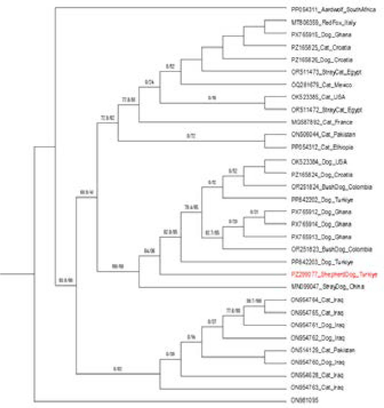
Fig. 1.
Maximum Likelihood phylogenetic tree based on a 363-bp COX1 alignment of Dipylidium caninum and related Dipylidiidae sequences.
The tree was reconstructed in IQ-TREE v3 using the GTR+I+G substitution model and rooted with Joyeuxiella pasqualei ON981095 as the outgroup. Branch values are shown as SH-aLRT support (%) / ultrafast bootstrap support (%) based on 1,000 replicates. The Turkish isolate generated in this study, PZ299077.1, is highlighted in red.
Previous molecular and genomic studies have shown that D. caninum contains largely host-associated canine and feline lineages, with marked genetic differentiation between these groups (Labuschagne et al., 2018; Rousseau et al., 2022; Jesudoss Chelladurai et al., 2023). However, host association is not strict, and dogs may carry parasites related to different genotype groups. Recent data from Ghana also reported both canine and feline–canine-associated genotypes, although most dog-derived isolates were assigned to the canine genotype (Addy et al., 2026). Therefore, the placement of PZ299077.1 near Ghanaian dog-derived sequences and other canine-host records supports its affinity with the canine-derived group. Considering the known mitochondrial diversity within the D. caninum complex, the relatively low COX1 identity values observed here do not preclude this placement. However, because the analysis was based on a partial COX1 fragment, the result should be interpreted as molecular placement within the canine-derived group rather than as a final genotype-level conclusion.
Earlier parasitological surveys conducted in different provinces of Türkiye have repeatedly reported D. caninum in dogs using copromicroscopic examination or necropsy-based approaches, with prevalence values varying widely depending on geographic region, dog population structure, and diagnostic methodology (Güralp et al., 1977; Taşan, 1983; Tınar et al., 1989; Doğanay & Öge, 1993; Aydenizöz, 1997; Ayçiçek et al., 1998; Yaman et al., 2006; Kozan et al., 2007; Yıldırım et al., 2007; Öter et al., 2011; Işık et al., 2014; Nas & Biçek, 2018; Karakuş & Denizhan, 2019; Ceylan et al., 2024). However, most of these investigations lacked molecular confirmation, leaving uncertainties regarding species identity and the extent of genetic diversity within Dipylidiidae. The national overview presented in Figure 2 supports this pattern. It shows that D. caninum has been reported from both the western and eastern regions of Türkiye, using both dog-based surveys and flea-based molecular detection. The 5 % molecular prevalence detected in Iğdır therefore adds PCR-based evidence from eastern Türkiye and supports the need for further molecular surveillance. Additionally, the Tokat flea-based PCR record was considered as supporting evidence for transmission rather than as directly comparable dog prevalence data (Özdemir et al., 2025).
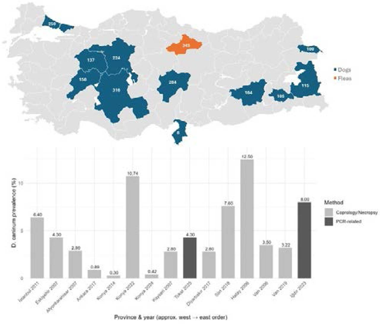
Fig. 2.
Geographic and temporal distribution of Dipylidium caninum in Türkiye.
The map and accompanying bar plot summarize published prevalence data from 2006 to 2025 across different provinces of Türkiye. Coprological and necropsy-based surveys are shown in light shading, whereas PCR-based studies are shown in dark shading. The figure illustrates the long-term and widespread circulation of D. caninum in both western and eastern regions of the country.
In conclusion, this study provides a new COX1-based molecular record of D. caninum in dogs from Iğdır Province, eastern Türkiye. It adds new mitochondrial data to the limited molecular dataset currently available from Türkiye. The analyzed sequence showed affinity with canine-derived D. caninum sequences in the phylogenetic analysis and contributes additional information to the growing body of evidence for genetic diversity within the D. caninum complex. In line with recent studies, the present findings also highlight the need for broader sampling and the inclusion of additional mitochondrial and nuclear markers from underrepresented regions to understand better the evolutionary structure and global diversity of this parasite. From a One Health perspective, continued molecular surveillance of dogs, together with monitoring of flea intermediate hosts, may contribute to an improved understanding of the transmission dynamics and zoonotic risk associated with canine dipylidiasis.
Acknowledgements
The authors thank the Department of Genetics, Faculty of Veterinary Medicine, Van Yüzüncü Yıl University, for providing technical support and access to laboratory facilities.
Notes
[1] Contributed by Author Contributions
Fatma Ertaş Oğuz: Study design, field sampling, laboratory work, and final review of the manuscript. Hasret Öztürk: GenBank data handling, data analysis, figure preparation, and manuscript writing and revision. Adnan Ayan: Laboratory work, data analysis, and final review of the manuscript.