Helminth infections continue to be a major worldwide health burden, especially in developing regions with inadequate access to healthcare and poor sanitation. Helminth infections are caused by parasitic worms such as nematodes, cestodes, and trematodes (Hotez et al., 2014). Also, parasitic nematodes that infect animals lead to significant economic losses worldwide (Ye et al., 2022). Helminth infections are predominantly transmitted through contaminated food, water, soil, or vector-borne mechanisms, depending on the species involved. They frequently afflict people living in underdeveloped areas with inadequate access to clean water, poor sanitation, and inadequate healthcare systems (Pullan et al., 2014). Depending on the type of worm involved, the severity of the infection, and the host’s immune system, helminth infections can present with a wide range of clinical symptoms. According to Bethony et al. (2006), symptoms can vary from minor gastrointestinal distress to serious consequences such anemia, malnutrition, growth retardation, cognitive decline, and in cases of severe infections, even death.
Anthelmintic drugs are essential for managing and controlling helminth infections. Nevertheless, there are difficulties, such as medication resistance, a lack of available treatments, and possible side effects. Therefore, safe and effective complementary or alternative therapies are needed (Geary et al., 2012). The potential of plant extracts as substitute anthelmintic has drawn more attention in recent years.
Throughout history, traditional medical systems have utilized plants as a means of treating a wide range of diseases. Alkaloids, tannins, flavonoids, and saponins are examples of bioactive substances found in many plant species that have anthelmintic qualities and can target various stages of the helminth life cycle (Hoste et al., 2012). Research has demonstrated that a variety of helminth species can be effectively inhibited by plant extracts through notable anthelmintic activity (Ondua et al., 2021; Albani et al., 2022; Wulandari et al., 2023; Deori et al., 2024). These extracts may exert their effects by disrupting the integrity of the worm’s cuticle, interfering with their metabolic processes, or modulating the host’s immune response to enhance parasite clearance (Rashwan et al., 2020).
Olive trees, or Olea europaea, are well known for their nutritional and therapeutic qualities. This is especially in Mediterranean regions, where olives have long been used in traditional medicine. The bioactive substances found in the tree’s leaves, fruits, and oil include hydroxytyrosol, oleuropein, and various flavonoids (Tekaya et al., 2022; Mir-Cerdà et al., 2024). These compounds have been well researched for their potential to improve heath (Manna et al., 1997; Sudjana et al., 2009).
Olive leaves have shown promise as a natural anthelmintic due to their potent bioactive compounds. Research reveals that oleuropein and hydroxytyrosol can damage the worm’s cuticle, prevent the hatching of eggs, and alter the host’s immune system to fight parasitic diseases (Pereira et al., 2007; Goulas et al., 2009). These substances have strong anti-inflammatory and antioxidant characteristics, which further support their potential application as all-natural anthelmintic drugs by lowering the oxidative stress and inflammation linked to helminth infections.
Moreover, numerous investigations have demonstrated the efficacy of O. europaea extracts and specific compounds in them in a range of in vivo and in vitro models of cancer. The application of cell culture models of disease has been the focus of these investigations. Although O. europaea’s exact cause of cytotoxicity is unknown, substances like verbascoside and oleuropein have been shown to have separate cytotoxic effects on animal models of cancer (Antoniou & Hull, 2021)
The purpose of this study was to evaluate the phytochemical profiling and cytotoxic and anthelmintic activity of O. europaea (stem and leaves) methanolic extract (in vitro), considering the pressing need for new anthelmintic treatment and encouraging early results on the efficacy of olive extracts.
The O. europaea leaves and stems were collected in Al-Baha, Saudi Arabia. A taxonomist from the Botany Department of King Saud University (Riyadh, Saudi Arabia) identified and confirmed the botanical identity of the plant. The Manikandan et al. (2008) procedure was used to prepare the extracts: leaves and stem of O. europaea were dried in the air at 40 °C, crushed into a powder (~150 g each), and then extracted for an entire day at 4 °C using 70 % methanol. The extract was filtered and concentrated under reduced pressure in a Yamato RE300 rotary vacuum evaporator (Japan) at 55ºC. To evaporate the alcohol and obtain a dry extract, it was placed at 37°C for 15 hours. The powder was dissolved in distilled water (H2O) for further steps.
The infrared spectra of the samples were obtained using a Fourier Transform Infrared (FT-IR) spectrometer, specifically the Nicolet 6700 model (Thermo Fisher Scientific, Waltham, MA, USA) (Abu Hawsah et al., 2023). This instrument is equipped with an infrared source, a KBr beam splitter, and a DTGS detector, allowing measurements in the middle infrared (MIR) region (4000 – 400 m−1). For the measurements, the KBr pressed disk technique was employed, utilizing approximately 1 mg of the sample combined with 200 mg of KBr. The spectra were recorded with a resolution of 4 cm−1. To eliminate most of the water adsorbed on KBr, the KBr pellets were heated for 24 hours at 110 °C. The manipulation of the spectra was performed using the OMNIC™ software package (version 8.3, Thermo Fisher Scientific, Waltham, MA, USA).
The procedure described by Zhao et al. (2008) was used to calculate iron reduction power. Briefly, 2.5 mL of 200 mmol/L phosphate buffer (pH 6.6), 500 μL of the extract (4 mg/mL), and 2.5 mL potassium ferricyanide (1 %) were mixed. The mixture was incubated at 50 °C for 20 minutes. 2.5 mL of trichloroacetic acid (10 %) was added, and the tubes were centrifuged at 10,000 rpm for 10 minutes. After that, 1 mL of ferric chloride (0.1 %), 5.0 mL of distilled water, and 5 mL of the top layer were combined, and the absorbance of the reaction mixture was measured at 700 nm. The ferric-reducing power test used ascorbic acid equivalents, measured in milligrams per gram of extract, as the unit of measurement.
With some modifications, Tang et al. (2020) reported that ABTS scavenging activity was measured using ABTS+ radical cation decolorization assay. 5 mL of 7 mmol/L ABTS and 88 μL of 140 mM potassium persulfate solution were combined to produce ABTS+. The combination was incubated for sixteen hours at room temperature in the dark. The generated ABTS+ solution was then diluted with methanol to achieve an initial absorbance of 0.7 at 734 nm. After mixing 500 μL of the extract with 1,000 μL of the diluted ABTS solution, the combination was left to stand at room temperature for ten minutes in the absence of light. The absorbance was measured at 734 nm.
Lung cancer cells A549 and breast cancer cells MCF-7 were seeded in 96-well plates (104/well) and cultured in a CO2 incubator at 37 °C for 24 hours. After that, different concentrations of plant extracts (0, 50, 100, 250, and 500 μg/ml) were added to the cells. The cells were grown for an extra 48 hours after the addition of plant extract or doxorubicin (positive control). After that, 10 μl of MTT (5 mg/mL in PBS) was added to each well. The MTT was removed, and the purple formazan result was dissolved in acidified isopropanol following an additional 4 hours of incubation at 37 °C. After that, the absorbance was calculated using a microplate reader (BioTek, USA) at 540 nm. Three replicates of each trial were conducted. The results were expressed as a percentage of cell viability (100 %) with untreated control cells. The dose-response curve’s IC50 values were extracted using OriginPro software.
A specialist from King Saud University’s College of Food and Agriculture Sciences identified forty earthworms (E. fetida) that were gathered from agricultural areas. The experiment is conducted on E. fetida due to their anatomical and physiological similarity to intestinal roundworm parasites of humans. Five almost equalsized worms were housed in each Petri dish. The negative control was distilled water, whereas the positive control was albendazole (10 mg/mL). In distilled water, the OESME and OELME extracts were prepared at concentrations of 200, 100, 50, and 25 mg/mL. According to Parida et al. (2010), the worms were considered paralyzed if they did not move except when they were shaken violently, and they were considered dead if they did not move at all when submerged in warm water (50 °C).
The earthworm body was fixed in 10 % buffered neutral formalin and made ready for paraffin embedding before sections 4μm thick were stained with haematoxylin and eosin (H&E) stain (Drury & Wallington, 1973). A digital camera (DP 73) attached to an Olympus B×61 microscope (Tokyo, Japan) was used to inspect and photograph sections.
One-way analysis of variance (ANOVA) was employed to analyse the data using SigmaPlot® version 11.0 (Systat Software, Inc., Chicago, IL, USA). The differences between the groups were considered significant at a p-value of less than 0.05.
Not applicable.
Major bands were detected at 3410.86, 2933.25, 1717.40, 1613.77, 1517.71, 1447.29, 1356.37, 1202.98, 1101.19, 1047.44, 863.12, 798.46, 761.97, and 650.63 cm−1 in the FT-IR analysis of OELME (Fig. 1 and Table 1). These bands verify the presence of 1,2,4-tri-substituted, 1,2,3-trisubstituted, and 1,2-disubstituted aliphatic primary amines, as well as C-H stretching for alkanes. O-H bending for alcohol, C-O stretching for secondary alcohol, CO-O-CO stretching for anhydride, C-O stretching for conjugated acid, C=C stretching for α,β-unsaturated ketone, N-O stretching for nitro compound, C-N stretching for vinyl ether, and C-I stretching for halo compound.
Infrared (IR) spectrum of OESME by frequency range.
| Absorption (cm−1) | Transmittance (%) | Appearance | Group | Compound class |
|---|---|---|---|---|
| 3410.86 | 1.911949 | medium | N-H stretching | aliphatic primary amine |
| 2933.25 | 5.776791 | medium | C-H stretching | alkane |
| 1717.40 | 8.022296 | strong | C=O stretching | conjugated acid |
| 1613.77 | 4.368951 | strong | C=C stretching | α,β-unsaturated ketone |
| 1517.71 | 8.653738 | strong | N-O stretching | nitro compound |
| 1447.29 | 5.797619 | medium | C-H bending | alkane |
| 1356.37 | 5.676562 | medium | O-H bending | alcohol |
| 1202.98 | 5.827868 | strong | C-N stretching | vinyl ether |
| 1101.19 | 5.561848 | strong | C-O stretching | secondary alcohol |
| 1047.44 | 4.379712 | strong, broad | CO-O-CO stretching | anhydride |
| 863.12 | 12.04349 | strong | C-H bending | 1,2,4-trisubstituted |
| 798.46 | 11.73666 | strong | C-H bending | 1,2,3-trisubstituted |
| 761.97 | 10.57862 | strong | C-H bending | 1,2-disubstituted |
| 650.63 | 9.6639 | strong | C-I stretching | halo compound |
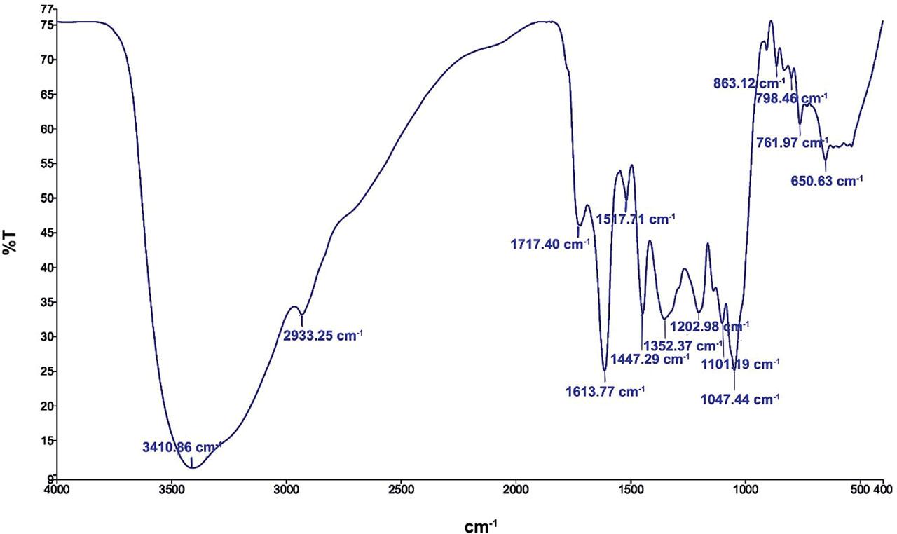
FT-IR of OESME in an aqueous medium showing the functional characteristic of the material.
The significant bands at 3397.55, 2933.45, 1719.84, 1610.96, 1515.67, 1447.61, 1354.99, 1202.49, 1099.89, 1046.74, 905.22, 863.38, 799.03, 762.46, and 650.57 cm−1 were revealed by the FT-IR analysis of OESME (Fig. 2 and Table 2). These bands verify the existence of C=O stretching for α,β-unsaturated ester, N-O stretching for nitro compound, C=C stretching for α,β-unsaturated ketone, and alkene. N-H stretching is present for aliphatic primary amine and amine salt. C-O stretching for vinyl ether and secondary alcohol, C-H bending for alkane, 1,2,4-trisubstituted, and 1,2-disubstituted, O-H bending for alcohol, CO-O-CO stretching for anhydride, C=C bending for alkene, and C-I stretching for halo compound.
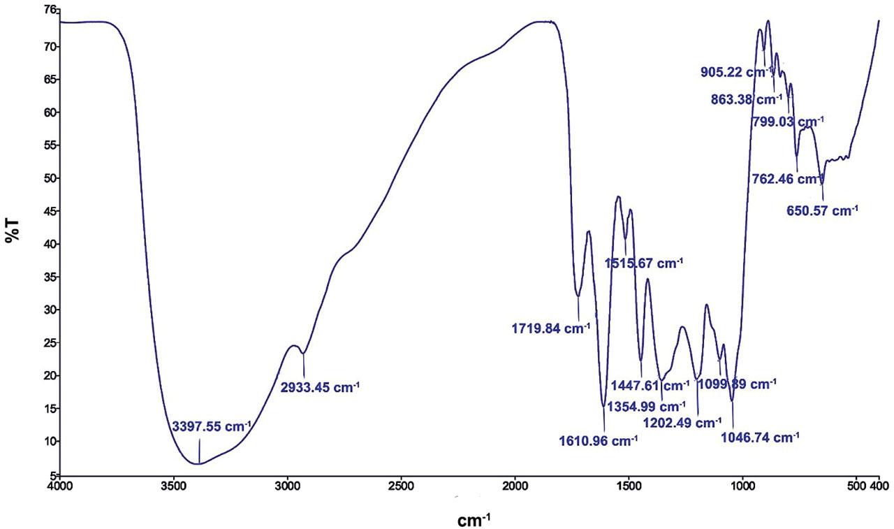
FT-IR of OELME in an aqueous medium showing the functional characteristic of the material.
Infrared (IR) spectrum of OELME by frequency range.
| Absorption (cm−1) | Transmittance (%) | Appearance | Group | Compound class |
|---|---|---|---|---|
| 3397.55 | 1.252463 | medium | N-H stretching | aliphatic primary amine |
| 2933.45 | 4.512668 | strong, broad | N-H stretching | amine salt |
| 1719.84 | 6.204029 | strong | C=O stretching | α.β-unsaturated ester |
| 1610.96 | 2.947124 | strong | C=C stretching | α.β-unsaturated ketone |
| 1515.67 | 7.879805 | strong | N-O stretching | nitro compound |
| 1447.61 | 4.298548 | medium | C-H bending | alkane |
| 1354.99 | 3.710983 | medium | O-H bending | alcohol |
| 1202.49 | 3.755297 | strong | C-O stretching | vinyl ether |
| 1099.89 | 4.343475 | strong | C-O stretching | Secondary alcohol |
| 1046.74 | 3.118347 | strong, broad | CO-O-CO stretching | anhydride |
| 905.22 | 13.43325 | strong | C=C bending | alkene |
| 863.38 | 12.6943 | strong | C-H bending | 1,2,4-trisubstituted |
| 799.03 | 12.05039 | medium | C=C bending | alkene |
| 762.46 | 10.30954 | strong | C-H bending | 1,2-disubstituted |
| 650.57 | 9.489779 | strong | C-I stretching | halo compound |
The OESME and OELME demonstrated varying antioxidant potentials about the antioxidant activity measured by ABTS. These potentials rose with increasing extract concentrations, peaked at 100 μg/mL, and then started to decline (Fig. 3). The findings demonstrated that, in comparison to OELME (94.9 ± 0.04 mg GAE/L), OESME had the highest percentage inhibition value of ABTS at 100 μg/mL (96.8 ± 0.02 mg GAE/L) (Fig. 3). Furthermore, the FRAP assay findings demonstrated a sharp rise in antioxidant activity, which peaked at 100 μg/mL and subsequently began to decrease based on concentration, amounting to 0.455 ± 0.002 mg GAE/L in OELME as opposed to 0.51 ± 0.003 mg GAE/L in OESME (Fig. 4).
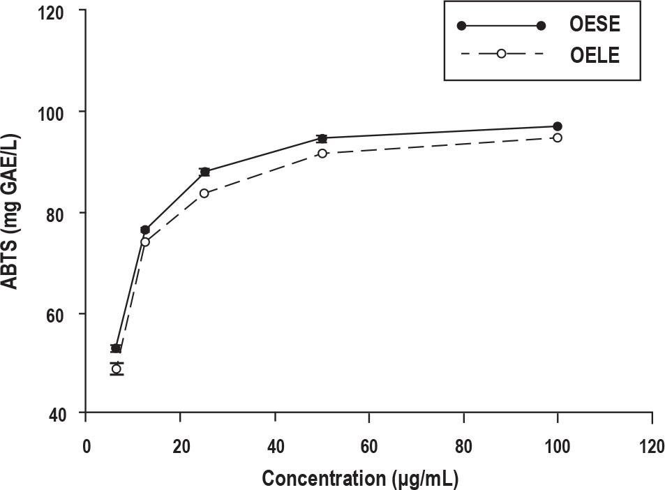
ABTS radical scavenging activity of the methanolic stem and leaves extracts of O. europaea. Results are presented as the means ± SD.
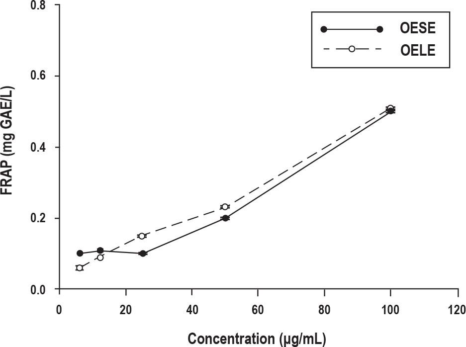
The ability of OESME and OELME extracts to reduce ferric iron. The ability of antioxidants to reduce the oxidative effects of reactive oxygen species is measured by the ferric reducing antioxidant power (FRAP) test. Results are presented as the means ± SD.
The cytotoxicity of OESME and OELME was investigated in vitro for 48 hours against the A549 and MCF-7 cell lines at different doses (0, 50, 100, 250, and 500 μg/mL). The lung (A549) cell line’s OESME IC50 was found to be 303.5 ± 3.25 μg/mL, while 252.5 ± 3.9 μg/ml in OELME (Table 3). Whereas, the breast (MCF-7) cell line’s IC50 for OESME and OELME were 326.5 ± 2.87 and 363.3 ± 3.04 μg/mL, respectively (Table 3).
Cytotoxicity in terms of IC50 dose of OESME and OELME against the lung (A549) cancer and human breast (MCF7) cell lines after 48 h of incubation by MTT Assay.
| Sample | Cell lines and IC50 (µg/ml) | |
|---|---|---|
| A549 | MCF-7 | |
| OESME | 303.5 ± 3.25 * | 326.5 ± 2.87 * |
| OELME | 252.5 ± 3.9 * | 363.3 ± 3.04 * |
| Doxorubicin | 1.5 ± 0.04 | 1.2 ± 0.06 |
Values are mean ± SD.
compared to Doxorubicin (p ≤ 0.001)
Our findings show that the amount administered, and the level of cell viability are positively correlated. The vitality of the OESME and OELME cells was significantly lower than that of the untreated cells after a 48-hour incubation period at various dosages with A549 and MCF-7 cell lines (Figs. 5 and 6).

Cell viability determined by MTT assay of OESME at various concentrations (µg/mL) against the lung (A549) cancer and human breast (MCF7) cell lines after 48 h of incubation. Every experiment was conducted thrice, and results are presented as the means ± SD.
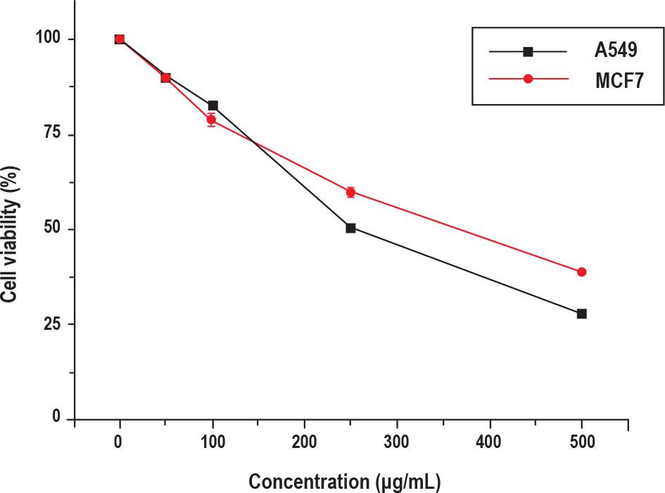
Cell viability determined by MTT assay of OELME at various concentrations (µg/mL) against the lung (A549) cancer and human breast (MCF7) cell lines after 48 h of incubation. Every experiment was conducted thrice, and results are presented as the means ± SD.
The anthelmintic activity of OESME and OELME against E. fetida was observed. The results indicate that at the highest effective dose of 200 mg/mL, OESME caused paralysis and death in 35.728 ± 2.396 and 36.848 ± 2.328 minutes, respectively. In contrast, OELME caused paralysis and death in 39.158 ± 4.068 and 48.336 ± 11.027 minutes, respectively. Moreover, paralysis and death times for mebendazole (10 mg/mL) were 13.91 ± 0.373 and 18.2 ± 0.980 minutes, respectively (Table 4). In histological changes, the tissues of the worms in the control group do not show any changes in the upper layer of the epidermis, while in the groups treated with plant extracts and mebendazole, they show destruction and disintegration of the upper layer to varying degrees (Fig. 7).
In vitro anthelmintic activity of Olea europaea extract (OESME and OELME).
| Test samples | Concentration (mg/ml) | Time is taken for paralysis (min.) | Percentage of worms paralyzed | Time is taken for death (min.) | Percentage of worms’ dead |
|---|---|---|---|---|---|
| Control (H2O) | -- | -- | -- | -- | -- |
| OELME | 25 mg/ml | 135.292 ± 9.247 *# | 20% | 164.548 ± 8.597 *# | 20% |
| 50 mg/ml | 63.074 ± 8.660 *# | 40% | 146.732 ± 5.756 *# | 40% | |
| 100 mg/ml | 84.808 ± 7.796 *# | 60% | 119.158 ± 9.198 *# | 60% | |
| 200 mg/ml | 39.158 ± 4.068 *# | 100% | 48.336 ± 11.027 *# | 100% | |
| OESME | 25 mg/ml | 145.368 ± 3.639 *# | 20% | 161.306 ± 7.641 *# | 20% |
| 50 mg/ml | 79.988 ± 8.372 *# | 40% | 106.456 ± 9.346 *# | 40% | |
| 100 mg/ml | 82.47 ± 8.231 *# | 60% | 94.066 ± 8.834 *# | 60% | |
| 200 mg/ml | 35.728 ± 2.396 *# | 100% | 36.848 ± 2.328 *# | 100% | |
| Mebendazole | 10 mg/ml | 13.91 ± 0.373 * | 100% | 18.2 ± 0.980 * | 100% |
Values are mean ± SD.
compared to control (H2O) (p ≤ 0.05),
compared to mebendazole (p ≤ 0.05).
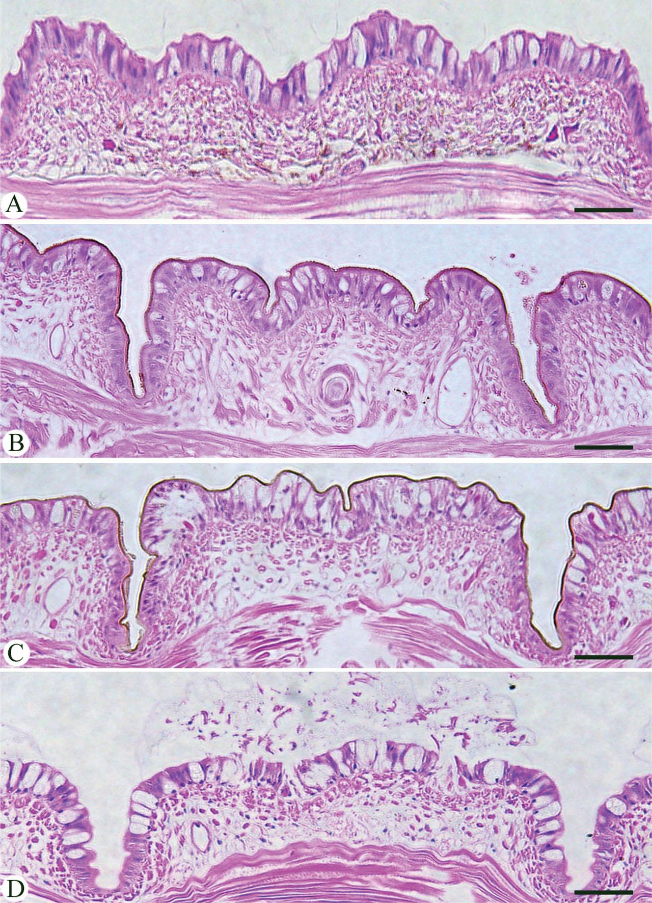
Cuticle thickness of E. fetida with various treatments. (A) worms in dist. H2O (control). (B) worms in OESME (200 mg/ml). (C) worms in OELME (200 mg/ml). (D) worms in mebendazole. Scale bar = 25 µm
One of the medicinal plants, O. europaea, has different pharmacological qualities for its fruit, oil, and leaves (Visioli et al., 2002). We assessed the plant’s stems and leaves for their cytotoxic and anthelmintic properties in the current investigation.
In line with Thenmozhi et al. (2011), who reported that the FT-IR spectrum was used to identify the functional groups of the chemical constituents based on the peak value and when run under IR region in the range of 4000 – 400 cm−1, absorption bands were assigned at different wavenumber regions for OESME and OELME in this study. The O. europaea leaves and stem’s FT-IR spectra displayed the same pattern but varied in the peak transmittance’s strength. According to Tsao (2010), who reported that the hydroxyl group can neutralise free radicals by donating an electron or hydrogen atom to prevent oxidation, the O-H group is one of the most functional groups in both OESME (at 1356.37 cm−1) and OELME (at 1354.99 cm−1) and is powerful for its antioxidant activity.
Based on the antioxidants’ capacity to scavenge free radicals, the antioxidant activities of OESME and OELME were assessed using ABTS and FRAP assays after the functional groups had been identified. Numerous extracts derived from the leaves and stem of O. europaea have demonstrated the potential for both ferric-reducing antioxidant capacity (Orak et al., 2019) and radical scavenging (Özcan & Matthäus, 2017). In this case, OESME’s ABTS-measured antioxidant activity was 96.8 ± 0.02 mg GAE/L, higher than OELME’s (94.9 ± 0.04 mg GAE/L). This is in line with the findings of Jellali et al. (2022), who reported that tested olive extracts exhibit their maximum of free radical scavenging activity, reaching up to 99 %. Also, Elnahas et al. (2021), revealed that phytochemicals with radical-scavenging properties have been identified as active components, including flavonoids, phenolics, tannins, and saponins. This, however, contradicts the findings of Osman and Tantawy (2017), who stated that among the various sections of olive trees, olive leaves were thought to have the highest scavenging activity.
Furthermore, it has been suggested by Yen et al. (1993) and Siddhuraju et al. (2002) that the antioxidant activity of bioactive substances is correlated with their ability to donate electrons. This study assessed and compared OESME and OELME’s capacity to decrease Fe+3 to Fe+2 to that of ascorbic acid. A concentration-dependent electron-donating capacity was demonstrated by OESME and OELME. Methanolic extract from leaves had the highest reducing power at 100 μg/mL, followed by those from the stem. This was in line with Beghdad et al. (2014), who noted that OELME, which contained the most total phenols — the most potent reducing agent—was the most active, while OESME, which contained the least amount of phenolics, performed the least well.
According to Ahamad et al. (2019), MTT can be used to assess the cytotoxic activity of medicinal herbs. To assess the cytotoxicity of OESME and OELME against the A549 and MCF-7 cell lines, an MTT test was used in this investigation. In line with Han et al. (2009), the viability of cells in this study exhibits a direct dose-dependent manner. Furthermore, according to Junkins et al. (2023) and Asghariazar et al. (2024), the presence of chemical constituents—particularly oleuropein—in O. europaea extracts increases their applicability and potential use as a future remedy to counteract the devastating effects of various human cancer cells that have little to no effect on normal cells. This study revealed that OESME and OELME, which contain oleuropein as major secondary metabolites, have antitumor activity in the studies cell lines. These results are partly consistent with the apoptosis mechanism described in MCF-7 cells treated with oleuropein (Han et al., 2009; Sirianni et al., 2010; Mijatovic et al., 2011). The IC50 of OESME and OELME on the investigated cell lines were recorded with varied concentrations. This is associated with findings of Lee et al. (2011) on the cytotoxicity of Phyllanthus plant extracts to inhibit MCF-7 (breast carcinoma) and A549 (lung carcinoma) cell growth with IC50 ranging from 50 – 180 μg/mL and 65 – 470 μg/mL for methanolic and aqueous extracts, respectively.
Drug-resistant parasites may be less likely to develop resistance if mass drug administration continues while synthetic anthelmintic use is limited (Bhattacharjee et al., 2018). Utilizing natural dietary components could further reduce the need for medication. This study evaluated the effects of OESME and OELME on the earthworm E. fetida, which shares similarities with human intestinal roundworms (George & Kousalya, 2018). OESME and OELME showed significant anthelmintic activity at 200 mg/mL in a dose-dependent manner compared to the control group. The finding of OESME is associated with findings of George and Kousalya (2018) about Basella alba and Amaranthus dubius combination, which showed the best anthelmintic activity, paralysis stage of E. fetida at 31 minutes and death at stage 36 minutes. Our results for OELME align with Kamran et al. (2023), who reported that an extract from O. europaea leaves had anthelmintic effects on mice infected with Aspiculuris tetraptra. Bhattacharjee et al. (2018) also highlighted the anthelmintic action of O. europaea leaf methanol extract on the adult earthworm Pheretima posthumus. Furthermore, Khokra et al. (2020) demonstrated the anthelmintic evaluation of Bombax ceiba stem extracts with paralysis time 30 ± 0.96 minutes and death 33.66 ± 0.71 minutes. Mebendazole (Pandey, 2012) caused hyperpolarization and decreased excitability, leading to muscular relaxation and flaccid paralysis. In contrast, OESME and OELME not only induced paralysis but also killed worms, particularly at a higher dose of 200 mg/mL. The study found that worms treated with OESME and OELME experienced significant cuticular shrinkage, supporting Kundu et al. (2012) on the cuticle as a target for anthelmintic. Sambodo et al. (2018) and Mrifag et al. (2021) noted that anthelmintic agents cause surface alterations that lead to paralysis and death. This can be attributed to phytochemicals like flavonoids, tannins, and saponins in the extracts, which are known for their anthelmintic properties, as mentioned by Mali and Wadekar (2008), Melzig et al. (2001), and Yoon et al. (2006).
The results of this investigation suggest that O. europaea leaves and stem extracts are a valuable source of cytotoxic, anthelmintic, and natural antioxidant agents. The in vitro findings of this study may pave the way for future research and the development of effective herbal medicines derived from the phytochemical compounds of O. europaea. Conducting in vivo studies will be essential for testing the efficacy of each active compound.