
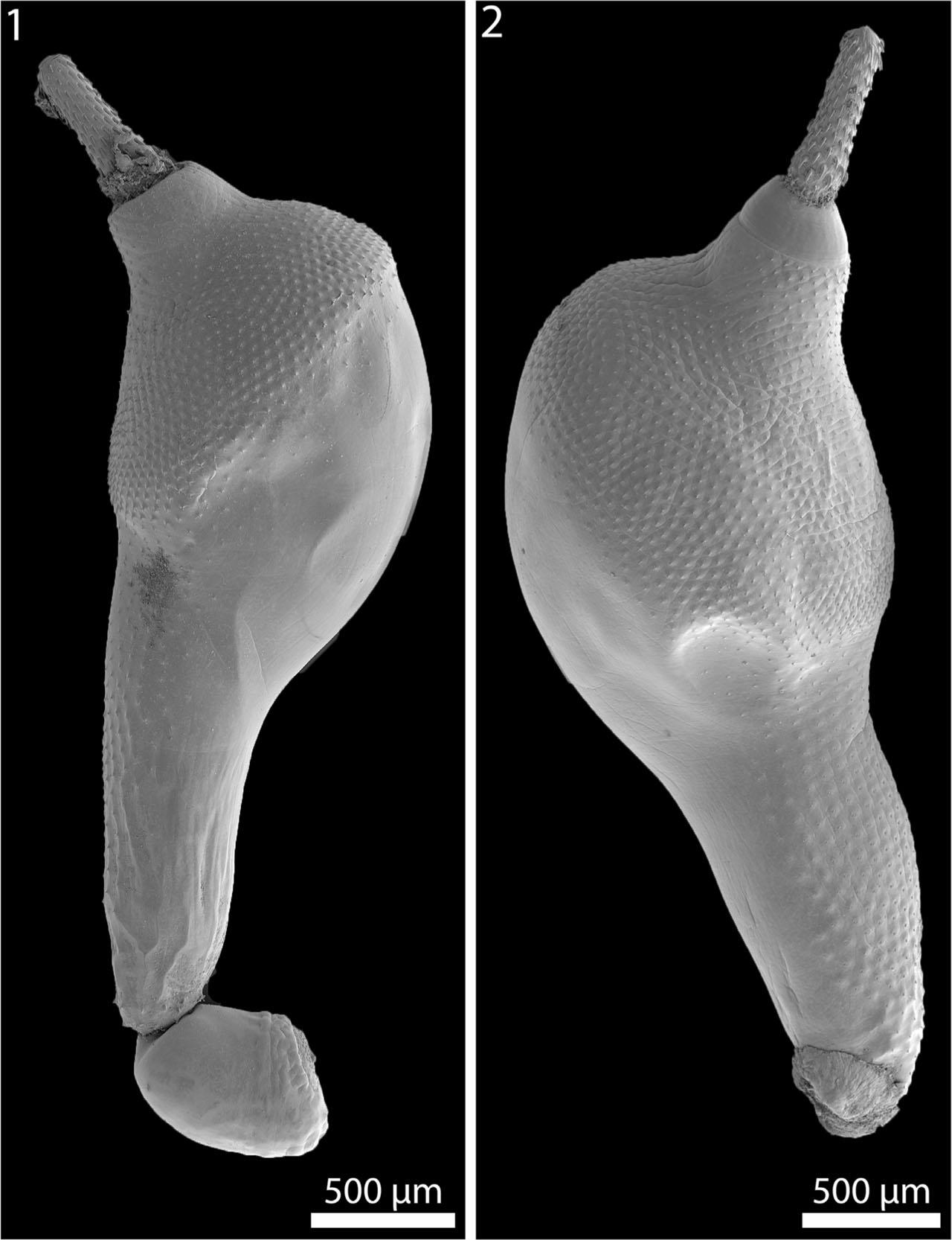
Figs. 1, 2.
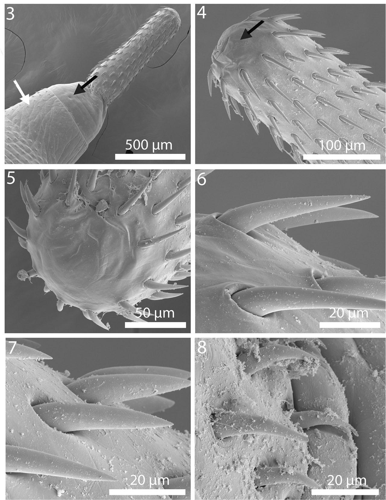
Figs. 3–8.
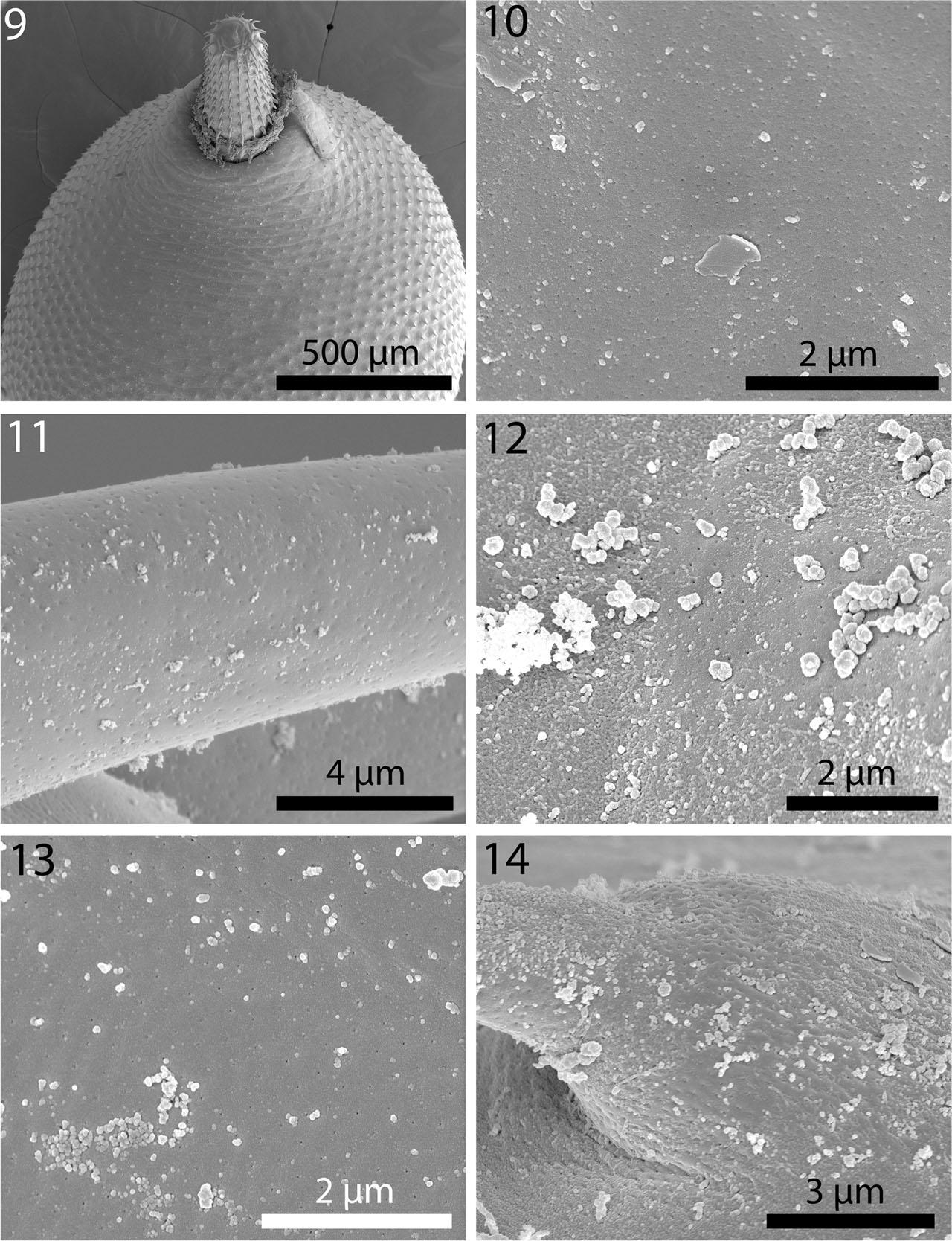
Figs. 9–14.
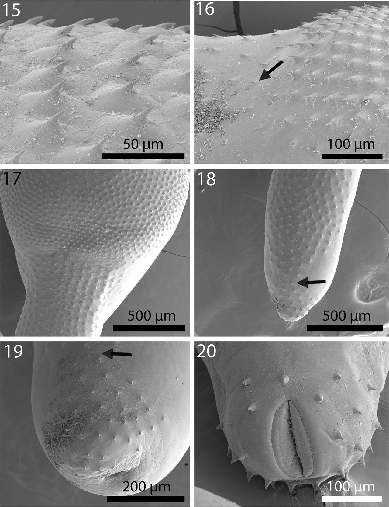
Figs. 15–20.
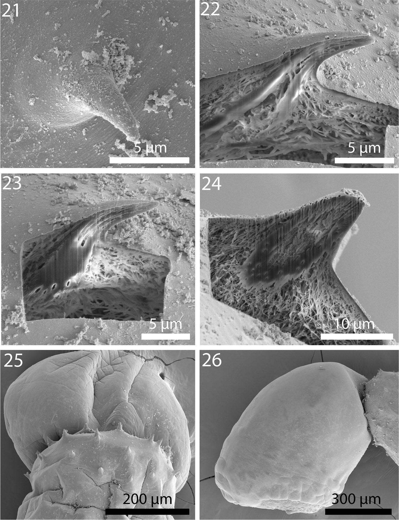
Figs. 21–26.
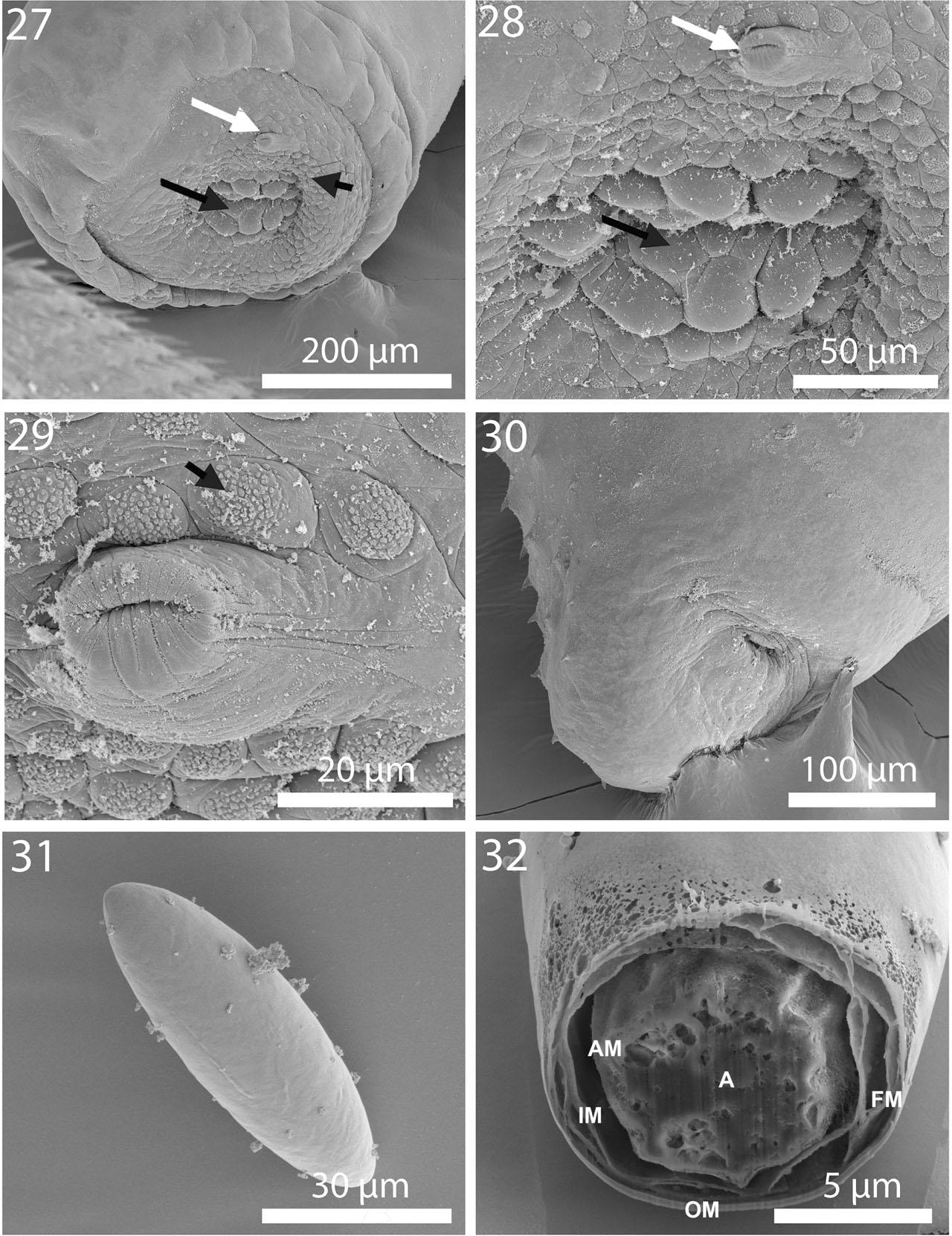
Figs. 27–32.
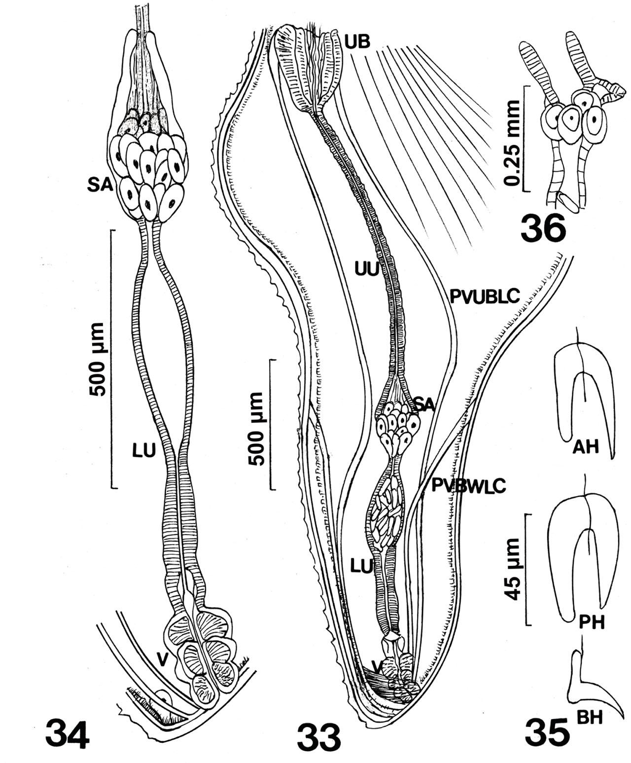
Figs. 33–36.
Figs. 37–39.
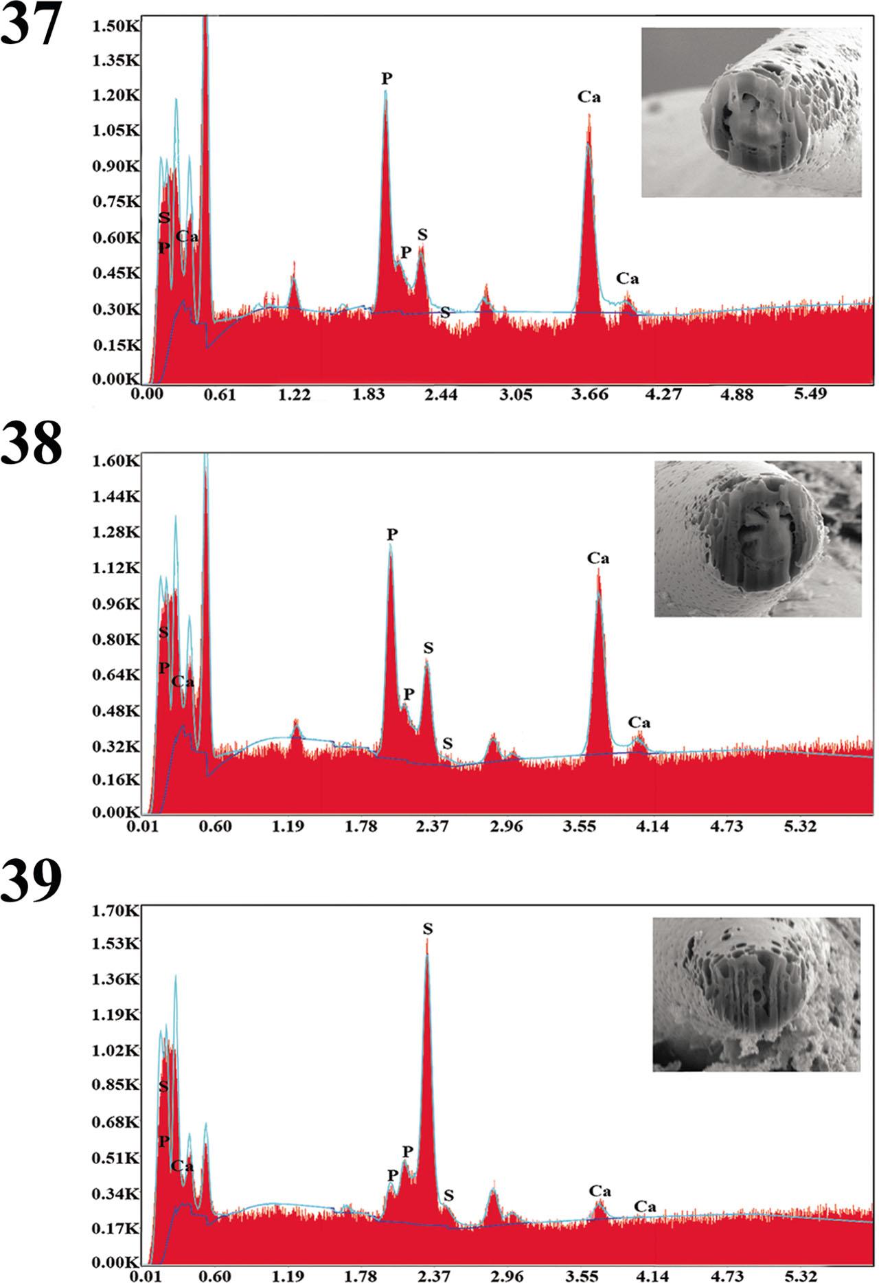
Fig. 40.
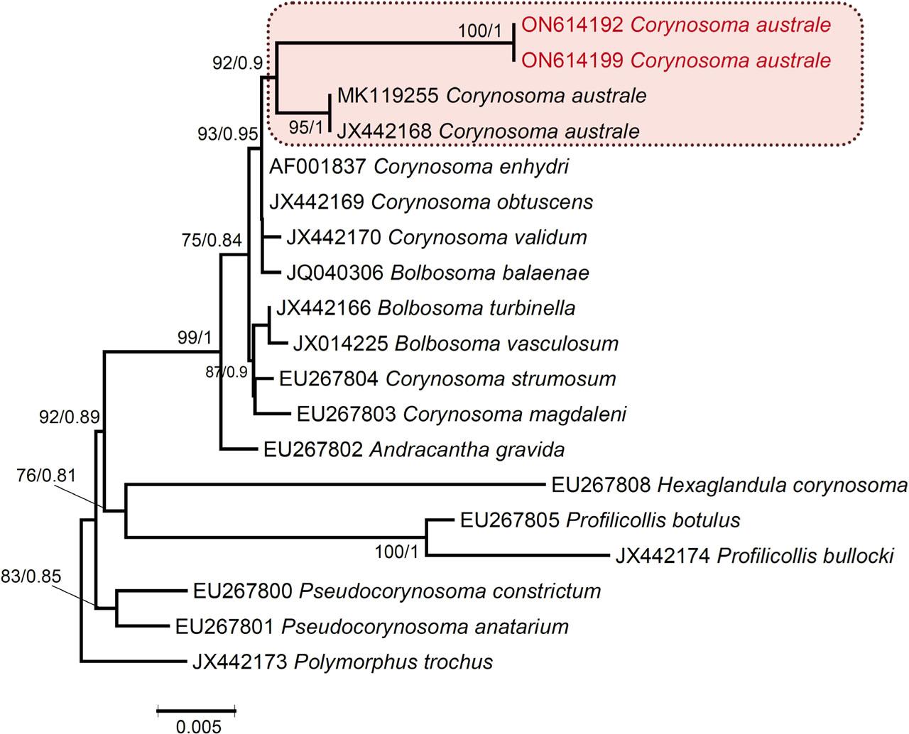
Fig. 41.
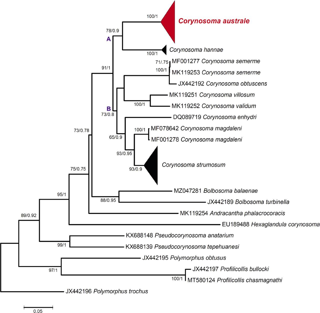
Fig. 42.
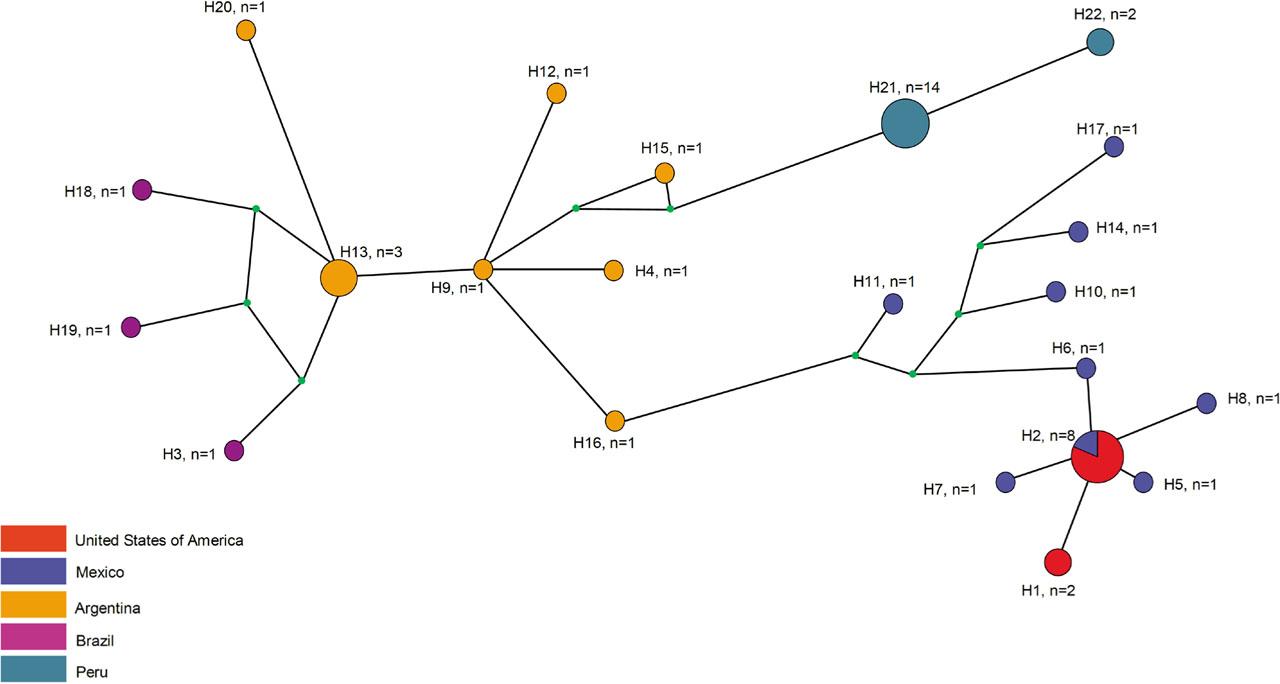
Fig. 43.
Chemical composition and localization of elements in hooks of Corynosoma australe from Zalophus californianus in California_
| Anterior hooks | Middle hooks | Posterior hooks | |||||||
|---|---|---|---|---|---|---|---|---|---|
| Element* | Tip x-section | Middle | Longitudinal section | Tip x-section | Tip edge | Middle x-section | Middle edge | Tip x-section | Middle edge |
| Magnesium (Mg) | 1.21 | 0.23–1.67** | 0.02–0.07 | 0.63 | 0.62 | 1.61 | 0.66 | 0.02 | 0.78–1.52 |
| Sodium (Na) | 0.25 | 0.00–0.04 | 0.00–0.03 | 0.00 | 0.03 | 0.08 | 0.00 | 0.02 | 0.05–0.07 |
| Phosphorous (P) | 9.95 | 15.74–18.71 | 12.78–20.35 | 10.64 | 11.00 | 21.32 | 16.10 | 2.82 | 14.91–20.49 |
| Sulfur (S) | 2.96 | 0.09–7.10 | 0.34–3.50 | 5.50 | 15.38 | 0.03 | 4.34 | 23.52 | 1.18–14.09 |
| Calcium (Ca) | 18.99 | 30.80–34.84 | 32.03–72.06 | 19.98 | 19.11 | 42.48 | 34.18 | 3.16 | 27.05–40.39 |
Chronological taxonomic history of Corynosoma australe from marine mammals, with special reference to ventral trunk spines in females_
| Author | Host | Locality | Described as | Stage | Ventral spines |
|---|---|---|---|---|---|
| Johnston (1937) | Neophoca cinerea (Péron) | South Australia | C. australe | Adults | Discontinuous |
| Lincicome (1943) | Zalophus californianis (Lesson) | San Diego, California | C. obtuscens | Adults | Continuous |
| Van Cleave (1953) | Mycteroperca pardalis Gilbert | Mazatalán, Mexico | C. obtuscens | Cystacanth | Continuous |
| Morini & Boero (1960) | Otaria flavescence Shaw | Argentina | C. otariae | Adults | Discontinuous |
| Zdzitowiecki (1984) | Hydrurga leptonyx (Blainville) | South Shetlands, Antarctica | C. australe | Adults | 25% continuous |
| Smales (1986) | Neophoca cinerea (Péron) | South Australia | C. australe | Adults (Figs. 10,11) | Discontinuous |
| Zdzitowiecki (1991) | Few “suitable” definitive & paratenic hosts | Antarctica | C. australe | Adults (Figs. 15 b, e) | Discontinuous |
| Sardella et al. (2005) | Arctocephalus australis (Zimmerman) | Argentina | C. australe | Adults | 85–100% continuous |
| Aznar et al. (2012) | Otaria flavescens (Shaw) | Argentina | C. australe | Adults (Fig. 1B) | Discontinuous |
| Hernández-Orts et al. (2017a) | Spheniscus magellanicus (Foster) | Brazil | C. australe | Adults (Fig. 2B) | Continuous & 82–89% continuous |
| Lisitsyna et al. (2018) | Zalophus californianus (Lesson) | Sausalito | C. obtuscens | Adults | Continuous |
| Lisitsyna et al. (2019) | Zalophus californianus (Lesson) | Sausalito | C. obtuscens & C. australe | Adults | 1% discontinuous |
| Present paper | Zalophus californianus (Lesson) | Sausalito | C. australe | Adults | Continuous with post. constriction |
List of acanthocephalan species used for phylogenetic analysis based on the mt Cox1 gene sequences_ Newly generated sequences are presented in bold_
| Species | Host | Host origin | GenBank accession nos. | References |
|---|---|---|---|---|
| Corynosoma australe | Zalophus californianus | USA | ON619618, ON614719 | present study |
| Zalophus californianus | USA | MK119245–MK119249 | Lisitsyna et al., 2019 | |
| Zalophus californianus | Mexico | MT676808–MT676818 | García-Varela et al., 2020 | |
| Merluccius hubbsi | Argentina | MT676819–MT676822 | García-Varela et al., 2020 | |
| Raneya brasiliensis | Argentina | MT676823–MT676824 | García-Varela et al., 2020 | |
| Paralichthys isosceles | Brazil | KU314822 | Fonseca et al., 2019 | |
| Stenella clymene | Argentina | MW724483 | Hernández-Orts et al., 2021 | |
| Arctocephalus australis | Argentina | MF497333 | Hernández-Orts et al., 2017a | |
| Spheniscus magellanicus | Brazil | MF497335 | Hernández-Orts et al., 2017a | |
| Otaria flavescens | Argentina | KX957714, MF497334 | Hernández-Orts et al., 2017a, b | |
| Paralichthys adspersus | Peru | MZ920052–MZ920055 | Mondragon-Martinez et al., 2021* | |
| Paralabrax humeralis | Peru | MZ920056–MZ920059 | Mondragon-Martinez et al., 2021* | |
| Cheilodactylus variegatus | Peru | MZ920060–MZ920063 | Mondragon-Martinez et al., 2021* | |
| Otaria byronia | Peru | MZ920064–MZ920067 | Mondragon-Martinez et al., 2021* | |
| Corynosoma hannae | Colistium guntheri | New Zealand | KX957724, KX957725 | Hernández-Orts et al., 2017b |
| Leucocarbo chalconotus | New Zealand | KX957718–KX957721, KX957723 | Hernández-Orts et al., 2017b | |
| Phalacrocorax punctatus | New Zealand | KX957722 | Hernández-Orts et al., 2017b | |
| Phocarctos hookeri | New Zealand | KX957715–KX957717, JX442191 | Hernández-Orts et al., 2017b; García-Varela et al., 2013 | |
| Peltorhamphus novaezeelandiae | New Zealand | KY909260–KY909263 | Anglade & Randhawa, 2018 | |
| Corynosoma semerme | Halichoerus grypus | Germany | MF001277 | Waindok et al., 2018 |
| Corynosoma semerme | Callorhinus ursinus | USA | MK119253 | Lisitsyna et al., 2019 |
| Corynosoma obtuscens | Halichoerus grypus | New Zealand | JX442192 | García-Varela et al., 2013 |
| Corynosoma villosum | Callorhinus ursinus | USA | MK119251 | Lisitsyna et al., 2019 |
| Corynosoma validum | Callorhinus ursinus | USA | MK119252 | Lisitsyna et al., 2019 |
| Corynosoma enhydri | Enhydra lutris | USA | DQ089719 | García-Varela & Nadler, 2006 |
| Corynosoma magdaleni | Phoca vitulina | Germany | MF078642 | Waindok et al., 2018 |
| Corynosoma nortmeri | Phoca vitulina | Germany | MF001278 | Waindok et al., 2018 |
| Corynosoma strumosum | Phoca vitulina | USA | EF467870 | García-Varela & Pérez-Ponce de León, 2008 |
| Pusa hispida botnica | Finland | EF467871 | García-Varela & Pérez-Ponce de León, 2008 | |
| Zalophus californianus | USA | MK119250 | Lisitsyna et al., 2019 | |
| Bolbosoma balaenae | Balaenoptera physalus | Italy | MZ047281 | Santoro et al., 2021 |
| Bolbosoma turbinella | Eschrichtius robustus | USA | JX442189 | García-Varela et al., 2013 |
| Andracantha phalacrocoracis | Zalophus californianus | USA | MK119254 | Lisitsyna et al., 2019 |
| Hexaglandula corynosoma | Nyctanassa violacea | Mexico | EU189488 | Guillén-Hernández et al., 2008 |
| Pseudocorynosoma anatarium | Bucephala albeola | Mexico | KX688148 | García-Varela et al., 2017 |
| Pseudocorynosoma tepehuanesi | Oxyura jamaicensis | Mexico | KX688139 | García-Varela et al., 2017 |
| Polymorphus obtusus | Aythya affinis | Mexico | JX442195 | García-Varela et al., 2013 |
| Profilicollis bullocki | Emerita analoga | Mexico | JX442197 | García-Varela et al., 2013 |
| Profilicollis chasmagnathi | Oligosarcus jenynsii | Argentina | MT580124 | Levy et al., 2020 |
| Polymorphus trochus | Fulica americana | Mexico | JX442196 | García-Varela et al., 2013 |
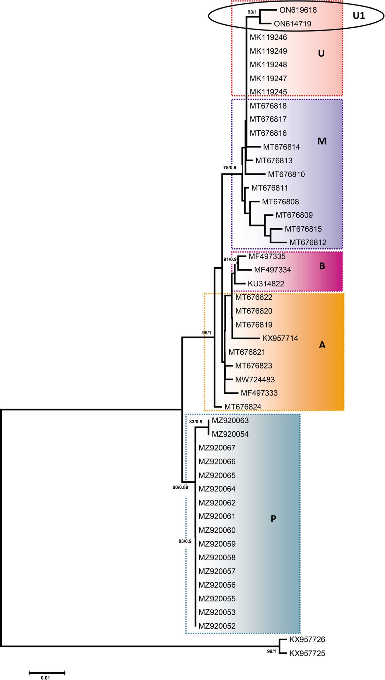
Data for the population of Corynosoma australe used in the haplotype networking using mt Cox1 gene_ Asterisk shows sequences unpublished on NCBI database_ Newly generated sequences are presented in bold_
| Geographical Locality* | Cox1 | ID in Fig. 46 | GenBank accession nos. | References |
|---|---|---|---|---|
| Pacific coast near San Francisco, California, USA | H1 | U1 | ON619618, ON614719 | Present study |
| Sausalito, California, USA | H2 | U | MK119245-MK119249 | Lisitsyna et al. 2019 |
| Baja California, Mexico | H2 | M | MT676816-MT676818 | García-Varela et al. 2021 |
| Rio de Janeiro, Brazil | H3 | B | KU314822 | Fonseca et al. 2019 |
| Chubut, Argentina | H4 | A | MW724483 | Hernández-Orts et al. 2021 |
| Baja California, Mexico | H5 | M | MT676813 | García-Varela et al. 2021 |
| Sonora, Mexico | H6 | M | MT676811 | García-Varela et al. 2021 |
| Baja California, Mexico | H7 | M | MT676814 | García-Varela et al. 2021 |
| Sonora, Mexico | H8 | M | MT676810 | García-Varela et al. 2021 |
| Northern Patagonia, Argentina | H9 | A | MT676821 | García-Varela et al. 2021 |
| Sonora, Mexico | H10 | M | MT676809 | García-Varela et al. 2021 |
| Baja California Sur, Mexico | H11 | M | MT676808 | García-Varela et al. 2021 |
| Northern Patagonia, Argentina | H12 | A | MT676823 | García-Varela et al. 2021 |
| Northern Patagonia, Argentina | H13 | A | MT676819, MT676820, MT676822 | García-Varela et al. 2021 |
| Baja California, Mexico | H14 | M | MT676815 | García-Varela et al. 2021 |
| Northern Patagonia, Argentina | H15 | A | MT676824 | García-Varela et al. 2021 |
| Northern Patagonia, Argentina | H16 | A | MF497333 | Hernández-Orts et al. 2021 |
| Baja California, Mexico | H17 | M | MT676812 | García-Varela et al. 2021 |
| Rio de Janeiro, Brazil | H18 | B | MF497335 | Hernández-Orts et al. 2021 |
| Northern Patagonia, Argentina | H19 | A | MF497334 | Hernández-Orts et al. 2021 |
| Northern Patagonia, Argentina | H20 | A | KX957714 | Fonseca et al. 2019 |
| Peru | H21 | P | MZ920052, MZ920053, MZ920055, MZ920056-MZ920062, MZ920064-MZ920067 | Mondragon-Martinez et al. 2021* |
| Peru | H22 | P | MZ920054, MZ920063 | Mondragon-Martinez et al. 2021* |
Chemical composition of trunk spines and eggs of Corynosoma australe from Zalophus californianus in California_
| Elements* | Spines (longitudinal sections) | Eggs (cross sections) | |||
|---|---|---|---|---|---|
| Anterior | Middle | Posterior | Edge (shell) | Center (acanthor) | |
| Magnesium (Mg) | 0.00 | 0.00 | 0.09 | 0.00 | 0.50 |
| Sodium (Na) | 0.00 | 0.00 | 0.00 | 0.00 | 0.00 |
| Phosphorous (P) | 1.39 | 0.00 | 1.16 | 0.91 | 7.73 |
| Sulfur (S) | 12.64 | 5.97 | 15.61 | 0 | 2.30 |
| Calcium (Ca) | 1.64 | 1.07 | 1.96 | 1.34 | 3.41 |
List of acanthocephalan species used for phylogenetic analysis based on the 18S rDNA gene sequences_ Newly generated sequences are presented in bold, NA=host name not available_
| Species | Host | Host origin | GenBank accession nos. | References |
|---|---|---|---|---|
| Corynosoma australe | Zalophus californianus | USA | ON614192, ON614199 | present study |
| Zalophus californianus | USA | MK119255 | Lisitsyna et al., 2019 | |
| Phocarctos hookeri | Mexico | JX442168 | García-Varela et al., 2013 | |
| Corynosoma obtuscens | Callorhinus ursinus | Mexico | JX442169 | García-Varela et al., 2013 |
| Corynosoma validum | Callorhinus ursinus | Mexico | JX442170 | García-Varela et al., 2013 |
| Corynosoma enhydri | NA | USA | AF001837 | Near et al., 1998 |
| Corynosoma magdaleni | Phoca hispida botnica | Mexico | EU267803 | García-Varela et al., 2009 |
| Corynosoma strumosum | Phoca vitulina | Mexico | EU267804 | García-Varela et al., 2009 |
| Bolbosoma balaenae | Nyctiphanes couchii | Spain | JQ040306 | Gregory et al., 2011* |
| Bolbosoma turbinella | Eschrichtius robustus | Mexico | JX442166 | García-Varela et al., 2013 |
| Bolbosoma vasculosum | Lepturacanthus savala | Indonesia | JX014225 | Verweyen et al., 2011 |
| Andracantha gravida | Phalacrocorax auritus | Mexico | EU267802 | García-Varela et al., 2009 |
| Hexaglandula corynosoma | Nyctanassa violacea | Mexico | EU267808 | García-Varela et al., 2009 |
| Pseudocorynosoma anatarium | Bucephala albeola | Mexico | EU267801 | García-Varela et al., 2009 |
| Pseudocorynosoma constrictum | Anas clypeata | Mexico | EU267800 | García-Varela et al., 2009 |
| Profilicollis bullocki | Emerita analoga | Mexico | JX442174 | García-Varela et al., 2013 |
| Profilicollis botulus | Somateria mollissima | Mexico | EU267805 | García-Varela et al., 2009 |
| Polymorphus trochus | Fulica americana | Mexico | JX442196 | García-Varela et al., 2013 |