Introduction
On Earth, humans are constantly impacted by a gravitational force of 1G that has been of paramount importance to human evolution and function. Micro-gravitational exposure during spaceflight poses many physiological challenges to different organ systems and overall health (Demontis et al., 2017). The pulmonary system is potentially vulnerable to the effects of microgravity (0G) as the lung tissue and surrounding blood vessels rely on gravitational forces on Earth to maintain its intrinsic structure, perfusion pressures, hydrostatic gradients, and ventilation-perfusion ratio (Prisk, 2019). Structural and functional changes to the lungs in microgravity may have implications towards the physiological well-being of the pulmonary system for current and future space travelers.
With spaceflight priorities now favoring long-duration and deep space travel, as well as flights of potentially “less-healthy” space tourists, understanding the effects of transient and sustained microgravity on the pulmonary system is of high importance. To date, participation of people with pre-existing chronic medical conditions in spaceflight missions is very limited. However, greater human spaceflight activity from the private sector increases the likelihood of people with a more diverse medical health status being exposed to the spaceflight environment. Additionally, current human spaceflight missions include returning to the moon and deeper space travel to explore Mars. Uncertainty remains regarding potential health impacts, specifically to the pulmonary system, from microgravity during such missions since humans have yet to spend extended periods of time in deep space. Therefore, understanding what we already know of the effects of microgravity in the spaceflight environment on the human lung and functional components of the pulmonary system is fundamental to preparing for the proper clinical management for all people in space.
While available data are limited, this systematic review provides a comprehensive analysis of the current state of pulmonary outcome measures in microgravity to understand the changes that occur to the human pulmonary system in space compared to Earth and the implications towards function. This functional measure study complements our work performed in a recent paper that explored lung shape, volume, and capacity outcomes in spaceflight (Ghani et al., 2023). The study showed that there were no observable changes in lung volume and capacity measurements between 0G and 1G in long-duration spaceflight, and either no or slight changes in short-duration spaceflight and parabolic flights. 1G has been used in this study to refer to gravity on Earth while 0G denotes microgravity conditions. In both settings no consistent effects were observed in lung shape and physical dimensions in 0G compared to 1G (Ghani et al., 2023). Our current paper focuses on additional outcome measures not previously reviewed, comprising a range of functional parameters. While our 2023 study focused on microgravity-induced structural pulmonary outcomes, this review investigates functional pulmonary parameters in microgravity, offering an in-depth and comprehensive perspective on the physiological adaptations of the pulmonary system in space. Although both structural and functional changes relate to pulmonary adaptation in space, the evidence base and biological implications differ substantially between the two areas. This study expands the scope to consider the implications of space tourism and pulmonary health in individuals with pre-existing conditions, which is an area previously underexplored.
Pulmonary function in space, particularly lung function as described in this study, has not been a highly researched area in recent years due to shifting priorities in research towards more moderate- and high-risk systems like cardiovascular and musculoskeletal, and consistent reports of the lungs’ resilience and adaptability in microgravity (Prisk et al., 2008). These risk classifications are decided according to the degree of potential functional importance during spaceflight and their persistence for at least six months after returning to Earth (Garrett-Bakelman et al., 2019). Despite this, there are risks which remain to be understood about the pulmonary system in microgravity. Risks regarding the current age of space tourism, people with pulmonary abnormalities flying to space, and upcoming plans for long-duration spaceflight missions were not heavily evaluated in earlier human spaceflight studies. A recent study contends that the current knowledge regarding the functional changes of the lungs and residing cells of the respiratory system in microgravity conditions is lacking (Smith et al., 2024). Additional recent studies highlight that gravitational changes alter the blood filling of the lungs and may modify the rate of gas uptake into the blood, affecting the transport of important gases in the body, which is relevant to today’s planetary exploration and long-duration mission plans (Karlsson et al., 2023; Karlsson et al., 2024).
Furthermore, the value of this review lies in its provision of a benchmark summary for experts in medicine and healthcare to understand what is known about lung changes. This will assist in their assessment of the risk profiles and proper management of astronauts, particularly citizen astronauts embarking on deep space missions. This work also provides a baseline and guideline for pulmonary computational modeling for prediction of longer exposure scenarios in spaceflight, especially in the new approaches in space omics.
Materials and Methods
This systematic review and meta-analysis were conducted in accordance with the Preferred Reporting Items for Systematic Reviews and Meta-Analyses (PRISMA) guidelines. The methodology closely follows that of our previously published review on lung volume, capacity, and shape in microgravity (Ghani et al., 2023). Adaptations were made to address a distinct set of outcomes related to pulmonary function.
Eligibility criteria
Studies were eligible for inclusion if they reported on functional pulmonary outcomes in human subjects exposed to microgravity (intervention) including parabolic flight (~20 seconds), short-duration spaceflight (≤30 days), and long-duration spaceflight (>30 days). PICOS (population, intervention, control, outcomes, and study design) criteria were followed (Amir-Behghadami and Janati, 2020). Control measures were taken pre-flight in normal gravity (1G) from the same population as the intervention group. All types of experimental studies were eligible.
Information sources and search strategy
Briefly, a comprehensive search of electronic databases PubMed, OVID, and Cochrane Library was performed. The search strategy was adapted from our previous review and updated to include terms specific to pulmonary function (e.g., “lung function,” “gas exchange,” “ventilation,” “microgravity,” “spaceflight”) (Supplementary Material 1). The final search was completed in June 2025. Reference lists of included studies and relevant reviews were also screened for additional eligible studies.
Study selection
All identified records were imported into Rayyan (https://rayyan.qcri.org/) for screening (Ouzzani et al., 2016). Two reviewers independently screened titles and abstracts, followed by full-text reviews of potentially eligible studies. Discrepancies were resolved through discussion and consensus.
Data extraction
An adapted version of the Space Biomedicine Systematic Review “Data extraction and analysis” spreadsheet (Winnard et al., 2021) was used for data extraction and management, as used in our previous study. A plot reader, WebPlotDigitizer, was used to extract data where studies only reported data in figures (WebPlotDigitizer v. 4.2, 2021). Extracted variables included participant characteristics, type and duration of microgravity exposure, pulmonary function outcomes, and key findings. At least two studies were required to explore an outcome measure for a meta-analysis, and hence, data which were reported in only one study were included in the review and analysis, but not in the meta-analysis (Cheung and Vijayakumar, 2016).
Risk of bias assessment
The risk of bias of included studies was assessed using the Risk of Bias Assessment Tool for Nonrandomized Studies (RoBANS). Each study was evaluated across six domains: selection of participants, confounding variables, measurement of exposure, blinding of outcome assessment, incomplete outcome data, and selective outcome reporting. Two reviewers independently assessed risk of bias, with disagreements resolved by consensus (Kim et al., 2013).
Data synthesis and statistical analysis
Meta-analyses were conducted using metafor package in R (version 3.6.1), consistent with our previous methodology (Ghani et al., 2023; A Language and Environment for Statistical Computing, 2020). The effect sizes were then bias corrected using weighted pooled standard deviations as per Hedges’ (1981) g method. For the effect size comparisons, a 95% confidence interval level was used.
Results
Study selection and description
PRISMA was used (Ghani et al., 2023). The selection process of the studies resulted in 28 studies included for the review (Fig. 2). An overview of the characteristics and outcome parameters found in each individual study is provided in Table 1. If a study collected measurements at different timepoints within a flight type, the mean value of that duration was taken.
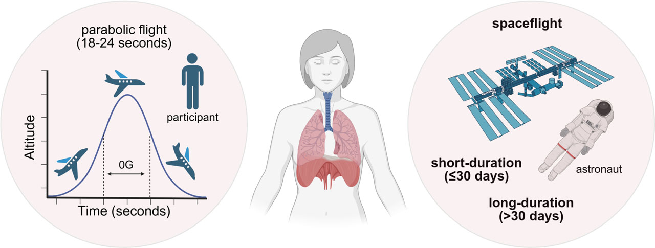
Figure 1.
Schematic diagram of systematic review focus on exploring functional pulmonary system outcome measures in space during parabolic flight, short- and long-duration spaceflight. Diagram shows parabolic flight path to create microgravity and space station in space for short- and long-duration spaceflight studies. Created with BioRender.com
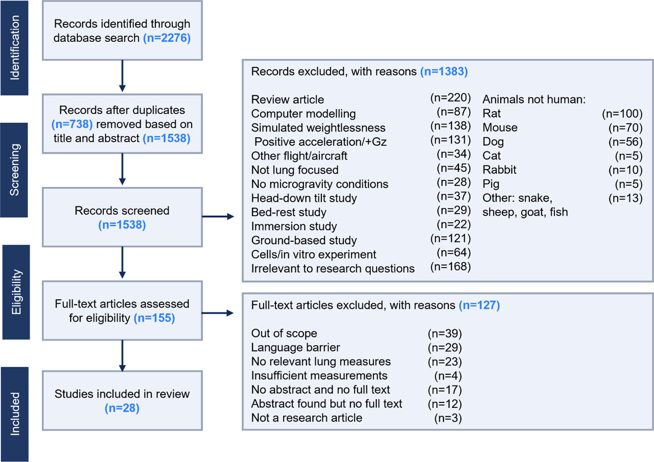
Figure 2.
PRISMA flow diagram demonstrating summary of literature search and screening process.
Table 1.
Characteristics and pulmonary outcome measures of included studies.
| Study | Number of participants (n) | Sex (M/F) | Flight type | 0G exposure duration | Outcome measures |
|---|---|---|---|---|---|
| Baevsky et al. 2007 | 8 | 8/0 | Long | 179 d | Respiration frequency |
| Bettinelli et al. 2002a | 6 | 4/2 | Parabolic | 20 s | Pressure, compliance |
| Bettinelli et al. 2002b | 4 | 3/1 | Parabolic | 20 s | Compliance |
| Conkin et al. 2017 | 8 | 8/0 | Short | 10 d | Respiration frequency |
| Dellacá et al. 2004 | 5 | 4/1 | Parabolic | 20 s | Breathing time, MIF, V̇E, compliance, respiratory work, elastic work |
| Di Rienzo et al. 2008 | 4 | 3/1 | Short | 16 d | Respiration frequency |
| Dutrieue et al. 2003 | 9 | 4/5 | Parabolic | 22 s | Breathing time, FEF |
| Dutrieue et al. 2005 | 7 | 4/3 | Parabolic | 18 s | Breathing time |
| Edyvean et al. 1991 | 5 | 5/0 | Parabolic | 18 s | Respiration frequency, pressure, compliance, resistance |
| Elliott et al. 1996 | 7 | 4/3 | Short | 9 d | PEFR |
| Elliott et al. 2001 | 5 | 4/1 | Short | 13 d | Respiration frequency |
| Estenne et al. 1992 | 5 | 5/0 | Parabolic | 20 s | Breathing time, MIF, respiration frequency |
| Frerichs et al. 2001 | 7 | 6/1 | Parabolic | 20 s | Respiration frequency |
| Frerichs et al. 2005 | 8 | 7/1 | Parabolic | 20 s | Respiration frequency |
| Guy et al. 1991 | 9 | 5/4 | Parabolic | 24 s | FEF, PEFR |
| Hahn et al. 2013 | 22 | 16/6 | Parabolic | 21 s | Respiration frequency, resistivity |
| Levine et al. 1996 | 6 | 4/2 | Short | 14 d | V̇O2, V̇E, work rate, economy |
| Limper et al. 2014 | 18 | 9/9 | Parabolic | 23 s | V̇O2 |
| Prisk et al. 1993 | 7 | 4/3 | Short | 9 d | Pulmonary diffusing capacity |
| Prisk et al. 1995 | 8 | 5/3 | Short | 23 d | Respiration frequency, V̇O2, V̇CO2, MIF, breathing time, pressure, V̇E |
| Prisk et al. 2000 | 5 | 4/1 | Short | 16 d | Respiration frequency, V̇E, pressure |
| Prisk et al. 2006 | 10 | 9/1 | Long | 163 d | Respiration frequency, V̇O2, V̇CO2, FEF, PEFR, pressure, V̇E |
| Sá et al. 2009 | 5 | 4/1 | Short | 17 d | Breathing time, MIF, V̇E |
| Stegemann et al. 1997 | 4 | 4/0 | Short | 10 d | V̇O2, V̇CO2, V̇E |
| Trappe et al. 2006 | 4 | 4/0 | Short | 17 d | V̇O2, V̇E |
| Väida et al. 1997 | 10 | 10/0 | Parabolic | 20 s | Pulmonary diffusing capacity |
| Verbanck et al. 1997 | 4 | 4/0 | Short | 10 d | Pulmonary diffusing capacity |
| Verheyden et al. 2010 | 11 | 11/0 | Long | 180 d | Respiration frequency |
[i] MIF - mean inspiratory flow, V̇E - minute ventilation, FEF - forced expiratory flow, PEFR - peak expiratory flow rate, V̇O2 - oxygen consumption, V̇CO2 - carbon dioxide production. A full list of definitions of pulmonary terms is found in Supplementary Material 2.
Methodological quality assessment
The methodological analysis of the quality of included studies was performed using the RoBANS assessment tool. Most studies were considered to have an overall low risk of bias. Twenty-four studies contained no missing outcome data or included reasons for missing outcome data and were considered low risk. Four studies were assigned as having an unclear overall risk of bias as they failed to provide sufficient details to allow a complete assessment of potential risk and/or contained missing or poorly reported information on which to base risk judgements. The blinding of outcome assessment was considered low risk since it is not possible to blind participants to microgravity. Finally, it was judged that the outcome is highly unlikely to be influenced by lack of blinding. The PRISMA guidelines and checklist were followed throughout this review (Supplementary Material 3).
Pulmonary outcomes assessed
Overview of functional pulmonary outcome measures available
Several different pulmonary variables were explored during this review. Gas exchange parameters including oxygen consumption (V̇O2) and carbon dioxide production (V̇CO2), respiration frequency, pulmonary flow rates like forced expiratory flow (FEF), mean inspiratory flow (MIF) and peak expiratory flow rate (PEFR), pulmonary diffusing capacity, minute ventilation (V̇E), breathing time like expiratory time, inspiratory time and breath-hold durations, pressure, compliance, resistivity, and respiratory work were analyzed.17–44 The greatest number of variables measured were found in parabolic flights while the least were found in long-duration spaceflight missions (Table 3).
Table 2.
Analysis of risk of bias of included studies using RoBANS.
| Study | Selection of participants | Confounding variables | Intervention (exposure) measurement | Blinding of outcome assessment | Incomplete outcome data | Selective outcome reporting | Overall risk of bias |
|---|---|---|---|---|---|---|---|
| Baevsky et al. 2007 | Low | Unclear | Low | Unclear | Low | Low | Low |
| Bettinelli et al. 2002a | Low | Unclear | Low | Unclear | Low | Low | Low |
| Bettinelli et al. 2002b | Low | Low | Low | Unclear | Low | Low | Low |
| Conkin et al. 2017 | Low | Low | Low | Low | Low | Low | Low |
| Dellacá et al. 2004 | Low | Unclear | Low | Unclear | Low | Low | Low |
| Di Rienzo et al. 2008 | Low | Unclear | Low | Unclear | Unclear | Low | Unclear |
| Dutrieue et al. 2003 | Low | Low | Low | Low | Low | Low | Low |
| Dutrieue et al. 2005 | Low | Low | Low | Low | Low | Low | Low |
| Edyvean et al. 1991 | Low | Unclear | Low | Unclear | Low | Low | Low |
| Elliott et al. 1996 | Low | Low | Low | Unclear | Unclear | Low | Low |
| Elliott et al. 2001 | Low | Low | Low | Low | Low | Low | Low |
| Estenne et al. 1992 | Low | Unclear | Low | Low | Low | Low | Low |
| Frerichs et al. 2001 | Low | Unclear | Low | Unclear | Low | Low | Low |
| Frerichs et al. 2005 | Low | Low | Low | Unclear | Low | Low | Low |
| Guy et al. 1991 | Low | Low | Low | Unclear | Low | Low | Low |
| Hahn et al. 2013 | Low | Unclear | Low | Unclear | Unclear | Low | Unclear |
| Levine et al. 1996 | Low | Unclear | Low | Low | High | Low | Unclear |
| Limper et al. 2014 | Low | Low | Low | Low | Low | Low | Low |
| Prisk et al. 1993 | Low | Low | Low | Low | High | Low | Low |
| Prisk et al. 1995 | Low | Low | Low | Low | Low | Low | Low |
| Prisk et al. 2000 | Low | Low | Low | Low | Low | Low | Low |
| Prisk et al. 2006 | Low | Low | Low | Unclear | Unclear | Low | Low |
| Sá et al. 2009 | Low | Low | Low | Low | Low | Low | Low |
| Stegemann et al. 1997 | Low | Low | Low | Low | Low | Low | Low |
| Trappe et al. 2006 | High | Unclear | Low | Unclear | Low | Low | Unclear |
| Väida et al. 1997 | Low | Low | Low | Low | Low | Low | Low |
| Verbanck et al. 1997 | Low | Low | Low | Low | Unclear | Low | Low |
| Verheyden et al. 2010 | Low | Unclear | Low | Low | Low | Low | Low |
Table 3.
Summary of pulmonary functional outcome measures in microgravity during parabolic flight, short- and long-duration spaceflight.
| Outcome measure | Parabolic | Short | Long |
|---|---|---|---|
| Respiration frequency | ✔ | ✔ | ✔ |
| Minute ventilation (V̇E) | ✔ | ✔ | ✔ |
| Volume of oxygen consumption (V̇O2) | ✔ | ✔ | ✔ |
| Volume of carbon dioxide production (V̇CO2) | – | ✔ | ✔ |
| Forced expiratory flow (FEF) | ✔ | – | ✔ |
| Mean inspiratory flow (MIF) | ✔ | ✔ | – |
| Peak expiratory flow rate (PEFR) | ✔ | ✔ | ✔ |
| Breathing time | ✔ | ✔ | – |
| Pulmonary diffusing capacity | ✔ | ✔ | – |
| Pulmonary capillary blood volume (VC) | – | ✔ | – |
| Pulmonary capillary blood flow (Qc) | – | ✔ | – |
| Pressure | ✔ | ✔ | ✔ |
| Compliance | ✔ | – | – |
| Resistance | ✔ | – | – |
| Resistivity | ✔ | – | – |
| Respiratory work | ✔ | – | – |
| Elastic work | ✔ | – | – |
| Exercise work rate | – | ✔ | – |
| Exercise economy | – | ✔ | – |
Main effects and effect sizes of pulmonary functional outcome changes from 1G to 0G
Thirteen studies demonstrated respiration frequency measurements in 1G and 0G (Fig. 3). The forest plot showed no change in respiration frequency between 1G and 0G across parabolic, short- and long-duration spaceflight, as demonstrated by both the common effect model and random effects model. However, a greater respiration frequency was observed in 0G than 1G during short-duration spaceflight breathing an inspired oxygen partial pressure (PiO2) of 148 mmHg20 and when standing at 1G (Prisk et al., 1995). A greater respiration frequency was also reported in 0G during short-duration spaceflight than in 1G when awake (Elliott et al., 2001), and during in-flight month 5 of a long-duration spaceflight (Baevsky et al., 2007).
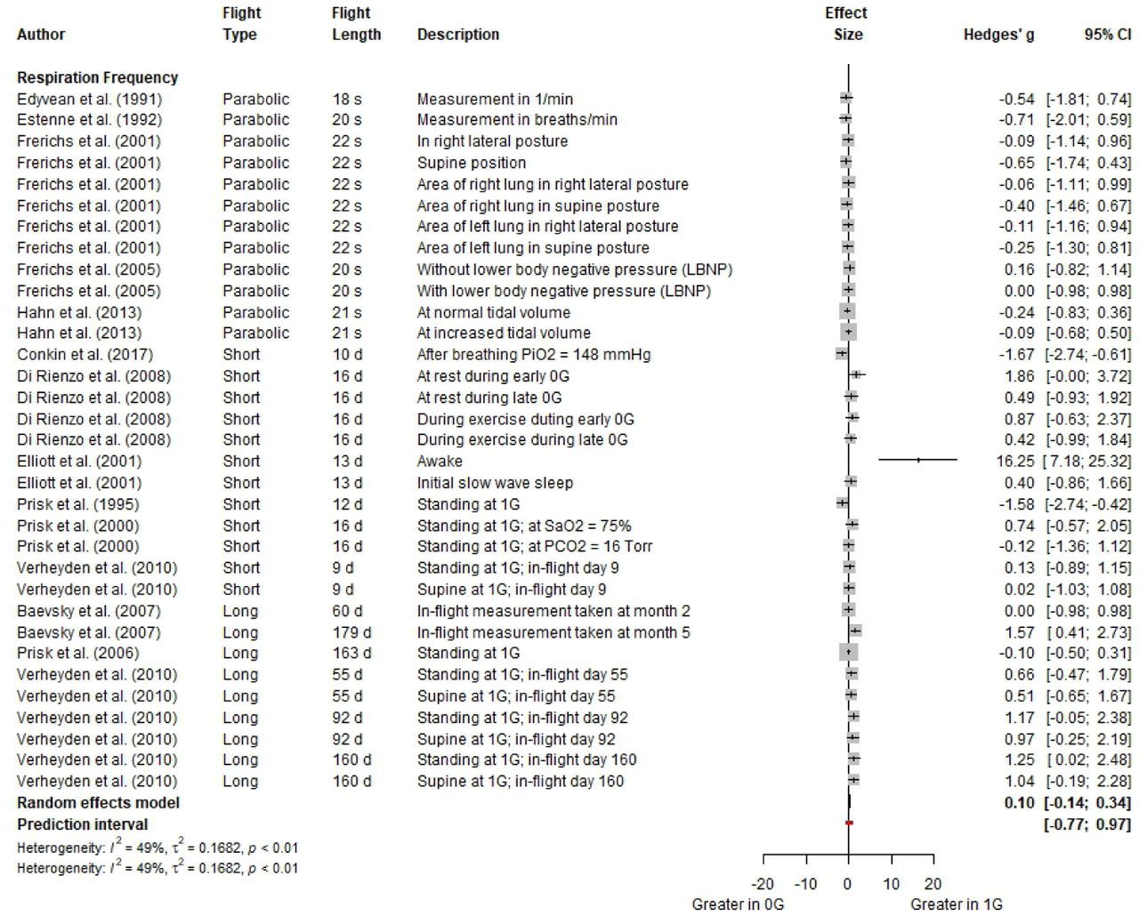
Figure 3.
Forest plot for respiration frequency in 0G compared to 1G (i.e., a right-ward shift indicated a larger measure in 1G). Effect sizes are reported as Hedge’s g with 95% confidence intervals.
Eight studies reported minute ventilation measurements in 1G and 0G across all flight types (Fig. 4). During parabolic flight and short-duration spaceflight, there was no overall change in minute ventilation in 0G compared to 1G. However, greater minute ventilation at 1G during short-duration (+1.16 [0.08; 2.25]) and long-duration (+0.58 [0.17; 0.99]) spaceflight was reported when standing at 1G (Prisk et al., 1995; Prisk et al., 2006). Additionally, the hypoxic ventilatory response (initiated by reducing SaO2 to 75%) produced a greater minute ventilation in 1G than 0G (+1.60 [0.08; 3.13]) (Prisk et al., 2000).
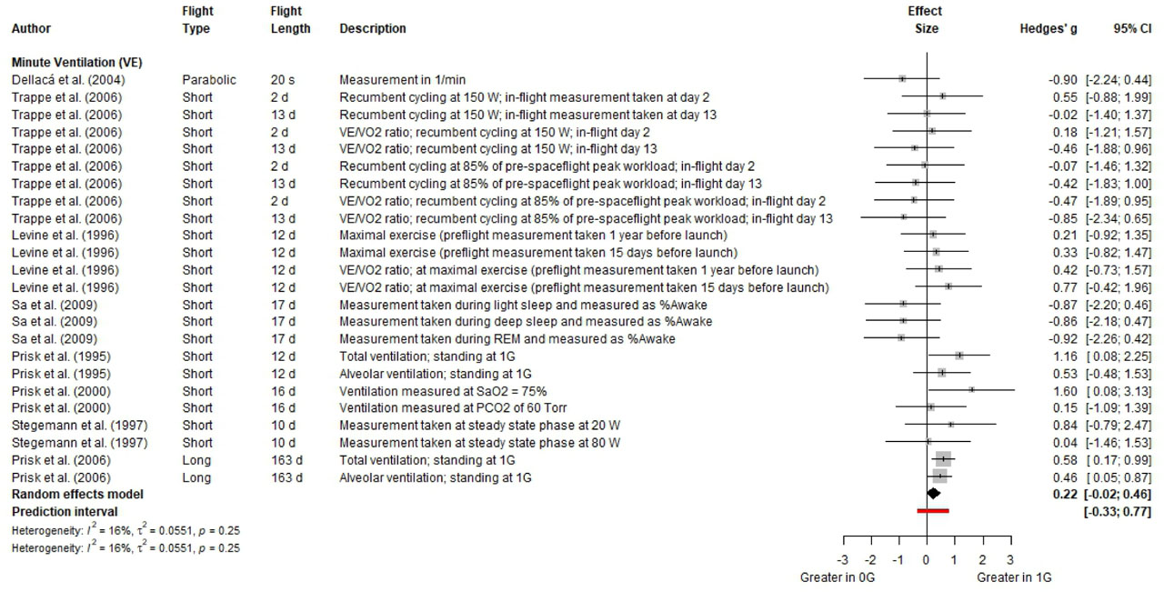
Figure 4.
Forest plot for minute ventilation in 0G compared to 1G (i.e., a right-ward shift indicated a larger measure in 1G). Effect sizes are reported as Hedge’s g with 95% confidence intervals.
Six studies measured V̇O2 in 0G and 1G (Fig. 5). No differences were observed between short-duration spaceflight in 0G and during recumbent cycling in 1G, in which the subject is in a laid-back reclining position on the bicycle (Trappe et al., 2006), at various work rates (Stegemann et al., 1997), and at maximal exercise (Levine et al., 1996). However, V̇O2 was greater at 0G than 1G in all measurements taken during parabolic flights (Limper et al., 2014). On the contrary, V̇O2 was greater at 1G than 0G for the measurements taken during long-duration spaceflight (Prisk et al., 2006). As for V̇CO2, three studies reported measurements in 1G and 0G, and there were no differences between 0G during short-duration spaceflight and 1G. However, a greater V̇CO2 in 1G during long-duration spaceflight is observed in comparison to 0G (Prisk et al., 2006).
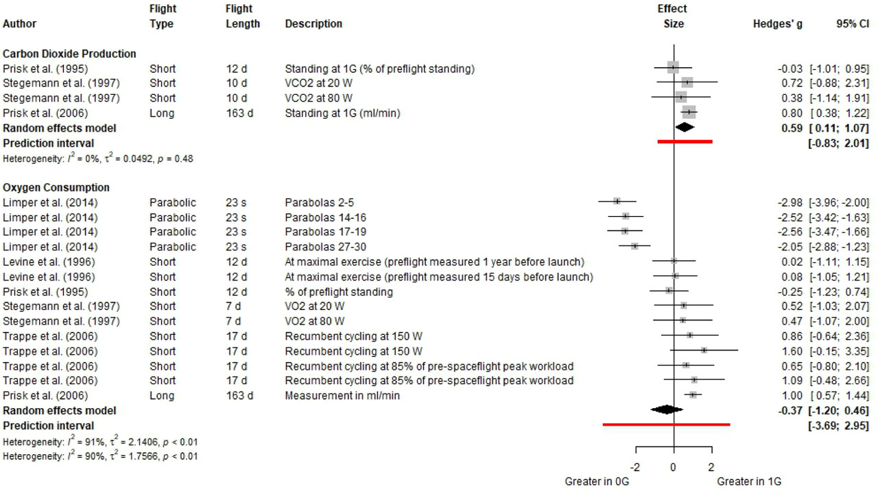
Figure 5.
Forest plot for parameters of pulmonary cellular respiration in 0G compared to 1G (i.e., a right-ward shift indicated a larger measure in 1G). Effect sizes are reported as Hedge’s g with 95% confidence intervals.
Three studies reported data on forced expiratory flow (FEF) in 0G and 1G and showed no difference between the two conditions for both parabolic and long-duration spaceflight (Fig. 6) (Prisk et al., 2006; Guy et al., 1991; Dutrieue et al., 2003). Four studies explored mean inspiratory flow (MIF) in 0G and 1G. Data obtained during parabolic flight and during light and deep sleep in short-duration spaceflight showed no changes between 0G and 1G (Dellaca et al., 2004; Estenne et al., 1992; Sa et al., 2009). However, another study’s data obtained during short-duration spaceflight showed a greater MIF in 1G than 0G (Prisk et al., 1995). On the other hand, a greater value in 0G than 1G was observed for MIF during REM sleep measurements (Sa et al., 2009). The common effects model generated shows an overall greater MIF in 0G than 1G. Three studies reported peak expiratory flow rate (PEFR) in 1G and 0G across all flight types and showed overall no differences between the two gravity conditions (Prisk et al., 2006; Guy et al., 1991; Elliott et al., 1996).
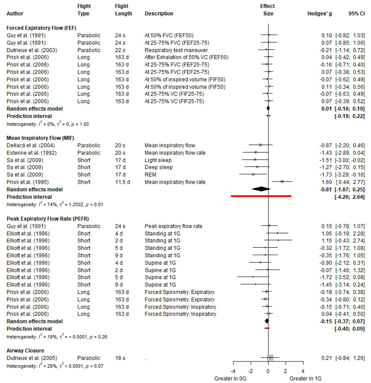
Figure 6.
Forest plot for parameters of pulmonary flow rates in 0G compared to 1G (i.e., a right-ward shift indicated a larger measure in 1G). Effect sizes are reported as Hedge’s g with 95% confidence intervals.
Six studies reported breathing time measurements in 1G and 0G (Fig. 7). A majority of measurements showed no difference between 0G during parabolic flight and short-duration spaceflight and 1G. However, one study showed greater breathing time in 1G than 0G during parabolic flight when a breath-hold was done at residual volume before test inspiration (Dutrieue et al., 2005). On the other hand, another study showed a greater breathing time in 0G in parabolic flight than 1G when inspiratory time is taken as a function of total breath time (% of preflight 1G measurement at the standing position) (Prisk et al., 1995).
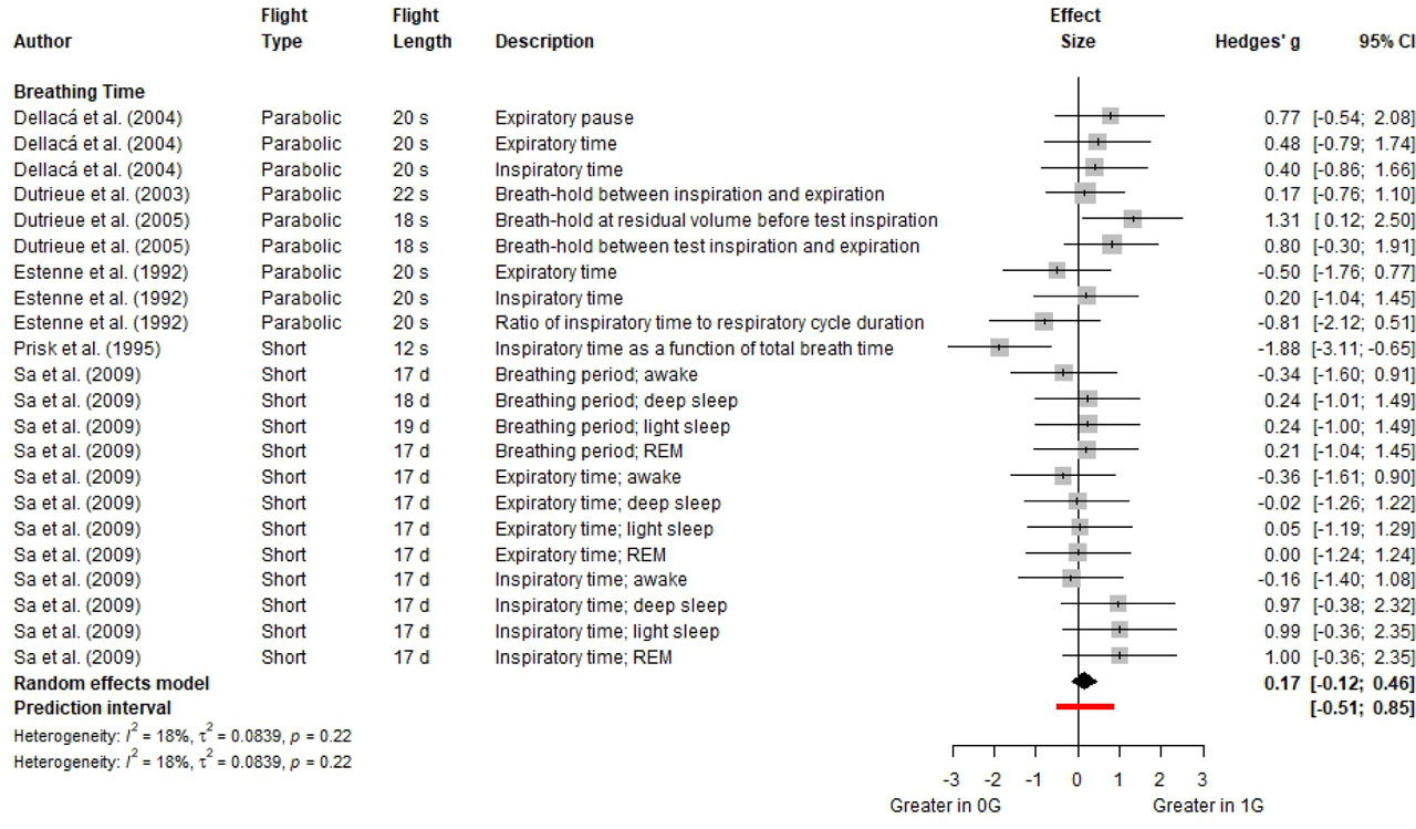
Figure 7.
Forest plot for breathing time in 0G compared to 1G (i.e., a right-ward shift indicated a larger measure in 1G). Effect sizes are reported as Hedge’s g with 95% confidence intervals.
Three studies investigated pulmonary diffusing capacity in 0G and 1G across parabolic flight and short-duration spaceflight (Fig. 8). Measurements reported include diffusing capacity for carbon monoxide (CO) or nitric oxide (NO) and diffusing capacity per unit alveolar volume. Pulmonary diffusing capacity was greater at 0G than at 1G during all parabolic flight measurements (Vaida et al., 1997). However, membrane diffusing capacity, and normoxic diffusing capacity for CO and per unit alveolar volume outcome measures were greater in 0G than 1G in short-duration spaceflight (Prisk et al., 1993). Lung CO-diffusing capacity and CO-diffusing capacity per unit alveolar volume showed no differences between 0G in short-duration spaceflight and 1G (Verbanck et al., 1997). Additionally, single measurements were reported for components of pulmonary diffusing capacity including alveolar volume, pulmonary capillary blood volume, and pulmonary capillary blood flow. No differences were observed in alveolar volume and pulmonary capillary blood flow between 0G in parabolic flight and 1G, while pulmonary capillary blood volume was increased in 0G relative to 1G in short-duration spaceflight (Prisk et al., 1993).
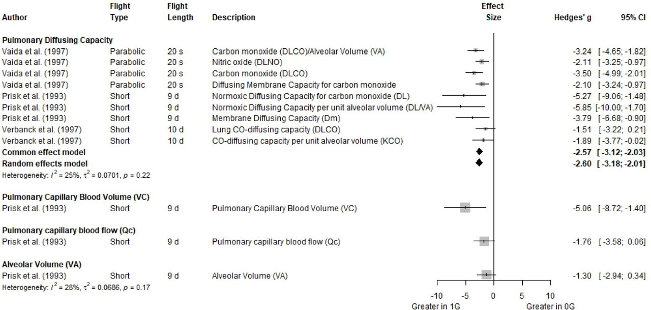
Figure 8.
Forest plot for pulmonary diffusing capacity in 0G compared to 1G (i.e., a right-ward shift indicated a larger measure in 1G). Effect sizes are reported as Hedge’s g with 95% confidence intervals.
Fig. 9 depicts measurements of pressure, compliance, elastic work, exercise economy, exercise work, resistivity, and respiratory work. Five studies measured pressure parameters within the pulmonary system in 1G and 0G across all flight types. Greater pulmonary pressure and transdiaphragmatic pressure were observed in 0G in parabolic flight than 1G when measured at end of expiration (Bettinelli et al., 2002; Bettinelli et al., 2002) while greater end-expiratory gastric pressure was found in 1G (Edyvean et al., 1991). No differences were seen for measurements taken at 1G and during short-duration spaceflight, except that end-tidal PCO2 when taken as % of preflight value at standing position was greater in 0G than in 1G (Prisk et al., 1995). However, another study noted no differences between 0G in short-duration spaceflight and 1G for that same measurement (Prisk et al., 2000). As for studies performed in long-duration spaceflight, maximum inspiratory pressure at functional residual capacity (FRC) is greater at 0G during both early and late in-flight timepoints than 1G. However, maximum inspiratory pressure at residual volume (RV) early and late in-flight and maximum expiratory pressure at total lung capacity (TLC) early in-flight are greater in 1G than 0G. There are no differences between 0G and 1G for maximum expiratory pressure at FRC during early and late in-flight, and maximum expiratory pressure at total lung capacity (TLC) late in-flight (Prisk et al., 2006).
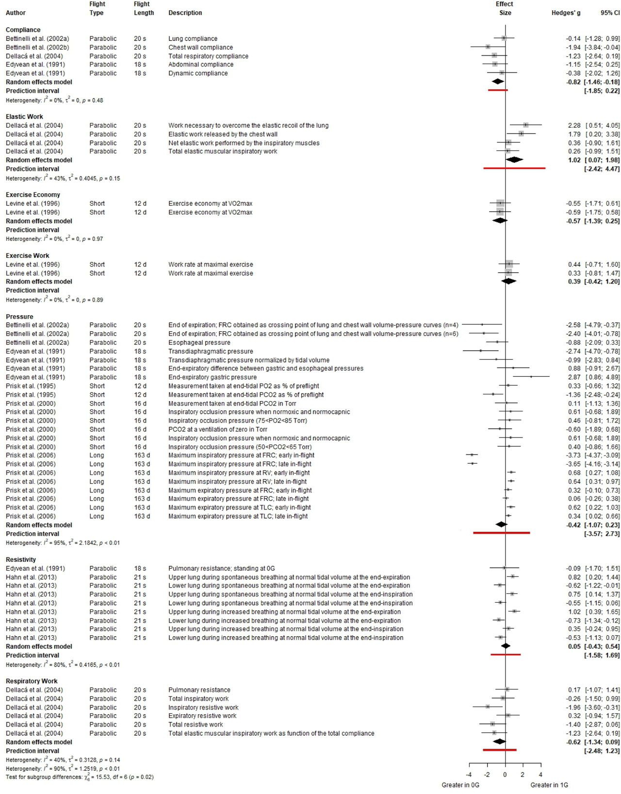
Figure 9.
Forest plot for pulmonary pressure, compliance, and resistance parameters in 0G compared to 1G (i.e., a right-ward shift indicated a larger measure in 1G). Effect sizes are reported as Hedge’s g with 95% confidence intervals.
Three studies measured various compliance measurements including lung, chest wall, abdominal and dynamic compliance during parabolic flight (Fig. 9). No differences were seen between 1G and 0G in the various compliance measurements. However, both common effect model and random effects models show an increase in 0G relative to 1G. Moreover, pulmonary resistance measurements show no differences between 0G during parabolic flight and 1G (Dellaca et al., 2004; Edyvean et al., 1991).
Various single studies reported other pulmonary measurements essential to understanding changes in pulmonary function in space, but these did not fit the criteria for a meta-analysis. This includes resistivity (Hahn et al., 2013), respiratory work, elastic work (Dellaca et al., 2004), exercise work rate, and exercise economy (Levine et al., 1996). Greater resistivity is observed in 1G during parabolic flight than in 0G in measurements taken of the upper lung during spontaneous at normal tidal volume at end-expiration and end-inspiration, and during increased breathing at normal tidal volume at end-expiration. However, greater resistivity is seen in 0G in measurements taken of the lower lung during increased breathing at normal tidal volume at the end-expiration (Hahn et al., 2013).
Several work parameters were explored in 0G during parabolic flight including respiratory work and elastic work, while work rate at maximal exercise and economy outcome measures in 0G were obtained during short-duration spaceflight. No differences between 0G and 1G were observed in total inspiratory work, expiratory resistive work, and total resistive work. However, inspiratory resistive work was greater in 1G than in 0G. As for measurements of elastic work, no differences were seen between 1G and 0G in short-duration spaceflight for net elastic work performed by the inspiratory muscles and total elastic muscular inspiratory work. However, work necessary to overcome the elastic recoil of the lung and elastic work released by the chest wall were greater in 1G than in 0G (Dellaca et al., 2004). No differences were seen between 1G and 0G in short-duration spaceflight in the work rate measured at maximal exercise and economy at maximal V̇O2 (Levine et al., 1996).
Meta-analysis
For most variables, the meta-analysis revealed no changes due to microgravity except for pressure parameters (Table 4). The majority of measurements had a high residual heterogeneity, suggesting a lot of variability between individual results. There was a significant change in oxygen consumption between the duration levels with greater levels in 0G during transient microgravity exposure and lower levels during sustained exposure. There was an effect of microgravity on pressure and pulmonary diffusing capacity.
Table 4.
Results of the meta-analysis for changes in pulmonary function outcome measures in microgravity, and between different time categories (i.e., parabolic, short, long) in microgravity.
| Parameter | Residual Het. | Effect of Microgravity | Effect of Time | |
|---|---|---|---|---|
| SMD (95% CI) | p-value | p-value | ||
| CO2 production | 0.3084 | 0.5622 (−0.0063, 1.1306) | 0.0526 | 0.1582 |
| O2 consumption | <0.0001 | −0.0927 (−1.1816, 0.9963) | 0.8676 | <.0001 |
| Respiratory Frequency | 0.0001 | −0.0152 (−0.5364, 0.506) | 0.9544 | 0.3242 |
| Pulmonary Diffusing Capacity | 0.1383 | −2.163 (−3.3896, −0.9364) | 0.0005 | 0.5730 |
| Minute Ventilation | 0.2065 | 0.227 (−0.2059, 0.6600) | 0.3041 | 0.1019 |
| Breathing Time | 0.0258 | −0.070 (−0.7533, 0.6133) | 0.8408 | 0.5350 |
| Compliance | 0.3256 | −0.8549 (−1.5946, −0.1152) | 0.0235 | N/A |
| Pressure | 0.2820 | −0.6487 (−0.9539, −0.3435) | <.0001 | 0.9617 |
| Resistivity | 0.9086 | −0.0058 (−0.5602, 0.5486) | 0.9835 | 0.9835 |
| FEF | 0.8955 | −0.0222 (−0.4115, 0.3672) | 0.9112 | 0.8860 |
| MIF | 0.0006 | −0.5258 (−2.0184, 0.9668) | 0.4899 | 0.4545 |
| PEFR | 0.8034 | −0.115 (−0.5273, 0.2972) | 0.5845 | 0.6251 |
Discussion
Spaceflight and pulmonary function
General summary of method and scope of papers
This systematic review gathers all the available published parabolic flight and spaceflight studies with functional pulmonary outcomes to provide an overview of the changes in pulmonary system function in space relative to normal gravity on Earth. There were 28 papers that covered 0-1G exposure times from 18 seconds to a maximum of 180 days. The average duration for each category in the review was 21 ± 2 s (parabolic; n=115 individuals), 14 ± 4 d (short-duration; n=67 individuals), and 174 ± 8 d (long-duration; n=29 individuals). In a previous study we focused on anatomical changes with low gravity exposure (Ghani et al., 2023). In this study, the focus was on functional lung measures.
General summary of overall key findings
The lung comprises both fluid and gas dynamics, therefore, changes in gravity could alter pulmonary function, particularly the ventilation-perfusion ratio, leading to impaired physical performance and potential health issues. However, this systematic review found that all the measures demonstrated either no change between 1G and 0G or the changes were small in magnitude and of uncertain clinical relevance. Of the small but statistically significant changes these included: reductions in CO2 production and elastic work, and increases in pulmonary diffusing capacity, pulmonary capillary blood volume, and pulmonary compliance in 0G compared to 1G.
The reduction in elastic work and improved pulmonary compliance result in easier breathing, as the respiratory muscles exert less effort (Fig. 9) (Cross et al., 2012). However, in individuals with pulmonary disease the implications of this can vary. Restrictive lung diseases (e.g., fibrosis, acute respiratory distress) are characterized by an increase in elastic work and a reduction in compliance (Mannino et al., 2003). Hypothetically, the effects of microgravity might help alleviate some of these issues by offsetting the underlying mechanisms. Conversely, in obstructive pulmonary diseases (e.g., asthma) where compliance is already high and elastic work low, microgravity might exacerbate air trapping (Devine, 2008). This can lead to hyperinflation of the lungs, increase the work of breathing, and potentially impair gas exchange by causing ventilation-perfusion mismatching (Rossi et al., 2015). While persons with overt restrictive or obstructive pulmonary disease are not going to be selected for current long duration space missions, prolonged exposure to lunar or Martian dust could cause airway inflammation that leads to obstructive pulmonary disease (Hnizdo and Vallyathan, 2003). This is of course a hypothetical risk and it is important to note that these measures have not been recorded during spaceflight and are currently limited to parabolic flight. As parabolic flight involves pre- and post-loading of >1G loads, further investigations are needed in sustained microgravity environments to observe the effects that this could have, particularly regarding people with obstructive pulmonary diseases.
An observable increase in pulmonary diffusing capacity in microgravity was present in all three studies that assessed it (Fig. 8). This would improve pulmonary gas exchange (Roughton and Forster, 1957). One study showed diffusing capacity to be supported by increased pulmonary capillary blood (Fig. 7) (Prisk et al., 1993). These factors act to improve oxygen uptake and CO2 removal, thus helping ventilation-perfusion matching across the lungs. This benefit could compensate for pulmonary impairments, allowing gas exchange with less respiratory effort. However, no long-duration spaceflight studies have looked at this, so it is unclear whether this increase in pulmonary diffusing capacity is maintained or reduced over time, with the longest study evaluated here being 10 d (Verbanck et al., 1997).
The observed random effects decrease in CO2 production was only significant in one of the four studies that assessed this (Fig 5) (Prisk et al., 2006). This could be consistent with a lower metabolic rate, supported by trends towards lower minute ventilation and oxygen consumption (Fig. 4; Fig. 5), with oxygen consumption lower with longer durations spent in space (Table 4). Indeed, the longer-duration study of Prisk et al. (2006) that evaluated these variables suggested the underlying reason could arise from astronauts doing relatively more activity in shorter-duration flights. It was also postulated, that while unlikely, a reduction in body mass over time could reduce metabolic rate, although this did not align with other measured parameters (Prisk et al., 2006).
Overall conclusion of the scientific discussion above
Overall, the study highlights the adaptability of the lung, including in the longest duration 0G exposure studies that have reported minimal changes compared to pre-flight. The changes that were observed mostly lead to improved pulmonary function, by improving gas exchange and potentially providing a benefit for those with any underlying restrictive lung component. Obstructive lung disease could meet with some challenges, but whether these are ever likely to be present in short-and long-duration 0G exposures remains to be determined. Likewise, it is unknown whether the negative effects are offset by concurrent improvements in gas exchange.
The findings of this review as well as the many foundational studies done during NASA Spacelab missions and ESA parabolic flights showed that lung function remains surprisingly robust in space, with no major impairments in gas exchange or ventilation (Prisk, 2019; Prisk et al., 2008; West et al., 1997; Puchkova et al., 2025). A recent study provided a detailed investigation how simulated lunar gravity via head-up bed rest and microgravity via head-down bed rest minimally affected lung function in healthy human subjects. There was clinically insignificant decrease in key respiratory parameters like FVC and FEV1 within the first hours of exposure and the changes were not pathological and remained within normal physiological ranges. Observed changes normalized over time, indicating the respiratory system’s ability to adapt. Importantly, no long-term impairments were observed, and physical training during the simulation helped accelerate recovery of lung function (Puchkova et al., 2025). These findings alongside the results of our review suggest that human lung function remains resilient in space-like environments, supporting the feasibility of extended missions with appropriate countermeasures like exercise.
Limitations of this systematic review
Number of studies
Spaceflight reviews are inherently limited by the number of studies conducted, and particularly by the availability of repeated analyses of specific similar outcome variables. This is a consequence of both funding and time constraints. As a result, studies are rarely repeated, leading to inherent large heterogeneity in the study designs. This showed up in the current meta-analyses due to the impact of subtle differences between how variables were measured as well as the individual responses (Table 4). Additionally, the limited number of long-duration studies are the only evidence available that would be most indicative for long-duration missions to the Moon and Mars. More data is required from truly long-duration missions.
Lack of recent investigations
There is a lack of recent investigations to re-examine the longstanding observation that microgravity produces little to no measurable effect on the lungs, meaning this conclusion remains based on limited and outdated evidence. Also, the most valuable information for preparation of manned missions to the Moon and Mars comes from long-duration missions, and there have been no recent investigations thoroughly examining pulmonary functional outcomes in long-duration human space travel. The age of the available literature highlights the urgent need for renewed research attention to pulmonary physiology in spaceflight. The lack of recent studies is a field-wide limitation.
Subjects of the studies
Consistent with current selection criteria for astronauts or parabolic flights, all subjects in the included studies were fit and healthy people. Such subjects were either crew mission members, who have undergone rigorous medical screening and tests, or terrestrial people with good health. Both sets were within a relatively tight age range and so do not reflect the general human population. Hence, future versions of these studies that can include data of civilian astronauts who are not subjected to rigorous medical screening prior to spaceflight may exacerbate the individual variability and lead to more substantial effects. Also, people with lung diseases like asthma or chronic obstructive pulmonary disease may show different outcomes in microgravity, and hence, information regarding effects of microgravity towards these conditions could be potentially clinically significant as more data is collected in the future, given the increase in space tourism activities.
Sex differences
The studies included in this systematic review present a major gap in male and female participation, making the roles of sex in pulmonary function in space not well understood. 43% of the included studies included no female participants, while females only made up 32% in those that did. Only one study recruited an equal number of male and female participants (Limper et al., 2014). Overall, there were n=162 males and n=49 females included in the analysis of this systematic review. Sex and gender differences in pulmonary health studies should be considered a future priority, especially since females typically have smaller lungs and airways than males, resulting in lower total alveoli and alveoli surface area, which can impact pulmonary function (Dominelli and Molgat-Seon, 2022). Further, during exercise in 1G it is known that females have lower diffusion capacity, membrane diffusing capacity, and pulmonary capillary volume than males, limiting exercise tolerance (Bouwsema et al., 2017; Guzman and Summers, 1973). Females are also more likely to experience pulmonary hypertension, which could be exacerbated by the cephalic fluid shift seen in microgravity (Hughes-Fulford et al., 2024). Conversely, males are terrestrially at higher risk of developing pulmonary fibrosis and obstructive sleep apnea. However, the exclusion of women from many studies makes comparisons based on sex challenging, masking the sex-specific effects of spaceflight on pulmonary function. Such information could be important in informing future long-duration expeditions to the moon and Mars.
Technical challenges of data collection
Human spaceflight research is limited by the financial and logistical costs associated with launching and conducting experiments in true microgravity, and technical constraints to research methods and data collection tools. Parabolic flights are composed of an average of 30 parabolas per flight, which includes periods of both microgravity and hypergravity that may act as a confounding variable (Golding et al., 2017). Therefore, the transient microgravity in parabolic flights may not be indicative of observations in sustained, stable microgravity. In addition to the inherent challenges of conducting human spaceflight experiments and recruiting participants, this systematic review is limited by the data available at different time-points in space (Table 3). These gaps make it difficult to confirm whether the reported changes are unique to each study or scientific fact. This is especially the case for long-duration spaceflight where almost two-thirds of the pulmonary functional variables included in this review were not measured during long-duration missions, which only accounted for three papers (Baevsky et al., 2007; Prisk et al., 2006; Verheyden et al., 2010).
Review focus
This review does not provide information about the functional changes that happen to the pulmonary system upon return to Earth, as it focuses on comparisons solely between 0G in-flight and 1G at pre-flight and not post-flight. Specifically, exploring possible changes to pulmonary function during early and late post-flight time points could reveal a great deal about the effects of space on the lungs and provide further insights in recovery durations, complementing the findings of this review. This is particularly relevant to assessing the overall risk of spaceflight on the pulmonary system; according to the NASA Human System Risk Assessment Process, a key factor to determining risk is the time to return to baseline (reconditioning). While microgravity can cause changes to lung function, several studies noted that these changes were temporary and reversible upon return to Earth’s gravity, even after 6 months of exposure to microgravity (Prisk, 2019; Prisk et al., 2008; Prisk et al., 2006).2,4,38 Therefore, since there were either no changes or the changes were reversible, the changes were unlikely to be clinically significant. However, if a person has a lung condition that alter mechanics, such as asthma, COPD, or idiopathic pulmonary fibrosis (IPF), then they are likely to have a different situation.
Implications for future space travel
The absence in the available data of clinically significant impairments to functional pulmonary outcome measures in LEO is reassuring and a testament to the pulmonary system’s robust nature. Even under challenging conditions, the pulmonary system can maintain homeostasis and ensure vital functions like gas exchange and oxygen delivery are kept the same or even improved. However, it is unknown what the impact of extended microgravity exposure (e.g., years) and its interaction with other hazards like dust exposure or radiation during deep space travel in the future may have on pulmonary function. Specifically, microgravity and space radiation encountered within deep space have the potential to alter molecular processes and functional variables within the pulmonary system through a variety of different mechanisms and potentially lead to damages in lung tissue and lung cancer (Prisk, 2019). Studies investigating effects of galactic cosmic radiation using high linear energy transfer (LET) ions and x-rays on genome-wide methylation patterns in human bronchial epithelial cells have shown that these high exposures affect sites relevant to human lung cancer like adenocarcinomas and squamous cell carcinomas (Kennedy et al., 2018). Therefore, perturbations in pulmonary structure and function during deep space may originate from the exposure to microgravity for long periods of time, space radiation, or the combined effects of both.
In addition to cancer risk, missions to the Moon and Mars bring additional challenges, like dust inhalation. The combination of altered pulmonary deposition of extraterrestrial dust and the potential for the dust to be highly toxic makes such prolonged exposures a plausible threat to the lung in planetary exploration (Prisk, 2019).
Pulmonary function in the age of space omics
Historically, pulmonary function research in space was grounded in direct physiological assessments. Between the 1990s and early 2010s, missions such as the NASA Spacelab and ESA parabolic flight campaigns conducted detailed measurements of respiratory parameters V̇O2, V̇CO2, FEF, PEFR, and pulmonary diffusing capacity. These foundational studies provided insights into how microgravity alters lung mechanics, ventilation-perfusion matching, and gas exchange efficiency. The consensus from this era was that, despite mechanical changes in the thoracic cavity and fluid shifts, pulmonary function remained largely preserved in microgravity, as supported by the findings of this systematic review. Recent space health research has pivoted towards more critical risks for long-duration missions like cardiovascular deconditioning, neuro-vestibular adaptation, immune system dysregulation, radiation exposure, and behavioral health and performance. Also, recent studies concerned with the field of pulmonary function in microgravity often use bed rest, dry immersion, and other microgravity simulations to study pulmonary function and health in space. These analogs are cheaper and safer, although they may not fully replicate spaceflight conditions.
Moreover, the past decade has seen a marked shift in the focus of space health research. With the end of the Space Shuttle program and the rise of commercial spaceflight, the logistical and scientific priorities of missions have evolved. Contemporary missions, such as Inspiration4 and those supported by the Axiom and Polaris programs, emphasize compact, high-throughput data collection methods. This has led to a pivot from traditional spirometry and metabolic testing toward molecular and systems biology approaches, exemplified by initiatives like the Space Omics and Medical Atlas (SOMA). SOMA integrates multi-omics data like genomics, transcriptomics, proteomics, and metabolomics with physiological and environmental metadata to create a comprehensive atlas of human adaptation to spaceflight. While some physiological data were collected during missions like Inspiration4 (heart rate, blood oxygen, ultrasound), there is no direct mention of pulmonary function testing, such as gas exchange or lung mechanics, in the SOMA datasets. However, SOMA’s multi-omics approach could indirectly inform pulmonary health by identifying inflammatory markers, oxidative stress pathways, and immune responses that may affect lung tissue or respiratory function.
While this transition has reduced the frequency of direct pulmonary measurements, it offers new opportunities to understand lung health at a deeper biological level. Omics data can reveal systemic changes such as inflammation, oxidative stress, and immune modulation, which may precede or accompany functional respiratory changes. Additionally, wearable biosensors and AI-driven analytics now enable continuous, non-invasive monitoring of vital signs, including respiratory rate and blood oxygen saturation, potentially detecting early signs of pulmonary compromise.
Two significant effects emerge from this change. On one hand, the lack of recent direct pulmonary function data limits the ability to compare new findings with historical baselines, potentially creating gaps in longitudinal understanding, as seen in this review with the most recent study included published over a decade ago (Limper et al., 2014). On the other hand, the integration of molecular insights with real-time physiological monitoring could lead to more predictive and personalized models of respiratory health in space. Future research that combines these approaches, where both legacy physiological tools and modern omics technologies can work together, will be essential to fully characterize pulmonary adaptation and resilience in microgravity, especially in the context of long-duration missions to the Moon and Mars. It also benefits our understanding of how different individuals are affected by space with the current increase in space tourism.
Conclusions
This systematic review and quantitative meta-analysis provide an overview of the effects of microgravity on various functional variables of the pulmonary system in humans. Overall, pulmonary function remains either the same as on Earth or may even improve for some measures, such as in the case of pulmonary diffusion capacity. However, changes in lung compliance and elastic work may pose issues for people with any underlying or acquired obstructive pulmonary disorders. Future pulmonary spaceflight research should focus on this area, specifically ways to prevent these effects from occurring. Further, the development of obstructive pulmonary disorders during spaceflight should be considered, particularly regarding how dust inhalation could contribute via airway inflammation and potential cancer risk. The new space age of commercial spaceflight and space tourism, and new space medicine approaches, including multi-omics and AI-driven prediction models, will change the way we study and understand the effects of microgravity on pulmonary function and other organ systems.
Notes
Notes
Supplementary Material 1. Search strategy
OVID Search
Lung terms
exp Respiratory Dead Space/ or exp Respiratory Function Tests/ or exp Respiratory Mechanics/ or exp Respiratory Rate/ or exp Respiratory System/ or exp Respiratory Physiological Phenomena/ or exp Respiratory Muscles/ or exp lung/ or exp Diagnostic Techniques, Respiratory System/ or exp Pulmonary Ventilation/ or exp Ventilation-Perfusion Ratio/ or exp Pulmonary Gas Exchange/ or exp Respiration/ or exp Pulmonary Diffusing Capacity/ or Lung.ab. Or Pulmonary.ab.
Space terms
exp spaceflight/ or exp gravitation/ or exp Weightlessness/ or exp Astronauts/ or exp Spacecraft/ or exp Extraterrestrial Environment/ or Microgravity.ab.
Table S.1.
Search strategy.
| Search number | Term | Keywords in search format |
|---|---|---|
| 1 | Lung | exp Respiratory Dead Space/ or exp Respiratory Function Tests/ or exp Respiratory Mechanics/ or exp Respiratory Rate/ or exp Respiratory System/ or exp Respiratory Physiological Phenomena/ or exp Respiratory Muscles/ or exp lung/ or exp Diagnostic Techniques, Respiratory System/ or exp Pulmonary Ventilation/ or exp Ventilation-Perfusion Ratio/ or exp Pulmonary Gas Exchange/ or exp Respiration/ or exp Pulmonary Diffusing Capacity/ or Lung.ab. Or Pulmonary.ab. |
| 2 | Space | exp spaceflight/ or exp gravitation/ or exp Weightlessness/ or exp Astronauts/ or exp Spacecraft/ or exp Extraterrestrial Environment/ or Microgravity.ab. |
| 3 | Combined Search | 1 AND 2 |
PubMed Search
(“respiratory dead space”[MeSH Major Topic] OR “respiratory function tests”[MeSH Major Topic] OR “respiratory mechanics”[MeSH Major Topic] OR “respiratory rate”[MeSH Major Topic] OR “respiratory system”[MeSH Major Topic] OR “respiratory system agents”[MeSH Major Topic] OR “respiratory physiological phenomena”[MeSH Major Topic] OR “respiratory muscles”[MeSH Major Topic] OR “intercostal muscles”[MeSH Major Topic] OR “Lung”[MeSH Major Topic] OR “diagnostic techniques, respiratory system”[MeSH Major Topic] OR “pulmonary ventilation”[MeSH Major Topic] OR “ventilation perfusion ratio”[MeSH Major Topic] OR “pulmonary gas exchange”[MeSH Major Topic] OR “respiration”[MeSH Major Topic] OR “cell respiration”[MeSH Major Topic] OR “pulmonary diffusing capacity”[MeSH Major Topic] OR “Lung”[Title/Abstract] or “pulmonary”[Title/Abstract]) AND (spaceflight[MeSH Major Topic] or gravitation[MeSH Major Topic] or Weightlessness[MeSH Major Topic] or Astronauts[MeSH Major Topic] or Spacecraft[MeSH Major Topic] or Extraterrestrial Environment[MeSH Major Topic] or Microgravity[Title/Abstract])
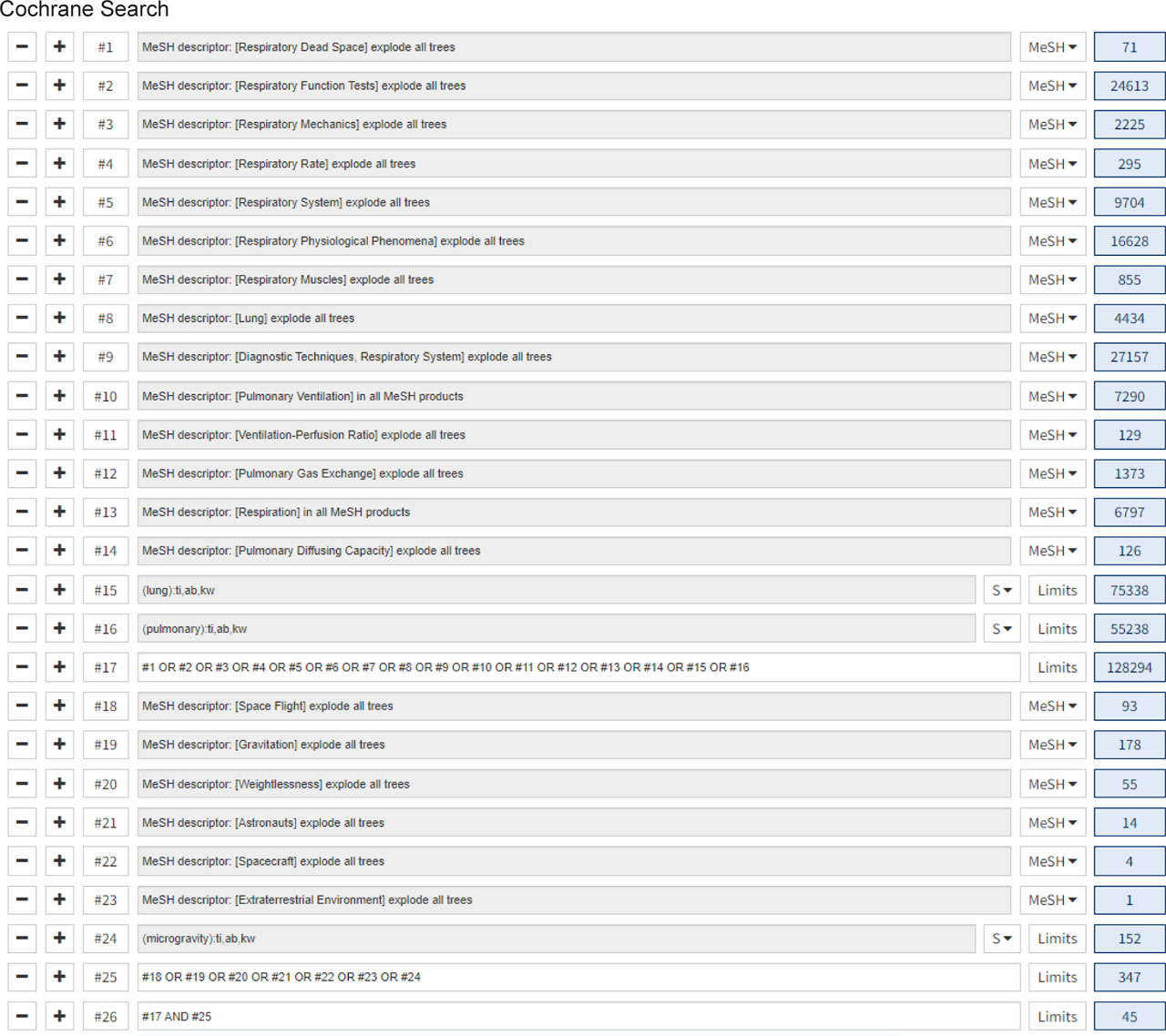
Figure S1.
Snapshot of Cochrane search strategy
Supplemental Material 2. List of definitions in the pulmonary system
Breathing time: the duration of a single respiratory cycle, which is the time taken to inhale and exhale once.
Elastic work: the energy stored in a tissue or structure due to its elastic properties, which is then released when the tissue is stretched or deformed, allowing it to return to its original shape.
Forced expiratory flow (FEF): the rate at which air is forcefully exhaled from the lungs during a specific period.
Mean inspiratory flow (MIF): the average airflow rate during the inhalation phase of a breath.
Minute ventilation (V̇E): the volume of gas inhaled (inhaled minute volume) or exhaled (exhaled minute volume) from a person’s lungs per minute.
Peak expiratory flow rate (PEFR): the maximum flow rate (expressed in Liters per minute [L/min]) generated during a forceful exhalation, starting from full inspiration.
Pressure:
Pulmonary capillary blood flow (Qc): the rate at which blood flows through capillaries within the lungs, where gas exchange occurs between the blood and alveolar air.
Pulmonary capillary blood volume (VC): the volume of blood in the pulmonary capillaries.
Pulmonary compliance: the measure of how easily the lungs can expand and change volume in response to a change in pressure.
Pulmonary diffusing capacity: a physiological measure that indicates the ability of the lungs to exchange gases, specifically oxygen, between the air in the alveoli and the blood within the pulmonary capillaries.
Resistance: the opposition to blood flow within the pulmonary circulation.
Respiration frequency: the number of breaths per minute.
Respiratory work: the energy expenditure required by the respiratory muscles to move air in and out of the lungs, essentially the effort needed to breathe, which involves overcoming the elastic forces of the lungs and the resistance of the airways to achieve gas exchange.
Volume of carbon dioxide production (V̇CO2): volume of carbon dioxide produced per unit of time.
Volume of oxygen consumption (V̇O2): volume of oxygen consumed per unit of time.
Supplementary Material 3. PRISMA checklist

| Section and Topic | Item # | Checklist item | Location where item is reported |
|---|---|---|---|
| TITLE | |||
| Title | 1 | Identify the report as a systematic review. | Included |
| ABSTRACT | |||
| Abstract | 2 | See the PRISMA 2020 for Abstracts checklist. | Seen and followed |
| INTRODUCTION | |||
| Rationale | 3 | Describe the rationale for the review in the context of existing knowledge. | Included |
| Objectives | 4 | Provide an explicit statement of the objective(s) or question(s) the review addresses. | Included |
| METHODS | |||
| Eligibility criteria | 5 | Specify the inclusion and exclusion criteria for the review and how studies were grouped for the syntheses. | Section 2.2 and 2.3 |
| Information sources | 6 | Specify all databases, registers, websites, organisations, reference lists and other sources searched or consulted to identify studies. Specify the date when each source was last searched or consulted. | Section 2.1 |
| Search strategy | 7 | Present the full search strategies for all databases, registers and websites, including any filters and limits used. | Section 2.1 and Supplementary Material |
| Selection process | 8 | Specify the methods used to decide whether a study met the inclusion criteria of the review, including how many reviewers screened each record and each report retrieved, whether they worked independently, and if applicable, details of automation tools used in the process. | Section 2.2 |
| Data collection process | 9 | Specify the methods used to collect data from reports, including how many reviewers collected data from each report, whether they worked independently, any processes for obtaining or confirming data from study investigators, and if applicable, details of automation tools used in the process. | Section 2.2 and 2.3 |
| Data items | 10a | List and define all outcomes for which data were sought. Specify whether all results that were compatible with each outcome domain in each study were sought (e.g. for all measures, time points, analyses), and if not, the methods used to decide which results to collect. | Section 2.3 and 2.4 |
| 10b | List and define all other variables for which data were sought (e.g. participant and intervention characteristics, funding sources). Describe any assumptions made about any missing or unclear information. | Section 2.3 and 2.4 | |
| Study risk of bias assessment | 11 | Specify the methods used to assess risk of bias in the included studies, including details of the tool(s) used, how many reviewers assessed each study and whether they worked independently, and if applicable, details of automation tools used in the process. | Section 2.2 |
| Effect measures | 12 | Specify for each outcome the effect measure(s) (e.g. risk ratio, mean difference) used in the synthesis or presentation of results. | Section 2.3 and 2.4 |
| Synthesis methods | 13a | Describe the processes used to decide which studies were eligible for each synthesis (e.g. tabulating the study intervention characteristics and comparing against the planned groups for each synthesis (item #5)). | Section 2.2 and 2.3 |
| 13b | Describe any methods required to prepare the data for presentation or synthesis, such as handling of missing summary statistics, or data conversions. | Section 2.4 | |
| 13c | Describe any methods used to tabulate or visually display results of individual studies and syntheses. | Section 2.3 | |
| 13d | Describe any methods used to synthesize results and provide a rationale for the choice(s). If meta-analysis was performed, describe the model(s), method(s) to identify the presence and extent of statistical heterogeneity, and software package(s) used. | Section 2.4 | |
| 13e | Describe any methods used to explore possible causes of heterogeneity among study results (e.g. subgroup analysis, meta-regression). | Section 2.4 | |
| 13f | Describe any sensitivity analyses conducted to assess robustness of the synthesized results. | Section 2.4 | |
| Reporting bias assessment | 14 | Describe any methods used to assess risk of bias due to missing results in a synthesis (arising from reporting biases). | Section 2.2 |
| Certainty assessment | 15 | Describe any methods used to assess certainty (or confidence) in the body of evidence for an outcome. | Section 2.4 |
| RESULTS | |||
| Study selection | 16a | Describe the results of the search and selection process, from the number of records identified in the search to the number of studies included in the review, ideally using a flow diagram. | Section 3.1 |
| 16b | Cite studies that might appear to meet the inclusion criteria, but which were excluded, and explain why they were excluded. | Section 3.1 | |
| Study characteristics | 17 | Cite each included study and present its characteristics. | Section 3.1 |
| Risk of bias in studies | 18 | Present assessments of risk of bias for each included study. | Section 3.2 |
| Results of individual studies | 19 | For all outcomes, present, for each study: (a) summary statistics for each group (where appropriate) and (b) an effect estimate and its precision (e.g. confidence/credible interval), ideally using structured tables or plots. | Section 3.3 |
| Results of syntheses | 20a | For each synthesis, briefly summarise the characteristics and risk of bias among contributing studies. | Section 3.2 |
| 20b | Present results of all statistical syntheses conducted. If meta-analysis was done, present for each the summary estimate and its precision (e.g. confidence/credible interval) and measures of statistical heterogeneity. If comparing groups, describe the direction of the effect. | Section 3.3 | |
| 20c | Present results of all investigations of possible causes of heterogeneity among study results. | Section 3.3 | |
| 20d | Present results of all sensitivity analyses conducted to assess the robustness of the synthesized results. | Section 3.3 | |
| Reporting biases | 21 | Present assessments of risk of bias due to missing results (arising from reporting biases) for each synthesis assessed. | Section 3.3 |
| Certainty of evidence | 22 | Present assessments of certainty (or confidence) in the body of evidence for each outcome assessed. | Section 3.3 |
| DISCUSSION | |||
| Discussion | 23a | Provide a general interpretation of the results in the context of other evidence. | Section 4.1 |
| 23b | Discuss any limitations of the evidence included in the review. | Section 4.2 | |
| 23c | Discuss any limitations of the review processes used. | Section 4.2 | |
| 23d | Discuss implications of the results for practice, policy, and future research. | Section 4.3 | |
| OTHER INFORMATION | |||
| Registration and protocol | 24a | Provide registration information for the review, including register name and registration number, or state that the review was not registered. | Review was not registered |
| 24b | Indicate where the review protocol can be accessed, or state that a protocol was not prepared. | Protocol was not prepared | |
| 24c | Describe and explain any amendments to information provided at registration or in the protocol. | N/A | |
| Support | 25 | Describe sources of financial or non-financial support for the review, and the role of the funders or sponsors in the review. | Included in ‘Funding’ section |
| Competing interests | 26 | Declare any competing interests of review authors. | Included in ‘Declaration of competing interest’ |
| Availability of data, code and other materials | 27 | Report which of the following are publicly available and where they can be found: template data collection forms; data extracted from included studies; data used for all analyses; analytic code; any other materials used in the review. | Included in ‘Data statement’ |
[i] From: Page MJ, McKenzie JE, Bossuyt PM, Boutron I, Hoffmann TC, Mulrow CD, et al. The PRISMA 2020 statement: an updated guideline for reporting systematic reviews. BMJ 2021;372:n71. doi: 10.1136/bmj.n71
[ii] For more information, visit: http://www.prisma-statement.org/