
INTRODUCTION
Avocado (Persea americana Mill.) belongs to the Lauraceae family and is recognised as a commercially and nutritionally valuable fruit species, commonly grown in tropical and subtropical regions across the world (Renner, 1999; Sora, 2023; Nascimento et al., 2025). In recent years, the consumption and economic value of avocado have risen considerably on a global scale, largely owing to its nutrient-rich composition, functional food potential and reported health benefits (Silva and Ledesma, 2014; Huang et al., 2023). Accordingly, the development of effective and reliable propagation techniques plays a crucial role in ensuring sustainable production in the avocado industry.
Avocado is a highly heterozygous species, which results in considerable genetic variation during seed propagation. As a result, seedlings do not maintain genetic uniformity and often exhibit distinct genetic traits (Bandaralage et al., 2017). Therefore, vegetative propagation is required for the production of genetically uniform plants (Bandaralage et al., 2015). Currently, propagation in the avocado industry is largely carried out using grafting methods, where the use of selected clonal rootstocks plays a key role. Due to its tolerance to Phytophthora cinnamomi, resistance to low temperatures, and high graft compatibility, ‘Duke 7’ stands out as one of the most widely used clonal rootstocks (Alberti et al., 2018). For nearly 45 years, the double-grafting technique developed by Frolich and Platt has been the standard method used for the propagation of these rootstocks (Frolich and Platt, 1972; Bandaralage et al., 2015). However, this approach is associated with high costs, substantial labour requirements and considerable time consumption (Bandaralage et al., 2015). In contrast to the problems encountered in conventional propagation techniques, micropropagation offers an efficient and scalable method for mass clonal propagation.
Avocado is known to be highly recalcitrant under in vitro culture conditions. Consequently, numerous challenges are encountered during the micropropagation process (Bandaralage et al., 2015). These limitations are also evident in ‘Duke 7’ rootstock, where sterilant-induced tissue necrosis, low regeneration capacity and poor rooting response significantly hinder the development of effective and reproducible micropropagation protocols (Nel, 1983; Zirari and Lionakis, 1994; Wessels, 1996). Furthermore, high levels of microbial contamination during the establishment stage reduce regenerative competence, inhibit growth and may ultimately lead to explant mortality (De La Viña et al., 2001; Barceló-Muñoz and Pliego-Alfaro, 2003). Therefore, various classical sterilising agents, including ethanol (EtOH), sodium hypochlorite (NaOCl), mercuric chloride (HgCL2) and calcium hypochlorite [Ca(OCl)2] are widely employed for the surface sterilisation of explants from different avocado genotypes (Nhut et al., 2008; Zulfiqar et al., 2009; Bandaralage et al., 2015; Osorio et al., 2018). However, a standardised protocol for these agents has not yet been established in avocado, as sterilisation efficiency varies depending on explant type, season of collection and the biological and physiological characteristics of the donor plant (Palaz et al., 2025). Recently, sodium merthiolate (Na-merthiolate) and sodium dichloroisocyanurate (NaDCC) have attracted attention as alternatives to classical sterilising agents. In particular, NaDCC has been reported to provide high sterilisation efficiency with lower phytotoxic effects during explant disinfection and to represent a more environmentally friendly option (Onica et al., 2025). On the other hand, Na-merthiolate has been reported to exhibit sterilisation efficiency comparable to NaOCl (Bacchetta et al., 2008). To the best of our knowledge, no in vitro study has investigated the use of Na-merthiolate and NaDCC as sterilising agents in explants of avocado genotypes. However, it has been reported that these compounds have been used in the in vitro sterilisation of some woody species (Bacchetta et al., 2008; Kirillov et al., 2024; Onica et al., 2025).
In recent years, interest in optimising mineral nutrition in micropropagation has increased. Several studies have demonstrated that adjusting the balance of macro- and micronutrients, modifying nitrogen sources and minimising ion interactions can significantly improve morphogenetic responses in woody species (Reed et al., 2013; Akin et al., 2017; Kovalchuk et al., 2017). In avocado micropropagation, Murashige and Skoog (MS) (Murashige and Skoog, 1962) medium is commonly used as the basal culture medium. In addition, modified MS formulations are frequently employed, with adjustments made to mineral salts and vitamins. Previous studies have reported that certain components of the standard MS formulation may negatively affect avocado plant growth in vitro conditions. Therefore, reducing the concentration of macronutrients in the MS medium, particularly ammonium nitrate (NH4NO3) and potassium nitrate (KNO3), has been reported to positively influence both shoot development and rooting in avocado (Cortés-Rodríguez et al., 2010; Bandaralage et al., 2017; Mansoor, 2018; García-Cabrera et al., 2025). In addition to the mineral composition of the culture medium, the type and concentration of plant growth regulators (PGRs) are also among the principal factors determining the success of avocado micropropagation.
In micropropagation, cytokinins are adeninederived compounds that regulate shoot induction and multiplication by stimulating cell division and differentiation (Faisal et al., 2018). Among the most commonly applied cytokinins, 6-benzylaminopurine (BAP) is a synthetic adenine derivative widely used in avocado micropropagation due to its chemical stability and strong promotive effect on shoot proliferation (Taah et al., 2009; Zulfiqar et al., 2009; Rohim et al., 2013; Ibarra-López et al., 2016; Rabuma et al., 2020; El-Fadl et al., 2022; Qasrawi and Sholi, 2022; Arbeláez Galvis et al., 2025; García-Cabrera et al., 2025). However, in avocado micropropagation, not only BAP but also other cytokinins with different chemical structures are used. Kinetin (KIN) and thidiazuron (TDZ) also display cytokinin-like activity, although they differ in chemical structure and mode of action (Arafa et al., 2021). Whereas KIN belongs to the adenine-type cytokinins, TDZ is a substituted phenylurea compound that can trigger pronounced morphogenetic responses even at relatively low concentrations (Arafa et al., 2021). Nevertheless, research on the application of KIN and TDZ in avocado micropropagation remains limited (Qasrawi and Sholi, 2022; Ashika et al., 2026). Therefore, the effectiveness of BAP, KIN and TDZ may vary according to species, genotype, concentration and culture stage, underscoring the importance of experimentally identifying the optimal cytokinin type and concentration for each genotype.
Following shoot proliferation, the next stage of micropropagation is root induction. During this stage, the hormonal control mechanism shifts from cytokines to auxins. Auxin application is the primary driver of root induction. According to the literature, indole-3-butyric acid (IBA) is the most commonly used auxin for both ex vitro and in vitro rooting in avocado. In addition, 1-naphthaleneacetic acid (NAA) and indole-3-acetic acid (IAA) have also been employed to promote root formation (Bandaralage et al., 2017). In recent years, the effects of different auxin types and concentrations on the rooting of ‘Duke 7’ rootstock have been evaluated, particularly under etiolation-based and ex vitro conditions (Martínez-Villagómez et al., 2025). However, the method of auxin application in vitro (continuous exposure or quick-dip method) significantly affects rooting success (Bandaralage et al., 2017). In vitro rooting studies, auxins have been maintained in the culture medium (1–30 days), and their prolonged presence can, in some cases, suppress adventitious root formation or lead to excessive callus formation (Premkumar et al., 2002; Barceló-Muñoz and Pliego-Alfaro, 2003; Dobránszki and Da Silva, 2010). Alternatively, the quick-dip method, which relies on short-term application of auxins, has also been used for root induction. However, only a few in vitro studies have examined the effectiveness of the quick-dip method in avocado (Cooper, 1987; Barrera-Guerra et al., 1998).
Overall, previous research has investigated sterilisation, shoot proliferation and rooting under in vitro conditions in avocado. However, an integrated evaluation of these stages specifically tailored to ‘Duke 7’ rootstock has not yet been reported. Accordingly, the present study aimed to develop an efficient and reproducible micropropagation protocol for ‘Duke 7’ by comprehensively evaluating (i) the effectiveness of different surface sterilising agents (NaOCl, HgCL2, Na-merthiolate and NaDCC), their concentrations and exposure durations; (ii) the effects of different cytokinin types (BAP, KIN and TDZ) and their concentrations on shoot proliferation; and (iii) the role of quick-dip auxin types (IBA and NAA) and their concentrations in root induction.
MATERIAL AND METHODS
Plant material
In this study, 2-year-old ‘Duke 7’ rootstocks propagated using the ‘Frolich and Platt method’ (Frolich and Platt, 1972) were used as mother plants. This rootstock is widely utilised in commercial avocado orchards in the United States due to its high yield performance with the ‘Hass’ cultivar, moderate resistance to P. cinnamomi, and tolerance to environmental stress conditions (Alberti et al., 2018). The mother plants were maintained under greenhouse conditions at the Plant Transformation Center, University of California, Riverside (33°57’N; 117°23’W), and a fungicide containing 6.65 mM CAPTAN® was applied at 3-week intervals. Axillary shoots (10–15 cm in length) were collected and used as an explant.
Preparation and surface sterilisation of explants
To enhance the effectiveness of sterilising agents during surface sterilisation, all leaves were excised and removed from the shoots using pruning shears. Subsequently, the shoots were sectioned into nodal segments measuring 1–2 cm in length, each comprising one or two axillary buds (Figure 1A). Then, the nodal segments were washed under running tap water for 20 min (~20°C) to remove surface dust and other impurities (Figure 1B). Prior to surface sterilisation, the explants were soaked in 70% (v/v) ethanol for 30 s under a laminar airflow cabinet (Figure 1C). They were subsequently rinsed three times with sterile distilled water to eliminate any residual phytotoxic effects of ethanol (Figure 1D).
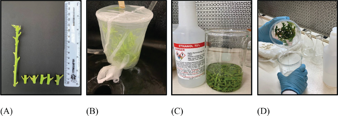
Figure 1.
Preparation of explants for surface sterilisation: the nodal segments measuring 1–2 cm in length (A), rinsing under running tap water (B), soaking in 70% ethyl alcohol (C) and rinsing with deionized distilled water (D).
Following the ethanol application, the explants were immersed in solutions of four different surface sterilising agents prepared at various concentrations for different exposure durations (Table 1). The sterilising agents used for surface sterilisation were NaOCl (Clorox®, containing approximately 5% available chlorine, USA), HgCL2 (7487-94-7, Sigma-Aldrich, USA), Na-merthiolate (T5125, Sigma-Aldrich, USA) and NaDCC (EfferSan®, containing 31.75% available chlorine, USA). During this period, to enhance sterilisation efficiency, 1–2 drops of Tween 20 (Sigma-Aldrich, USA) were added to all solutions, and a horizontal shaker operating at 250 rpm was used to ensure uniform contact between the sterilising agents and the explant surfaces. Following all treatments with the exception of NaDCC applications, the explants were washed three times with sterile water, each for 5 min, to minimise the phytotoxic effects of the sterilising agents. All sterilisation procedures were conducted under aseptic conditions within a laminar airflow cabinet.
Table 1.
Types, concentrations and durations of sterilising agents used for the sterilisation of nodal segments.
| Types of sterilising agents | Concentration (%) | Duration (min) |
|---|---|---|
| NaOCl | 5.0, 10.0, 20.0 (v/v) | 10, 20 |
| HgCI2 | 0.1, 0.5, 1.0 (w/v) | 5, 10 |
| Na-merthiolate | 0.01, 0.05, 0.5 (w/v) | 5, 10 |
| NaDCC | 0.15, 0.25, 0.35 (w/v) | 10, 20 |
Preparation of in vitro culture media and growing conditions
Based on the studies of Bandaralage et al. (2017) and García-Cabrera et al. (2025), modified MS medium (M561 and M557, PhytoTech Labs, USA) was selected as the basal medium during in vitro establishment, shoot proliferation and rooting stages. The medium contained half-strength NH4NO3, halfstrength KNO3 and full-strength micronutrients and vitamins. All culture media were supplemented with 3% (w/v) sucrose (Thermo Fisher Scientific, USA) and solidified with 0.7% (w/v) agar (A038, Caisson Lab, USA). BAP (B800, PhytoTech Labs, USA), KIN (K750, PhytoTech Labs, USA), TDZ (T888, PhytoTech Labs, USA) and gibberellic acid (GA3) (Sigma-Aldrich, USA), which were added to the proliferation medium, were incorporated into the culture medium before autoclaving. In contrast, indole-3-acetic acid (IAA) (I885, PhytoTech Labs, USA) was sterilised by filtration through 0.22 μm Millipore membrane filters and subsequently added to the culture medium under aseptic conditions after autoclaving. During the rooting stage, IBA (I460, PhytoTech Labs, USA) and NAA solutions (N600, PhytoTech Labs, USA) were prepared by mixing 96% ethanol and then diluting with sterile deionised water at a 1:1 (v/v) ratio under aseptic conditions in a laminar airflow cabinet. The pH was adjusted to 5.65 before the addition of agar and autoclaving. All culture media were autoclaved at 121°C for 15 min. During the sterilisation experiment, the culture medium was distributed into test tubes (120 × 25 mm), each containing 10 ml of culture medium. During the shoot proliferation and rooting experiments, the culture medium was dispensed into 250 ml jars, each containing 50 ml of medium.
During the establishment and proliferation stages, cultures were incubated in a growth room at 25°C ± 1°C under 16 hr light/8 hr dark photoperiod provided by cool white fluorescent lamps (35 μmol · m−2 · s−1). During the rooting stage, cultures were initially maintained in complete darkness for 3 days (Bandaralage et al., 2017) and subsequently transferred to the growth room under the same light intensity (35 μmol · m−2 · s−1).
Establishment stage
In the establishment culture, modified MS medium without PGRs and 0.01% (w/v) activated charcoal (AC) (C325, Sigma-Aldrich, USA) (Table 2). Sterilisation efficiency was assessed based on bacterial contamination rate (%), fungal contamination rate (%), tissue necrosis rate (%) and survival rate (%). These parameters were recorded for all treatments after 30 days of culture. Bacterial and fungal contamination, together with tissue necrosis, were considered negative responses, whereas a higher survival rate was regarded as a positive indicator of effective sterilisation.
Shoot proliferation stage
In this stage, three different cytokinin types and their respective concentrations [BAP (2.22, 4.44 and 8.88 μM), KIN (2.32, 4.65 and 9.29 μM) and TDZ (0.45, 2.27 and 4.54 μM)] were evaluated, along with a basal medium with no PGRs (control). The tested concentrations were determined based on earlier experimental trials conducted to define the effective activity ranges of the cytokinins (Qasrawi and Sholi, 2022). The modified MS medium, except for the control treatment, was additionally supplemented with 0.87 μM GA3 and 0.06 μM IAA (Table 2). The nodal segments that exhibited healthy development 4 weeks after the establishment stage were used as the explant source. The explants were subcultured three times at 4-week intervals and transferred to fresh modified MS medium with the same composition at each subculture.
Growth parameters were recorded 12 weeks after the initiation of the shoot proliferation stage. The evaluated parameters included the number of shoots per explant, shoot length (mm), number of leaves per explant and shoot growth index (0–4). The shoot growth index was assessed for each nodal explant using a 0–4 scale (0 = no development, 1 = weak, 2 = moderate, 3 = good, 4 = strong). This scale represents a modified version of the 0–5 shoot quality scale developed by Bandaralage et al. (2015) and was adapted for the present study.
Table 2.
Composition of the nutrient media used at different stages of micropropagation of ‘Duke 7’ rootstock.
| Different stages | Basal medium | PGR and additives | Carbohydrate source | Gelling agent |
|---|---|---|---|---|
| Establishment | Modified MS | 0.01% AC | 3% sucrose | 0.7% agar |
| Proliferation | Modified MS | BAP (0.00, 2.22, 4.44, 8.88 μM) or KIN (0.00, 2.32, 4.65, 9.29 μM) or TDZ (0.00, 0.45, 2.27, 4.54 μM) with GA3 (0.87 μM) and IAA (0.06 μM) | 3% sucrose | 0.7% agar |
| Rooting | Modified MS | – | 3% sucrose | 0.7% agar |
Rooting stage
In the rooting experiment, two different auxins (IBA and NAA) at different concentrations (0.0, 5.0, 10.0, 15.0 and 20.0 mM) were evaluated using the quickdip method. In this experiment, modified MS medium without PGRs was used as the culture medium (Table 2). Microcuttings measuring 1–2 cm in length were excised from microshoots derived from the third subculture of the shoot proliferation stage. Under aseptic conditions, the different concentrations of IBA and NAA were applied to the basal ends of the microcuttings using the quick-dip method for 10 s, after which the explants were transferred to the rooting medium.
Rooting parameters were recorded 6 weeks after transfer to the rooting medium. At the end of the experiment, the rooting rate (%), callus diameter (mm) and callus growth index (0–4) were determined. Additionally, the callus growth index was assessed using a 0–4 scale (0 = none, 1 = low, 2 = moderate, 3 = high, 4 = very high) for each micro cutting. This scale is a modified version of the 0–5 callus formation scale developed by Mansoor (2018), adapted for the study.
Experiment design and statistical analysis
The experimental layout followed a completely randomised design (CRD). Separate statistical analyses were conducted for the sterilisation, proliferation and rooting trials. In the sterilisation trial, treatments consisted of different combinations of sterilising agent type (NaOCl, HgCL2, Na-merthiolate and NaDCC), concentration and exposure duration. Each agent × concentration × exposure duration combination was considered an independent treatment. Each treatment consisted of 40 replications, with each glass tube considered one replication and containing a single explant. In the proliferation trial, treatments consisted of different cytokinin types (BAP, KIN and TDZ) and their respective concentrations. Similarly, the rooting trial involved two auxin types (IBA and NAA) and their respective concentrations. For both trials, the jar was considered the experimental unit. Each treatment consisted of five replications, with each jar considered one replication. Each replication (jar) contained six microshoots. For all experiments, the data were analysed using one-way analysis of variance (ANOVA). Treatment means were compared using Duncan’s multiple range test at p < 0.05. All statistical analyses and graphical visualisations were performed using R software (version 4.5.1) within RStudio (R Core Team, 2025).
RESULTS
Surface sterilisation experiment
The effects of sterilising agent type × concentration × exposure duration on explant sterilisation were statistically significant (p < 0.05) (Figure 2). Treatment of nodal segments with 0.25% NaDCC for 20 min showed the best overall performance, resulting in the highest survival rate (87.50% ± 16.23%) and a very low fungal contamination rate (5.00% ± 3.17%), with no bacterial contamination (Figure 3A). When the concentration of NaDCC was reduced to 0.15%, the explants exhibited no tissue necrosis, regardless of the exposure duration. At this concentration, survival rates ranged between 52.50% ± 16.23% and 62.50% ± 16.23%, indicating that NaDCC effectively maintained explant viability while minimising tissue damage. Conversely, high concentrations of NaOCl (20.0%) applied for 20 min and HgCL2 (1.0%) applied for 10 min led to severe tissue necrosis, with 80.00% ± 13.70% and 75.00% ± 13.70%, respectively. Notably, treatment with 0.50% Na-merthiolate for 10 min resulted in the highest tissue necrosis rate (95.00% ± 13.70%), completely inhibiting shoot development (Figure 3B). In contrast, treatment with 0.5% HgCL2 for 10 min resulted in a survival rate of 57.50% ± 16.23%. However, its sterilisation efficiency remained limited due to the persistence of bacterial (7.50% ± 1.64%) and fungal (15.00% ± 3.17%) contamination, as well as tissue browning observed at a rate of 20.00% ± 13.70%. Treatment with 5.0% NaOCl for 10 min resulted in the highest bacterial contamination (37.50% ± 1.64%) (Figure 3C, 3D) and fungal contamination (42.50% ± 3.17%) rates observed in nodal segments (Figure 3E).
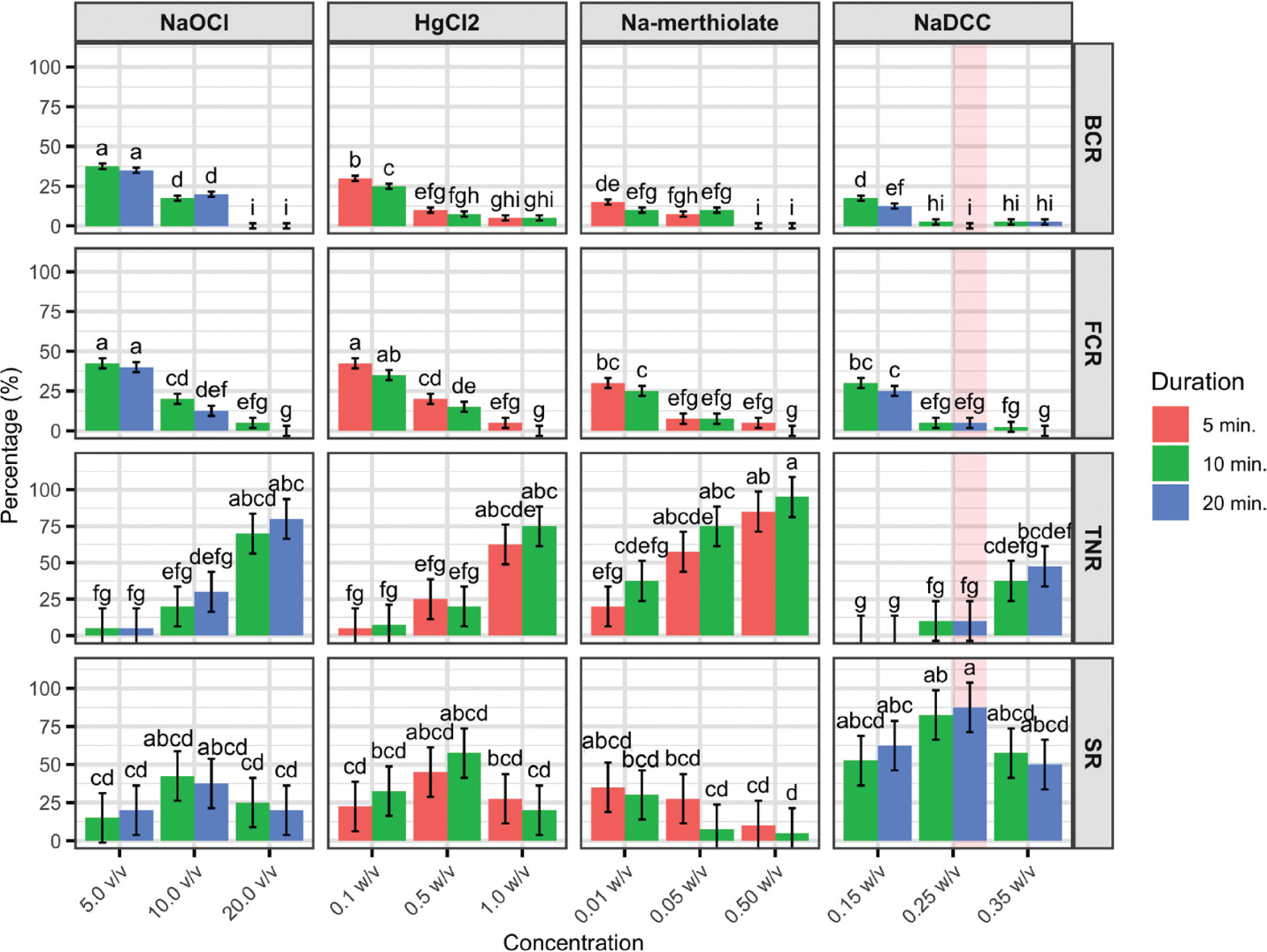
Figure 2.
Mean values of sterilisation traits (BCR, FCR, TNR and SR) for different agent-concentration-duration combinations are presented. Bars with different letters indicate statistically significant differences among all treatment combinations based on Duncan’s multiple range test (p ≤ 0.05), and the whiskers represent the standard error values. Red shading highlights the most desirable agent-concentration-duration combination for overall sterilisation traits. BCR, bacterial contamination (%); FCR, fungal contamination (%); TNR, tissue necrosis rate (%) and SR, survival rate (%).
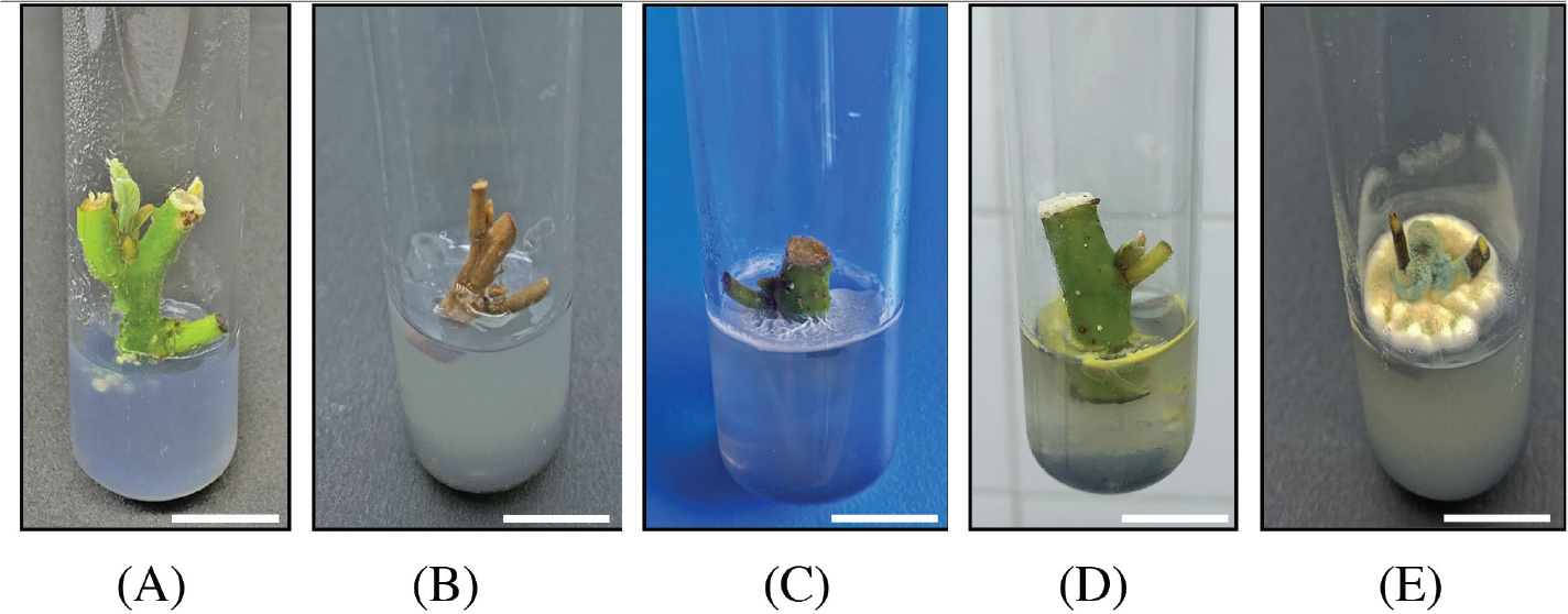
Figure 3.
Representative responses of ‘Duke 7’ nodal segments following different surface sterilisation treatments: healthy nodal segment (A), nodal segment necrosis (B), bacterial contamination (C, D), and fungal contamination (E) on modified MS medium. Scale bar = 1 cm.
Shoot proliferation experiment
In this study, the effects of different concentrations of BAP, KIN and TDZ on in vitro shoot development and proliferation of ‘Duke 7’ rootstock were investigated, and significant differences among treatments were determined (p < 0.05) (Figure 4). The highest number of shoots (2.07 ± 0.07 per explant) was recorded in the medium containing 8.88 μM BAP, followed by 4.44 μM BAP (1.50 ± 0.07 per explant) and 4.65 μMKIN (1.37 ± 0.07 per explant) (Figure 4). Although an increase in shoot number was observed at 4.65 μM KIN, treatmentscontaining BAP were generally more effective for shoot proliferation. In contrast, treatments containing TDZ did not result in a statistically significant difference in shoot number compared with the control. In the control treatment, the shoot number remained at 1.00 ± 0.07 per explant (Figure 5A). In terms of shoot length, the highest value (21.00 ± 1.05 mm) was obtained at 8.88 μM BAP, producing shoots approximately three times longer than those in the control group (7.17 ± 1.05 mm) (Figure 5B). This was followed by 9.29 μM KIN (18.40 ± 1.05 mm) and 4.65 μM KIN (16.30 ± 1.05 mm), respectively (Figure 5C). The highest number of leaves per explant was recorded on medium supplemented with 8.88 μM BAP (5.27 ± 0.26 per explant). This was followed by the 4.44 μM BAP (4.40 ± 0.26) and 2.27 μM TDZ (4.47 ± 0.26) (Figure 5D) treatments. Increasing the TDZ concentration above 2.27 μM led to a marked decline in leaf number, which decreased to 3.30 ± 0.26 per explant. Treatments containing KIN showed lower leaf numbers compared with BAP and TDZ treatments. The lowest number of leaves was recorded in the 2.32 μM KIN treatment (1.90 ± 0.26 per explant), which was not statistically different from the control (1.80 ± 0.26 per explant). Shoot growth index values (0–4 scale) showed a similar pattern. The highest score (3.13 ± 0.29, corresponding to ‘good’ development) was observed with the 8.88 μM BAP concentration. In contrast, shoot growth remained generally low in TDZ-containing treatments, with the lowest value observed at 4.54 μM TDZ (0.83 ± 0.29). In KIN-containing treatments, shoot growth was stimulated only within a limited concentration range, whereas concentrations below 4.65 μM were insufficient to promote shoot growth and remained below the control.
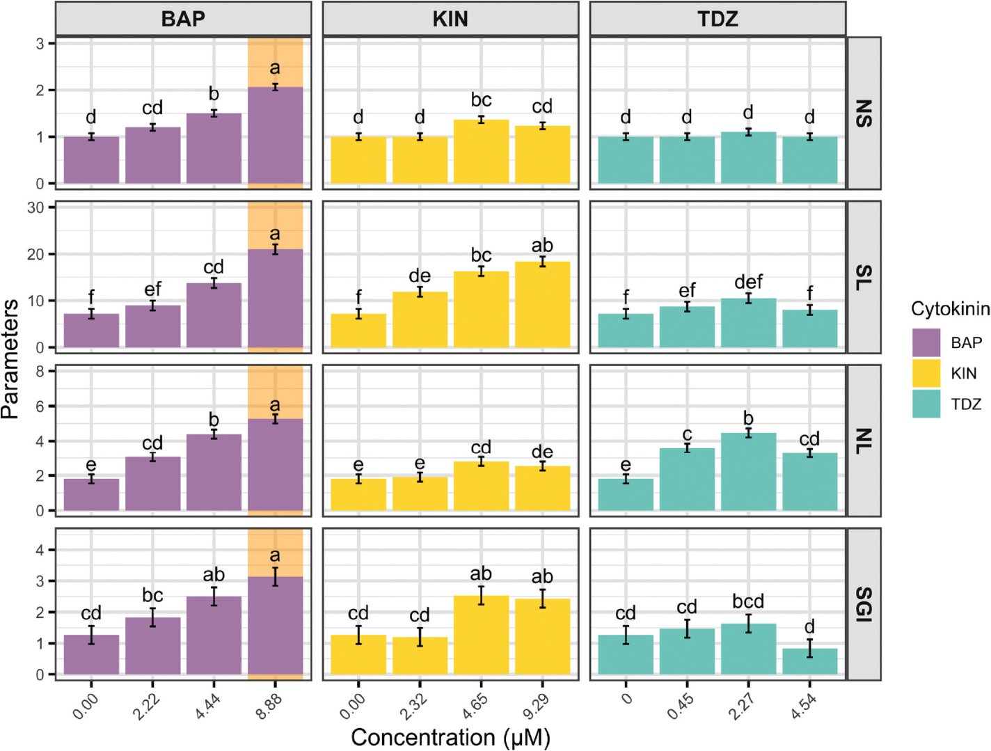
Figure 4.
Mean values of proliferation traits (NS, SL, NL and SGI) for different cytokinin–concentration combinations are presented. Bars with different letters indicate statistically significant differences among all treatment combinations based on Duncan’s multiple range test (p ≤ 0.05), and the whiskers represent the standard error values. Orange shading highlights the most desirable cytokinin-concentration combination for overall proliferation traits. NS, number of shoot (no.); SL: shoot length (mm); NL, number of leaves (no.) and SGI, shoot growth index (0–4).
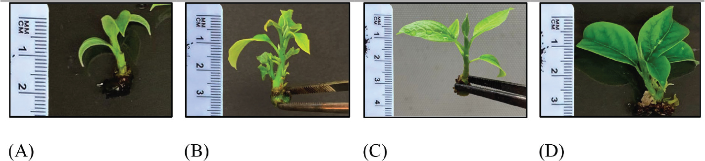
Figure 5.
Representative shoot development at the end of the 12th week of the proliferation stage under different treatments: control (A), 8.88 μM BAP (B), 9.29 μM KIN (C) and 2.27 μM TDZ (D).
Rooting experiments
The results demonstrated that different auxin treatments had a statistically significant effect on the rooting of ‘Duke 7’ microcuttings in the quick-dip method (p < 0.05), The highest rooting rate was recorded at 10.0 mM IBA (13.33% ± 0.79%), followed by 15.0 mM IBA (6.67% ± 0.79%). In contrast, no rooting occurred in any of the NAA treatments (Figure 6). According to the statistical analysis, the treatments showed important differences in callus diameter and callus growth index (p < 0.05). A statistically significant increase in callus diameter was detected with increasing concentrations, irrespective of the type of auxin applied. In terms of callus diameter, the largest callus was observed in the 15.0 mM NAA treatment (12.42 ± 0.40 mm), followed by 20.0 mM NAA (11.75 ± 0.40 mm) and 20.0 mM IBA (11.40 ± 0.40 mm). Based on the callus growth index, the highest score (4.00 ± 0.14, very high) was achieved with the 20.0 mM IBA treatment, whereas among the NAA treatments, the highest score NAA (3.67 ± 0.14, very high) was recorded with 15.0 mM NAA (Figure 6). In the control treatment (without auxin application), neither rooting nor callus formation was observed.
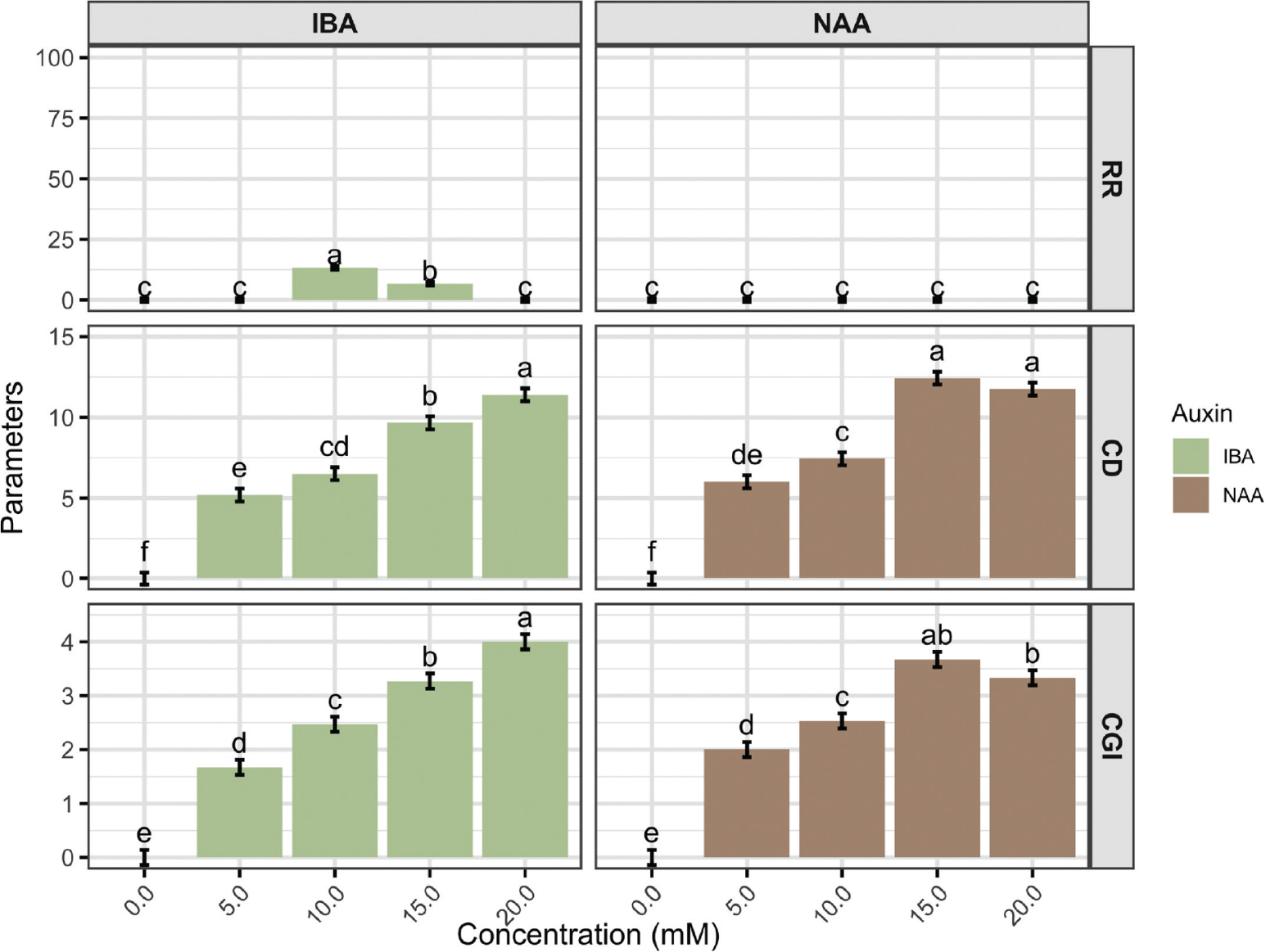
Figure 6.
Mean values of rooting traits (RR, CD and CGI) for different auxin-concentration combinations are presented. Bars with different letters indicate statistically significant differences among all treatment combinations based on Duncan’s multiple range test (p ≤ 0.05) and the whiskers represent the standard error values. RR, rooting rate (%), CD, callus diameter (mm) and CGI, callus growth index (0–4). IBA, indole-3-butyric acid; NAA, naphthalene acetic acid.
DISCUSSION
Contamination is one of the primary obstacles in micropropagation, as it negatively affects culture success and often results in substantial losses (Hesami et al., 2017). Therefore, optimising sterilisation protocols is essential to preserve explant viability and enhance the efficiency of the regeneration process. This study is the first to demonstrate that NaDCC is an effective sterilising agent for the in vitro establishment of avocado. Compared with other treatments (NaOCl, HgCL2 and Na-merthiolate), NaDCC resulted in lower contamination rates while preserving tissue viability. Moreover, the absence of a rinsing step shortens the procedure and reduces labour requirements, thereby enhancing its suitability for routine laboratory and commercial production systems.
In previous avocado micropropagation studies, sterilisation protocols using NaOCl (Barrera-Guerra et al., 1998; Taah et al., 2009; Zulfiqar et al., 2009; Chamandi and Peiris, 2024) and HgCL2 (Nhut et al., 2008; Shenoda et al., 2025) were reported to reduce contamination at the establishment stage; however, they were not always effective in preventing latent endophyte development or fully preserving tissue integrity. Na-merthiolate has not previously been reported for use in the micropropagation of avocado, although it has been applied as a disinfectant in Prunus and Corylus (Damiano et al., 2005; Bacchetta et al., 2008; Kushnarenko et al., 2025). In the present study, however, Na-merthiolate treatment resulted in the highest tissue necrosis rate and completely inhibited shoot development, suggesting that its suitability for avocado explant sterilisation may be limited.
In contrast, NaDCC application in the present study improved culture performance by maintaining tissue viability and supporting normal shoot development. The application of 0.25% NaDCC for 20 min was identified as the most effective sterilisation protocol. Similarly, in other woody species, 0.30% NaDCC applied for 3 hr in Pistacia (Lewis, 2021), 0.50% NaDCC for 35 min in sycamore maple (Karfik et al., 2026) and 2.00% NaDCC for 1 hr in Ulmus glabra (Välimäki et al., 2022) were reported to be effective. These findings indicate that the optimal concentration and exposure duration of NaDCC may vary depending on plant species, explant source and initial microbial load. Additionally, the costeffectiveness, practical applicability and environmental safety of NaDCC support its consideration as a promising sterilisation agent (Shetty and Narasimhan, 2021).
In micropropagation, in addition to the nutritional composition of the culture medium, PGRs play a critical role in determining in vitro developmental responses. In the micropropagation of avocado, BAP is the most commonly used cytokinin and strongly promotes shoot induction (Bandaralage et al., 2017). In this study, the highest shoot development in the ‘Duke 7’ rootstock was obtained at a BAP concentration of 8.88 μM. This result is consistent with the finding of Nel (1983), but differs from those of Cooper (1987) and Wessels (1996), who reported adequate development at lower concentrations. This indicates that even within the same genotype, the optimal cytokinin level depends on the explant source and culture conditions. Indeed, variable proliferation responses depending on BAP concentration have been reported in different avocado genotypes (Zulfiqar et al., 2009; Mansoor, 2018; El-Fadl et al., 2022; Qasrawi and Sholi, 2022).
The effectiveness of the other cytokinins (KIN and TDZ) on shoot development and proliferation was more limited compared with BAP. Although kinetin stimulated cell division, it resulted in lower proliferation. This is thought to be associated with its shorter persistence in the tissue and its limited capacity to stimulate lateral bud activation (Sakakibara, 2006; George et al., 2008; Werner and Schmülling, 2009). The findings obtained in this study are consistent with those reported by Qasrawi and Sholi (2022) and Ashika et al. (2026). In contrast to kinetin, TDZ is known to induce strong morphogenetic responses at low concentrations; however, in the present study, shoot development remained limited. Similarly, Qasrawi and Sholi (2022) reported that low TDZ levels promoted shoot formation in avocado but restricted shoot elongation. Moreover, high TDZ concentrations have been associated with oxidative stress and phytotoxicity, both of which suppress growth (Peñaloza-Remigio et al., 2020). This effect is thought to be related to the persistent cytokinin-like activity of TDZ in plant tissues and the resulting phenolic accumulation and oxidative stress (Huetteman and Preece, 1993; Guo et al., 2011). Overall, the superiority of BAP is associated with its greater stability in tissues and its ability to generate a sufficiently strong cytokinin signal without disrupting the auxin–cytokinin balance (Sakakibara, 2006; George et al., 2008; Werner and Schmülling, 2009). In the present study, the use of constant IAA and GA3 levels yielded results consistent with Ibarra-López et al. (2016). Low auxin levels are known to enhance cytokinin-induced proliferation, while GA3 promotes shoot elongation (Jagiełło-Kubiec et al., 2021).
In vitro rooting studies of avocado generally apply auxins directly to the culture medium; however, in some studies, the quick-dip method has been evaluated as an alternative approach. It has been reported that this method can be more effective than continuous auxin application under certain conditions (Cooper, 1987; Barrera-Guerra et al., 1998; Bandaralage, 2018). This effect may result from the brief auxin exposure provided by the quick-dip treatment, which is sufficient to initiate root meristem formation, whereas prolonged auxin exposure may instead favour callus development and suppress root differentiation. In the present study, IBA was more effective than NAA in promoting root induction. This effect may be associated with the ability of IBA to promote cell wall loosening and to enhance the expression of genes involved in adventitious root primordium formation. Such mechanisms favour direct root differentiation rather than unorganised cell proliferation (Ludwig-Müller, 2000). These findings are consistent with the results reported by Cooper (1987). Cooper (1987) reported that a 1-s quick-dip application of 15 mM IBA resulted in 89.00% rooting in juvenile explants. In contrast, Barrera-Guerra et al. (1998) reported that rooting was limited to 8.30% following a 5-s quick-dip method using 20 mM IBA. These results differ from the findings obtained in the present study using a 10-s quick-dip application of 5–20 mM IBA for the in vitro rooting of mature explants. Similarly, in other woody species, the response to IBA concentrations applied through the quick-dip method has been reported to vary considerably depending on the species and treatment conditions (Aygun and Dumanoglu, 2015; Osman and Dumanoglu, 2020; Lawson et al., 2023; Hejazi et al., 2025).
However, high auxin concentrations suppressed root development and promoted callus formation. In the present study, both IBA and NAA at concentrations of 15–20 mM promoted callus formation while suppressing root development. The observation by Barrera-Guerra et al. (1998) that the highest degree of callus formation occurred at 29.50 mM IBA is consistent with our findings. Moreover, increased callus development under high IBA concentrations has also been reported in olive and pear species (Denaxa et al., 2012; Aygun and Dumanoglu, 2015). This suggests that although high doses of auxins stimulate cell division, they may inhibit root cell differentiation (De Klerk et al., 1999). Moreover, excessive callus development has been reported to negatively affect root formation and overall plantlet growth (Gaba, 2005).
When these findings are considered collectively, the low rooting rates observed in the present study, despite the application of the quick-dip method, cannot be attributed solely to auxin concentration. The rooting response seems to depend on the combined influence of genotype, physiological stage (juvenile or mature), and culture conditions, which collectively determine the physiological status of the explants. In particular, the reduced morphogenetic capacity of mature tissues and the induction of excessive callus formation at higher auxin doses may have hindered the transition from root induction to the formation of organised root structures. Therefore, the present rooting rates indicate that the protocol is not yet sufficient for direct large-scale application, and further optimisation of the rooting stage is required.
CONCLUSION
This study provided meaningful advances in the sterilisation and shoot proliferation stages of the micropropagation protocol for ‘Duke 7’ avocado rootstock, while highlighting that rooting remains a critical constraint requiring further optimisation. Among the sterilising agents evaluated, NaDCC demonstrated higher sterilisation efficacy without inducing tissue necrosis and did not require a post-treatment rinsing step. These characteristics may offer practical advantages for commercial-scale production. During shoot proliferation, BAP exerted the most decisive influence on shoot development and multiplication, with optimum results achieved at 8.88 μM BAP incorporated into modified MS medium. Although quick-dip application of 10 mM IBA produced the highest rooting response, the overall rooting rate remained markedly limited, indicating that further optimisation at this stage is essential before the protocol can be considered suitable for large-scale implementation.
ACKNOWLEDGEMENTS
This research was financially supported by the Plant Transformation Research Centre, University of California, Riverside, and was conducted in the centre’s tissue culture laboratory.
Abbreviations
- AC
activated charcoal
- BAP
6-benzylaminopurine
- BCR
bacterial contamination rate
- Ca(OCl)2
calcium hypochlorite
- CD
callus diameter
- CGI
callus growth index
- CRD
completely randomised design
- FCR
fungal contamination rate
- GA3
gibberellic acid
- HgCL2
mercuric chloride
- IAA
indole-3-acetic acid
- IBA
indole-3-butyric acid
- KIN
kinetin
- KNO3
potassium nitrate
- NAA
naphthaleneacetic acid
- NaDCC
sodium dichloroisocyanurate
- Na-merthiolate
sodium Merthiolate
- NaOCl
sodium hypochlorite
- NH4NO3
ammonium nitrate
- NL
number of leaves
- NS
number of shoots
- PGRs
plant growth regulators
- RR
rooting rate
- SGI
shoot growth index
- SL
shoot length
- SR
survival rate
- TDZ
thidiazuron
- TNR
tissue necrosis rate.
Notes
[2] Contributed by AUTHOR CONTRIBUTION
G.G., R.B. and G.P. investigation, data curation, formal analysis and writing – original draft preparation. R.B. and G.P. statistical analysis and data interpretation. M.O.C. and M.L.A. resources, funding acquisition and writing – review and editing. H.G. supervision, project administration and writing – review and editing.
[3] Conflicts of interest CONFLICTS OF INTEREST
The authors declare no competing interests.