
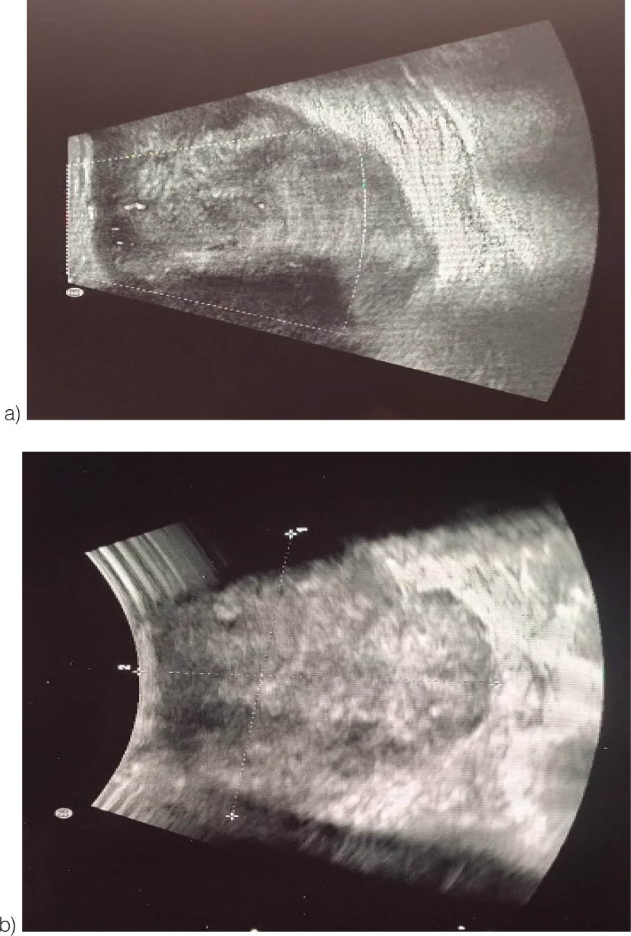
Figure 1:
Ultrasonography of the bilateral breast (a and b) showing an irregular hypoechoic solid mass with microlobulated margins, measuring ~6.5×5.1 cm, occupying the upper and lower inner quadrants. The left breast was categorized as BIRADS-IV.
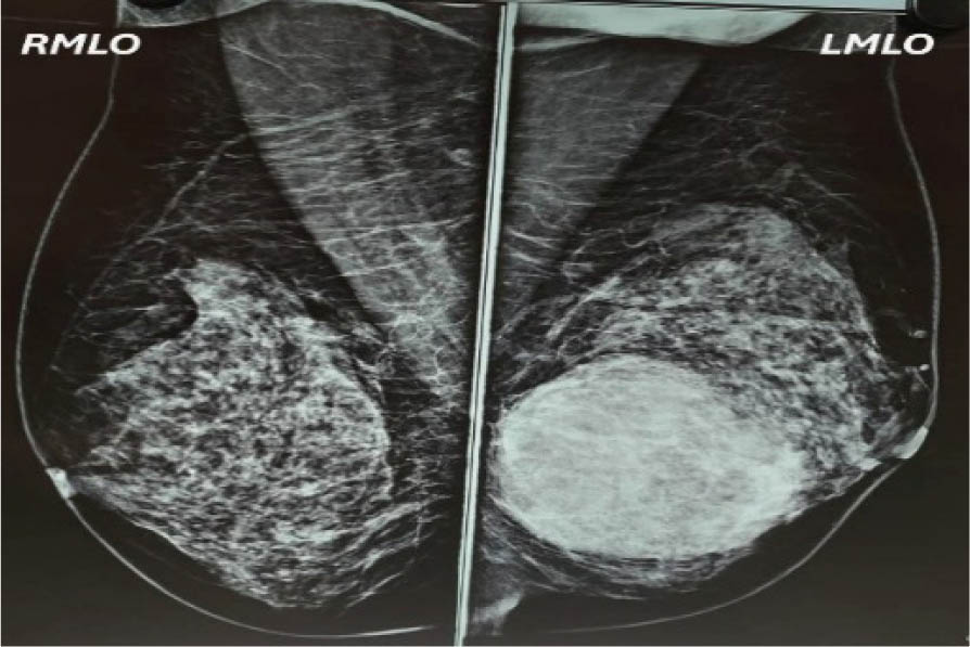
Figure 2.1:
Mammography image showing right mediolateral oblique view and left mediolateral oblique view.
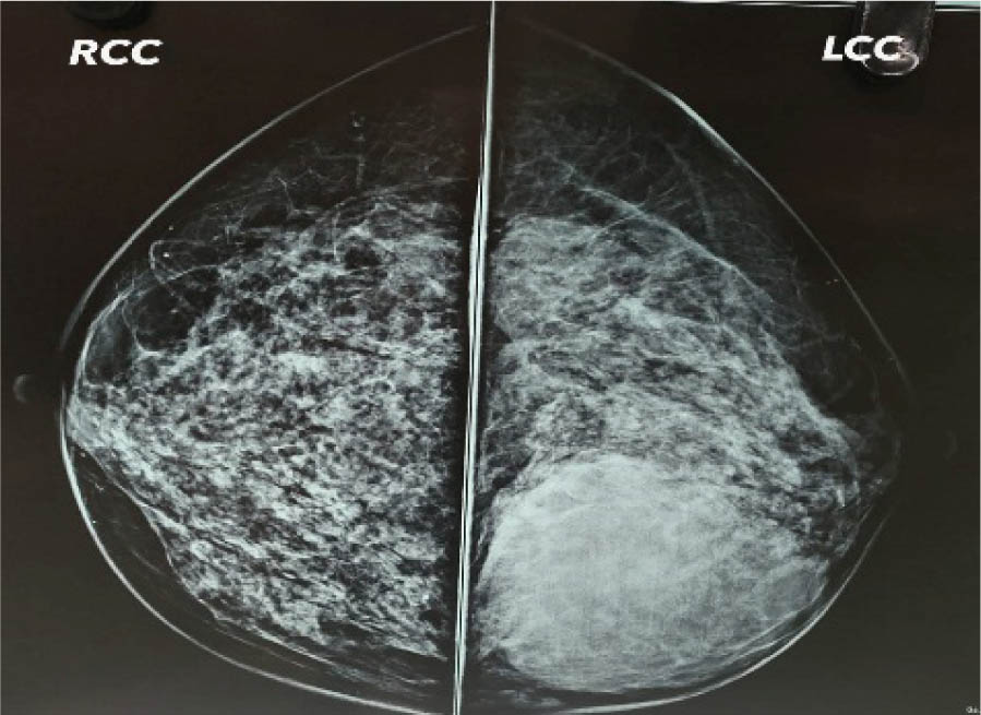
Figure 2.2:
Mammography image showing right cranial caudal view and left cranial caudal view.
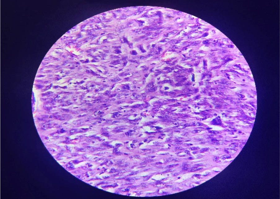
Figure 3:
Section examined shows fibro-collagenous tissue with few benign acini. Stroma is relatively cellular with mild pleomorphism and nuclear hyperchromasia. Mitotic figures were, however, not identified. 40× magnification.
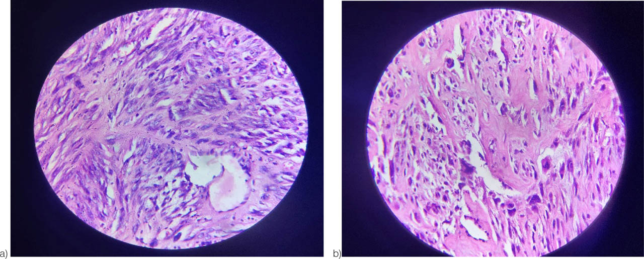
Figure 4:
(a) Image showing high cellularity with moderate atypia and areas of stromal overgrowth. The haphazard, infiltrative appearance of the stroma also suggests a permeative border. 40× magnification. (b) The image shows the spindle-shaped cells arranged in bundles or fascicles (common for smooth muscle or myofibroblastic tumors). Stromal hypercellularity comprising of highly atypical cells with nuclear pleomorphism and high N:C ratio, 40× magnification.
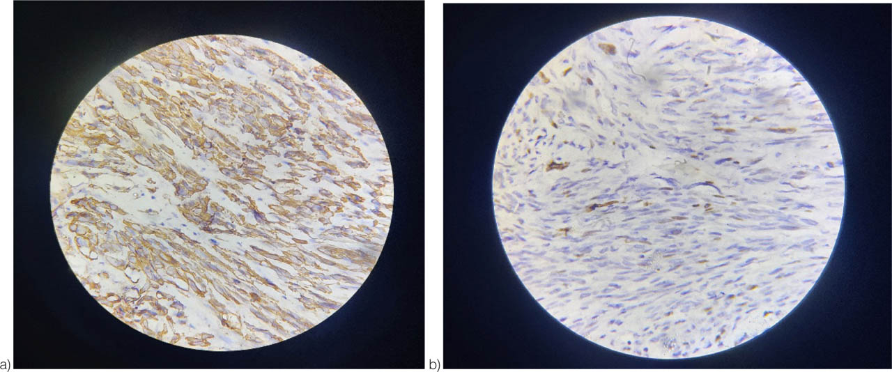
Figure 5:
(a) The image showing brown staining along the cytoplasmic and membranous areas is characteristic of SMA positivity, showing positivity on IHC, 40× magnification. (b) Ki-67, the image is showing the proliferative activity of the cells. Brown-stained nuclei represent Ki-67-positive cells. 40× magnification.
Table 1:
Reported cases (till 2019) based on the size of the tumor(8)
| Size (cm) | Number of cases | Age range (years) | Sex (F/M) | Common side (L/R) | Mitotic rate (/10HPF) | Lymph node/vascular invasion | Common treatment | Outcome |
|---|---|---|---|---|---|---|---|---|
| <2 | 7 | 19–86 | 6F/1M | 4L/3R | 0–24 variable | 1/7 had invasion | Excis-4 | 6 alive, 1 death |
| SM-2 | ||||||||
| MRM-1 | ||||||||
| 2–5 | 28 | 24–83 | 27F/1M | 15L/10R | 1–29 median:10 | 8/28 had invasion | MRM-12 | 24 alive |
| SM-8 | 3 death | |||||||
| Excis-6 | 1 Recu. | |||||||
| 5–10 | 19 | 18–86 | 18F/1M | 12L/5R | 2–40 (median:12) | 9/19 had invasion | MRM-14 | 15 alive |
| SM-3 | 2 Mets. | |||||||
| WLE-2 | 2 death | |||||||
| >10 | 14 | 19–73 | 14F | 8L/4R | 10–40 High | 7/14 had invasion | MRM-11 | 8 alive |
| Excis-2 | 4 Mets. | |||||||
| Pallia-1 | 2 death |
[i] Note: cm, centimeter; HPF, high-power fields; M, male; F, female; R, right; L, left; WLE, wide local excision; SM, simple mastectomy; MRM, modified radical mastectomy; NA, not available; NR, not reported; RT, radiotherapy; CT, chemotherapy; B/L, bilateral; Mets., metastases; Excis., excision; Pallia., palliative; Recu., recurrence.
Table 2:
| Year | Author | Age | Sex | Side | Size (cm) | Mitosis/10HPF | Lymph/vascular invasion | Treatment | Outcome |
|---|---|---|---|---|---|---|---|---|---|
| 2020 | Iliyas et al9 | 52 | F | Left | 6 | 2–50 | Yes | SM | Alive, 1 year |
| 2020 | Liu et al10 | 28 | F | Left | 1.6 | 10 | NR | WLE | Alive, 1 year |
| 2020 | Kumar et al11 | 53 | F | Right | 8 × 6 | Frequent | NR | WLE | Mets in lungs, bone, kidney |
| 2020 | Horton et al12 | 61 | F | Right | 1.6 | NR | No | WLE | NA |
| 2020 | Burger et al13 | 54 | F | Right | 3 | Up to 3 | No | BCS | Alive, 24 months |
| 2021 | Ely Cheikh et al14 | 65 | M | B/L | 7 (each) | 15 | NR | L-WLE | Alive, 11 months |
| R-SM +RT | |||||||||
| 2023 | R. Masadah et al8 | 30 | F | Left | 12 × 8 | 10 | NO | WLE | Alive, 8 months |
| 2025 | Present case | 60 | F | Left | 7.8 × 6 | 5–9 | NO | MRM | Alive, 1 month |