
1. Introduction
Primary leiomyosarcoma of the breast is a rare subtype of breast cancer. It was first reported by Waterworth in 1968.[1] To the best of our knowledge, this is the sixth documented case of primary leiomyosarcoma of the breast in a 60-year-old woman reported from India.
2. Case Presentation
A 60-year-old woman without any comorbidity presents with a lump in her left breast 10 years back, which was gradually progressive in nature and was not associated with pain or discharge. The patient underwent lumpectomy 10 years back at another institution for an underlying condition; however, the histopathological records of the previous surgery were not available for review. The patient remained disease free for 10 years, and now, the patient is again having a painless lump in her left breast retroareolar region.
Ultrasonography of bilateral breasts shows an irregular hypoechoic solid mass in the left breast, measuring approximately 6.5 × 5.1 cm, with microlobulated margins and internal vascularity on color Doppler (Figure 1a and 1b: left breast: BIRADS-IV).
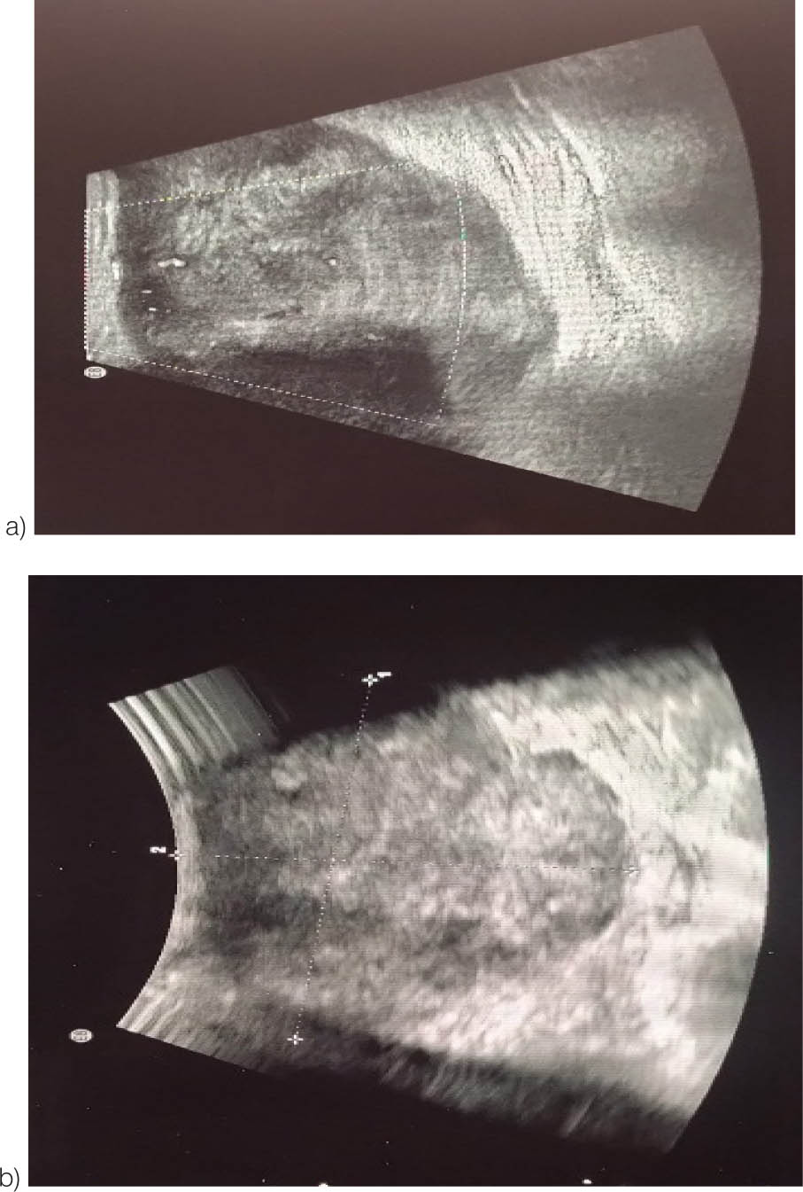
Figure 1:
Ultrasonography of the bilateral breast (a and b) showing an irregular hypoechoic solid mass with microlobulated margins, measuring ~6.5×5.1 cm, occupying the upper and lower inner quadrants. The left breast was categorized as BIRADS-IV.
Mammography shows a large well-defined round shape high-density mass lesion, measuring 6 × 6.8 cm in the lower inner quadrant (Figure 2.1 and 2.2).
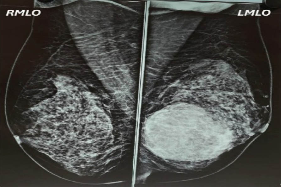
Figure 2.1:
Mammography image showing right mediolateral oblique view and left mediolateral oblique view.
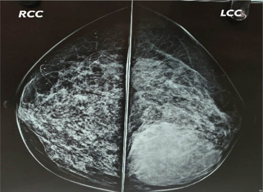
Figure 2.2:
Mammography image showing right cranial caudal view and left cranial caudal view.
True-cut biopsy of lump showed fibro-collagenous tissue with few benign acini. Stroma was cellular with mild pleomorphism with nuclear hyperchromasia as shown in Figure 3. On IHC (immunohistochemistry), CK was negative and Ki-67 was <5% (low).
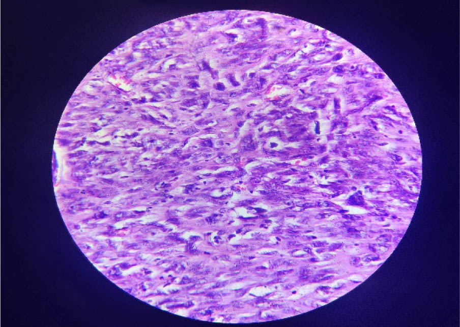
Figure 3:
Section examined shows fibro-collagenous tissue with few benign acini. Stroma is relatively cellular with mild pleomorphism and nuclear hyperchromasia. Mitotic figures were, however, not identified. 40× magnification.
The patient underwent a left modified radical mastectomy under general anesthesia in our institution. Intraoperative findings revealed a 7 × 5 cm irregular retroareolar lump with hard consistency adherent to breast tissue, free from muscle, and overlying skin. Postoperative patient was having a horizontal scar of approximately 15 cm on the left chest wall, and the scar was in a healing phase without any complications.
Histopathological findings (postoperative) demonstrated that a 7 × 6 cm mass was excised from the left breast of the patient. On gross examination, the tumor was centrally located, measuring 7.8 × 6 × 6 cm, and was noted to extend up to the underlying bone. The tumor was located 1.2 cm from the skin, 7 cm from the superior surface, 6 cm from the inferior surface, and 5 cm from the lateral margin and reached up to the medial margin. Grossly, the upper, lateral, and inferior margins were free from tumor involvement.
Microscopically, the tumor was diagnosed as a malignant phyllodes tumor. It exhibited marked stromal cellularity, moderate stromal atypia, and evidence of stromal overgrowth. The mitotic activity was noted to be 5–9 mitoses per 10 high-power fields. A malignant heterologous component in the form of leiomyosarcoma was identified within the tumor (the presence of heterologous element directly upgrade to malignant category). The tumor demonstrated a diffusely permeative border and was found to be reaching the base and medial surgical margins, while the remaining margins were free of tumor involvement.
No regional lymph nodes were submitted for evaluation. Immunohistochemistry showed positivity for smooth muscle actin (SMA), supporting the diagnosis of the leiomyosarcomatous component (Figure 5a), and the Ki-67 proliferation index was estimated at 7%–8% (Figure 5b). Based on the findings, the pathological stage of the tumor was designated as pT3NxMx.
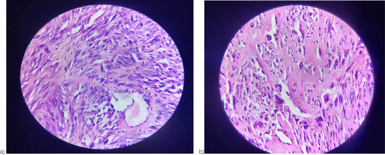
Figure 4:
(a) Image showing high cellularity with moderate atypia and areas of stromal overgrowth. The haphazard, infiltrative appearance of the stroma also suggests a permeative border. 40× magnification. (b) The image shows the spindle-shaped cells arranged in bundles or fascicles (common for smooth muscle or myofibroblastic tumors). Stromal hypercellularity comprising of highly atypical cells with nuclear pleomorphism and high N:C ratio, 40× magnification.
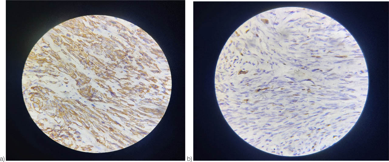
Figure 5:
(a) The image showing brown staining along the cytoplasmic and membranous areas is characteristic of SMA positivity, showing positivity on IHC, 40× magnification. (b) Ki-67, the image is showing the proliferative activity of the cells. Brown-stained nuclei represent Ki-67-positive cells. 40× magnification.
The patient underwent the left modified radical mastectomy under general anesthesia. The patient now planned to start on four courses of Doxorubicin 60 mg/m2 IV and Cyclophosphamide 750 mg/m2 IV after cardiac clearance. 2D ECHO revealed normal left ventricular systolic function (EF=50%). Patient completed one course of chemotherapy with Inj. Doxorubicin and Inj. Cyclophosphamide and tolerate chemotherapy well. The patient did not had any complains at present.
3. Discussion
Leiomyosarcoma of breast is a highly uncommon type of cancer that originates from smooth muscle cells within the breast tissue.[2] Unlike typical breast cancers, which usually arise from glandular or ductal epithelial cells, this tumor develops from the connective tissue elements, making it subtype of breast sarcoma.[3] Given the limited amount of smooth muscle in the breast, it is believed that these tumors often arise from the muscular wall of blood vessels.[2] It constitute less than 1% of all breast malignancies.[4] Due to its rarity, exact prevalence rates are difficult to determine, but fewer than 100 cases have been reported in the medical literature worldwide.[2] It typically affects women between the age of 40 and 70 years.[3] Patient with leiomyosarcoma of the breast usually present with painless, firm lump that grows gradually over time.[5] Because its appearance can resemble more common benign breast conditions, diagnosis often requires a detailed histopathological examination.[6] Immunohistochemistry tests are typically used to confirm the presence of smooth muscle markers like SMA (smooth muscle actin), desmin, vimentin, and h-caldesmon.[2]
Due to its rarity, there is no universally established treatment guideline, but surgical removal with clear margins is generally the preferred approach.[4] The role of radiation and chemotherapy remains uncertain and is usually considered based on individual case factors. Early diagnosis and complete surgical excision are key to achieving better outcomes.[4] Although rare, leiomyosarcoma of the breast can lead to several serious complications primarily due to its aggressive behavior and potential for recurrence. One of the most significant concerns is local recurrence, even after surgical removal with clear margins.[5] This makes close follow-up essential. Another major complication is the potential for distant metastasis, particularly to the lungs, liver, or bones.[7] Unlike typical breast cancers, leiomyosarcomas spread through the bloodstream rather than the lymphatic system, which can lead to late detection of metastatic disease.[7]
Delayed diagnosis is also a common issue, as the tumor often mimics benign lumps and grows slowly, which may lead to initial misdiagnosis and postponed treatment.[5] In some cases, wound healing problems and infection may occur after surgical intervention, especially in larger tumors that require extensive resection.[6] Additionally, if chemotherapy or radiotherapy is used, patients may face related side effects such as fatigue, immune suppression, or tissue damage.[4] Due to its rarity and unpredictable behavior, management of leiomyosarcoma of the breast can be complex, and complications may arise from both the disease itself and the treatment approach.[4] Total cases reported till 2019 were 68 (Table 1).
Table 1:
Reported cases (till 2019) based on the size of the tumor(8)
| Size (cm) | Number of cases | Age range (years) | Sex (F/M) | Common side (L/R) | Mitotic rate (/10HPF) | Lymph node/vascular invasion | Common treatment | Outcome |
|---|---|---|---|---|---|---|---|---|
| <2 | 7 | 19–86 | 6F/1M | 4L/3R | 0–24 variable | 1/7 had invasion | Excis-4 | 6 alive, 1 death |
| SM-2 | ||||||||
| MRM-1 | ||||||||
| 2–5 | 28 | 24–83 | 27F/1M | 15L/10R | 1–29 median:10 | 8/28 had invasion | MRM-12 | 24 alive |
| SM-8 | 3 death | |||||||
| Excis-6 | 1 Recu. | |||||||
| 5–10 | 19 | 18–86 | 18F/1M | 12L/5R | 2–40 (median:12) | 9/19 had invasion | MRM-14 | 15 alive |
| SM-3 | 2 Mets. | |||||||
| WLE-2 | 2 death | |||||||
| >10 | 14 | 19–73 | 14F | 8L/4R | 10–40 High | 7/14 had invasion | MRM-11 | 8 alive |
| Excis-2 | 4 Mets. | |||||||
| Pallia-1 | 2 death |
[i] Note: cm, centimeter; HPF, high-power fields; M, male; F, female; R, right; L, left; WLE, wide local excision; SM, simple mastectomy; MRM, modified radical mastectomy; NA, not available; NR, not reported; RT, radiotherapy; CT, chemotherapy; B/L, bilateral; Mets., metastases; Excis., excision; Pallia., palliative; Recu., recurrence.
Table 2:
| Year | Author | Age | Sex | Side | Size (cm) | Mitosis/10HPF | Lymph/vascular invasion | Treatment | Outcome |
|---|---|---|---|---|---|---|---|---|---|
| 2020 | Iliyas et al9 | 52 | F | Left | 6 | 2–50 | Yes | SM | Alive, 1 year |
| 2020 | Liu et al10 | 28 | F | Left | 1.6 | 10 | NR | WLE | Alive, 1 year |
| 2020 | Kumar et al11 | 53 | F | Right | 8 × 6 | Frequent | NR | WLE | Mets in lungs, bone, kidney |
| 2020 | Horton et al12 | 61 | F | Right | 1.6 | NR | No | WLE | NA |
| 2020 | Burger et al13 | 54 | F | Right | 3 | Up to 3 | No | BCS | Alive, 24 months |
| 2021 | Ely Cheikh et al14 | 65 | M | B/L | 7 (each) | 15 | NR | L-WLE | Alive, 11 months |
| R-SM +RT | |||||||||
| 2023 | R. Masadah et al8 | 30 | F | Left | 12 × 8 | 10 | NO | WLE | Alive, 8 months |
| 2025 | Present case | 60 | F | Left | 7.8 × 6 | 5–9 | NO | MRM | Alive, 1 month |
Total cases reported till 2019 were 68.
3.1. Key Observations
Size distribution:
Most tumors (28/68) were 2–5 cm.
Large tumors (>10 cm) were less common (14/68) but had worse outcomes.
Demographics
Predominantly female (94%), with rare male cases (4/68).
Median age: ~50 years (range: 18–86)
Mitotic activity
Higher mitotic rate (>10/10 HPF) correlates with large tumors and metastases.
Treatment
MRM (modified radical mastectomy) was most frequently for tumors more than 2-cm size
Excision or SM (simple mastectomy) is used for smaller tumors.
Outcome
<2 cm: 86% survival (6/7 alive)
>10 cm: 57% survival (8/14 alive)
Deaths due to lung or liver metastasis or recurrence.
➢ Smallest tumor: 0.3 cm of size and alive.
➢ Largest tumor: 16 × 10 cm and alive post-MRM/RT/CT.
➢ Worst prognosis: Tumor >10 cm with mitotic rates >20/10 HPF (recurrence and lung metastasis)
4. Conclusion
Primary leiomyosarcoma of the breast is an exceedingly rare neoplasm, with limited cases documented in medical literature worldwide. This case emphasizes the diagnostic complexities and management considerations associated with such uncommon presentations. The patient's recurrence after a decade-long disease-free interval illustrates the tumor's unpredictable nature and the necessity of prolonged clinical monitoring. Diagnosis was established through a combination of imaging, histopathology, and immunohistochemical analysis, which revealed a malignant phyllodes tumor with heterologous leiomyosarcomatous differentiation. Surgical intervention via modified radical mastectomy remains the cornerstone of treatment, although the role of adjuvant chemotherapy continues to be defined on a case-by-case basis due to the lack of standardized guidelines. This report contributes valuable insight to the limited existing data and highlights the importance of individualized therapeutic strategies and long-term surveillance in managing rare breast sarcomas.