
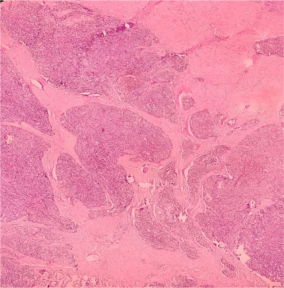
Figure 1:
Histological section of thymoma demonstrating lobular architecture with cellular lobules intersected by sharply demarcated fibrocollagenous bands (H&E stain, 40× magnification).
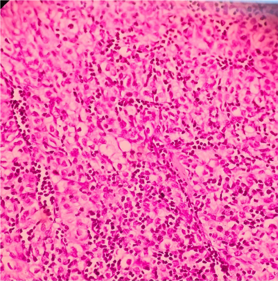
Figure 2:
Histological section of thymoma showing characteristic histomorphology with neoplastic polygonal epithelial cells and abundant thymocytes (H&E stain, 40× magnification).
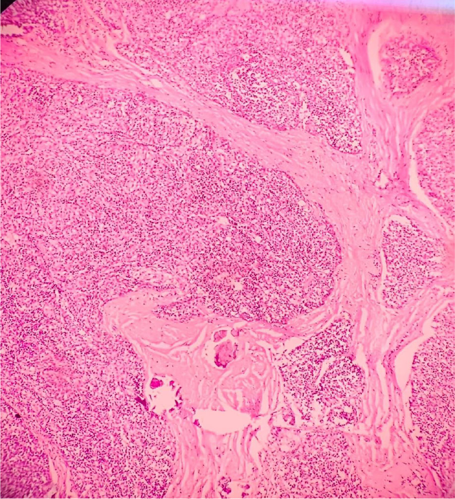
Figure 3:
Histological section of thymoma demonstrating lobular architecture with cellular lobules separated by fibrous bands (H&E stain, 10× magnification).
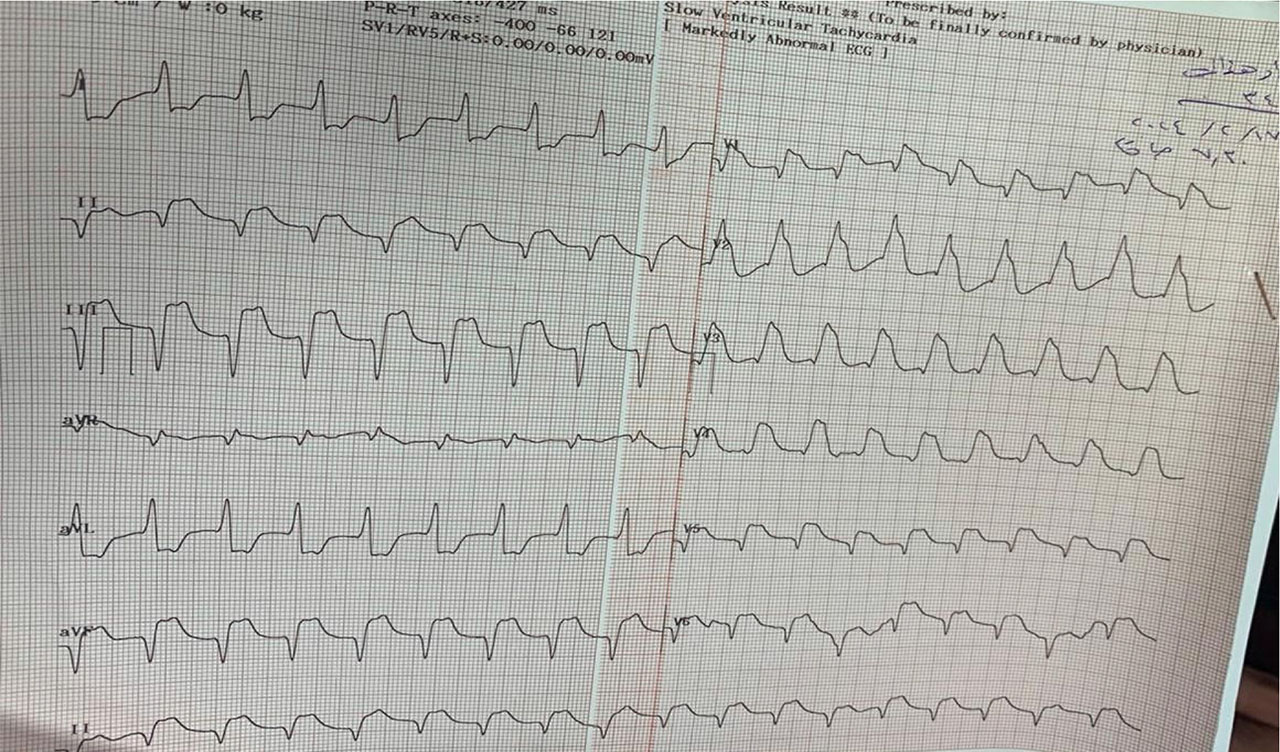
Figure 4:
ECG of the patient showing diffuse ST-T elevation.