
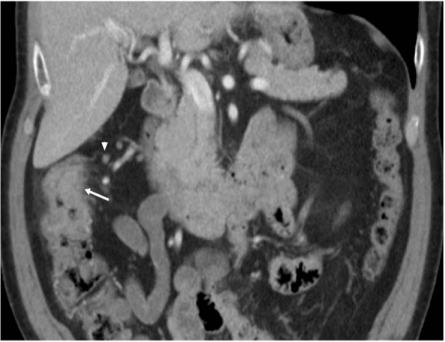
Figure 1:
Circumferential thickening and heterogenous attenuation of the ascending colon just proximal to the level of the hepatic flexure.
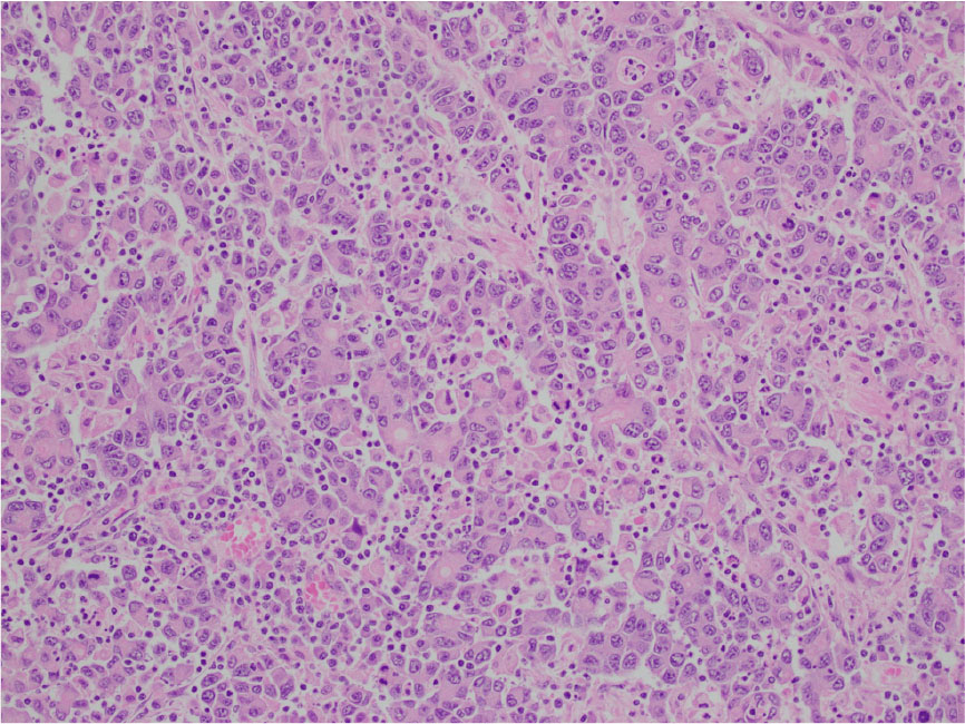
Figure 2:
Higher magnification reveals poorly differentiated cells with medium to large-sized vesicular nuclei, nucleoli, and moderate eosinophilic cytoplasm. The larger carcinomatous cells are interspersed by the smaller tumor infiltrating lymphocytes.
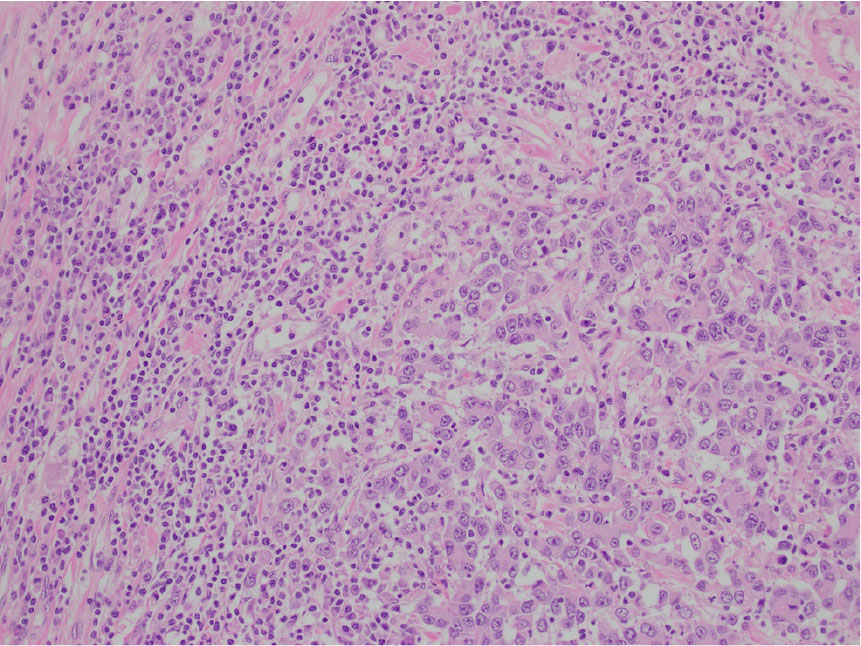
Figure 3:
The medullary carcinoma is composed of poorly to undifferentiated cells in a vaguely trabecular to solid pattern. It lacks the typical glandular formation and mucin content seen in traditional colonic adenocarcinomas. The carcinoma has a circumscribed, pushing border, with a brisk intraepithelial and peritumoral lymphocytic infiltration.