
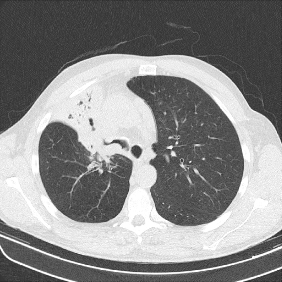
Figure 1:
CT scan at diagnosis showing a right upper lobe lesion with right upper and medium lobes collapse.
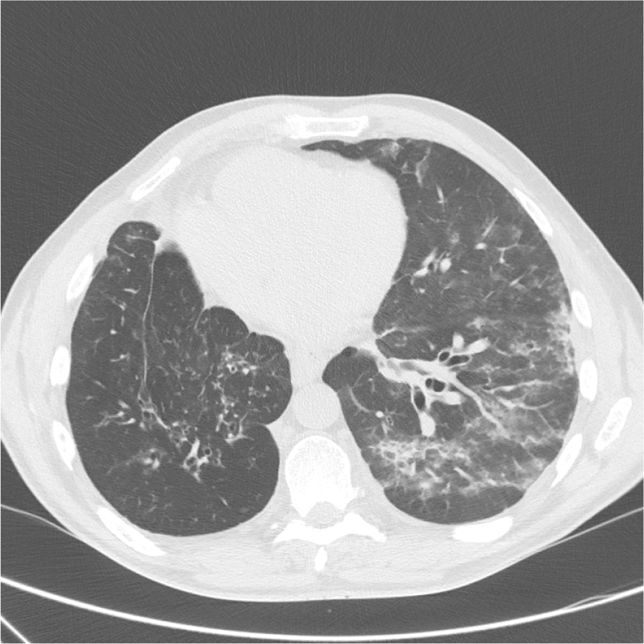
Figure 2:
CT scan after treatment with amivantamab showing scattered ground glass opacities in the left lung.
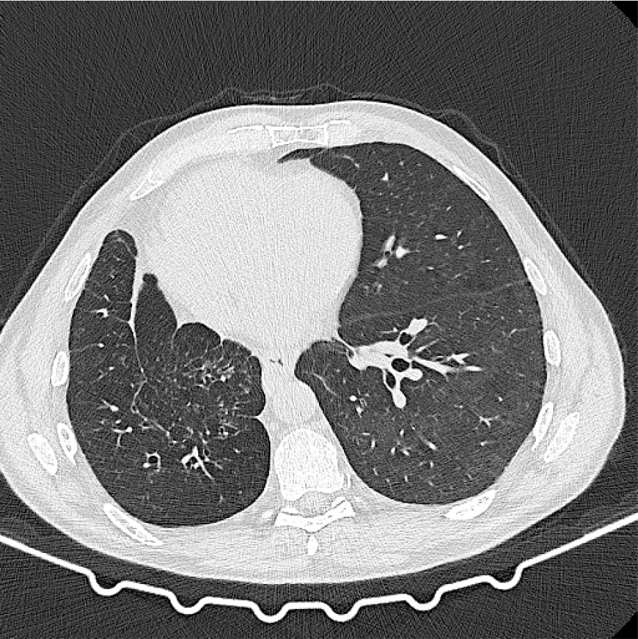
Figure 3:
CT scan after corticosteroid treatment showing resolution of the previous opacities.
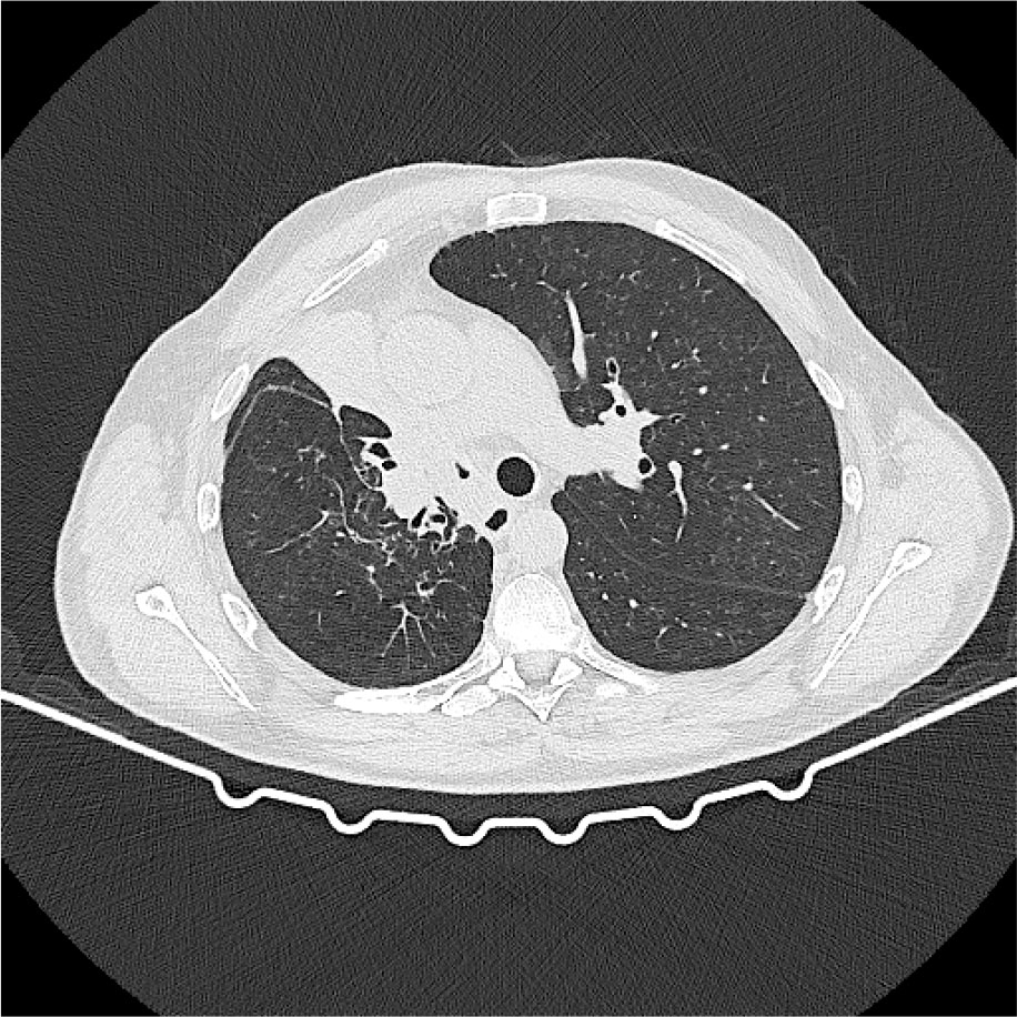
Figure 4:
CT scan after reintroduction of amivantamab, without evidence of toxicity and stable disease.