
Table 1:
Summary of the patients’ demographics and disease and treatment characteristics for the three different radiotherapy techniques utilized in this study.
| Characteristics | Treatment technique | Significance | ||
|---|---|---|---|---|
| 3D-CRT (n = 14) (%) | HT-IMRT (n = 8) (%) | VMAT (n = 6) (%) | ||
| Sex | p = 0.7 | |||
| Female | 1 (7.1) | 1 (12.5) | 0 (0.0) | |
| Male | 13 (92.9) | 7 (87.5) | 6 (100.0) | |
| Age at diagnosis | p = 0.1 | |||
| <60 years | 3 (21.4) | 1 (12.5) | 4 (66.7) | |
| ≥60 years | 11 (78.6) | 7 (87.5) | 2 (33.3) | |
| Performance status | p = 0.1 | |||
| ECOG 0 | 14 (100.0) | 6 (75.0) | 6 (100.0) | |
| ECOG 1 | 0 (0.0) | 2 (25.0) | 0 (0.0) | |
| Prognostic stage groups | p = 0.3 | |||
| Stage 0 | 2 (14.3) | 0 (0.0) | 0 (0.0) | |
| Stage I | 11 (78.6) | 5 (63.5) | 4 (66.7) | |
| Stage II | 1 (7.1) | 3 (37.5) | 2 (33.3) | |
| Tumor location/target treatment volume | p = 0.4 | |||
| Both vocal cords | 4 (28.6) | 3 (37.5) | 1 (16.7) | |
| Right vocal cord | 4 (28.6) | 2 (25.0) | 0 (0.0) | |
| Left vocal cord | 6 (42.9) | 3 (37.5) | 5 (83.3) | |
| Mean ± SD total radiation dose (Gy) | 64.9 ± 1.7 | 62.4 ± 5.4 | 64.4 ± 1.5 | p = 0.2 |
| Mean ± SD no. of fractions received | 31.2 ± 2.5 | 29.3 ± 4.3 | 29.8 ± 2.5 | p = 0.3 |
| Mean ± SD dose per fraction (Gy) | 2.1 ± 0.1 | 2.2 ± 0.2 | 2.2 ± 0.1 | p = 0.4 |
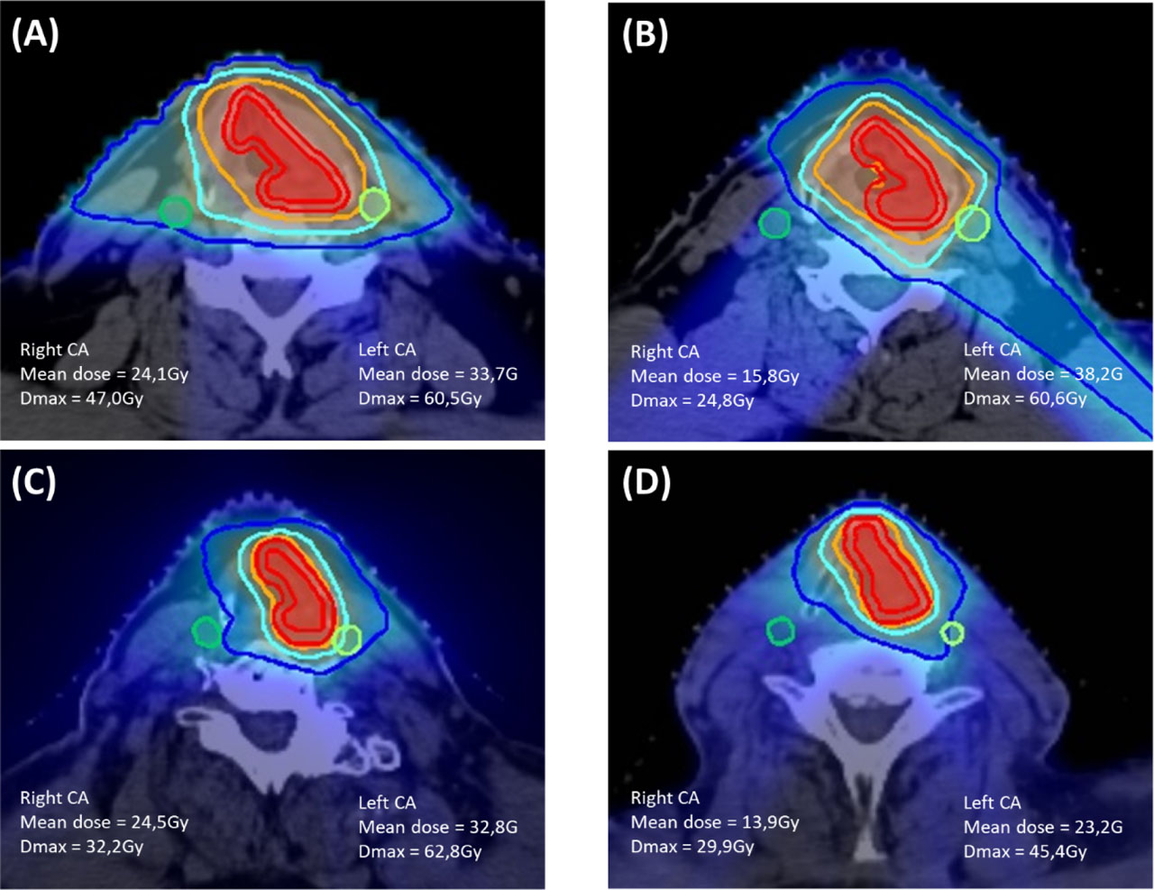
Figure 1:
Dose distribution with prescribed isodose curves (50% in dark blue, 80% in light blue, and 95% in orange) for four patients with T1–2 left glottic squamous cell cancer, who underwent radical radiotherapy at a dose of 63–65.25 Gy in 28–29 fractions over 5.6–5.8 weeks. The red volumes represent the clinical target volume (CTV) and the planning target volume (PTV). Axial slices of representative cases were planned using (A) 3D-conformal radiotherapy with opposed lateral fields, (B) 3D-conformal radiotherapy with oblique beams, (C) helical tomotherapy-intensity-modulated radiotherapy, and (D) volumetric modulated arc therapy.
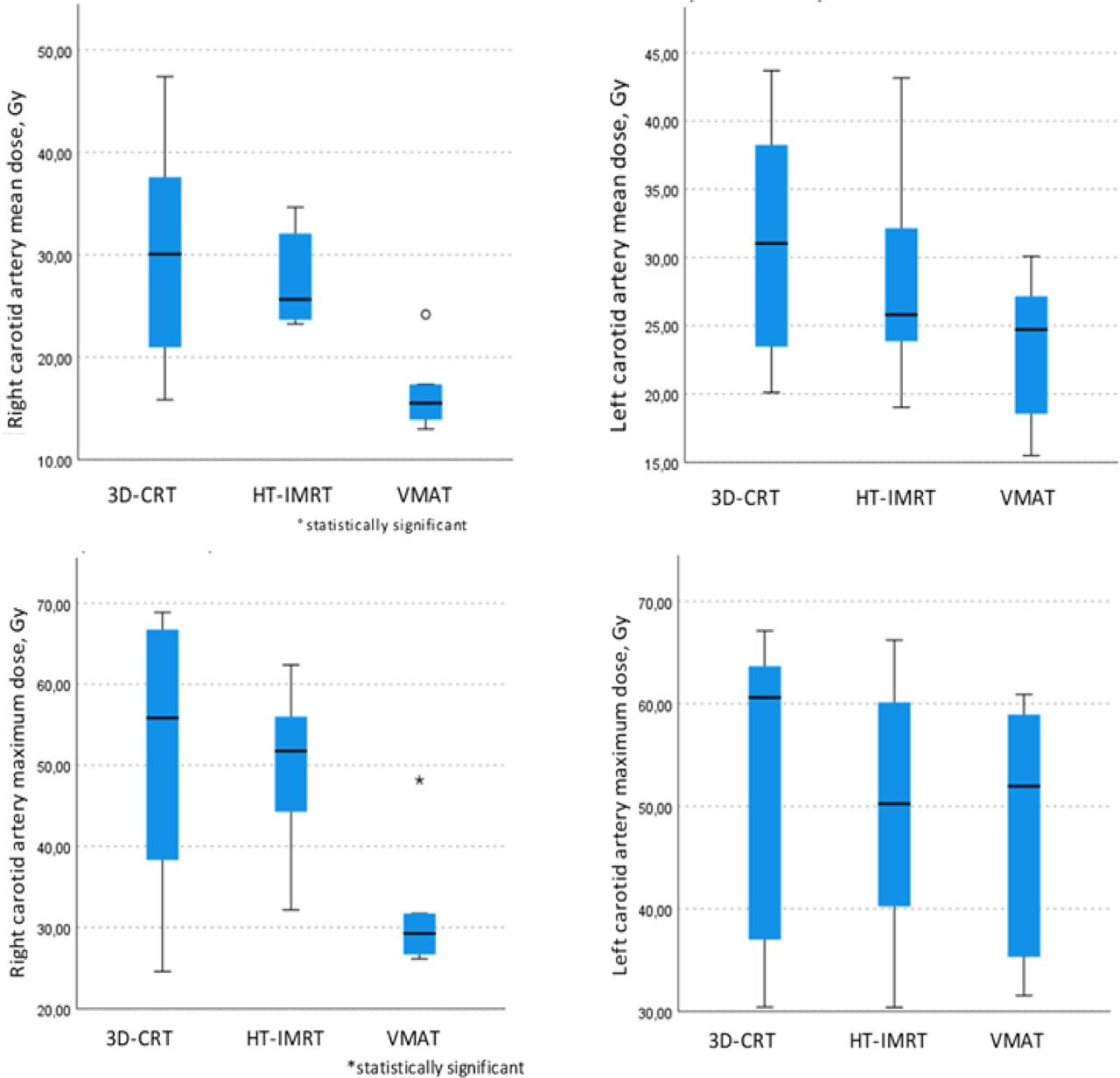
Figure 2:
Verification of differences in mean and maximum doses (Gy) to carotid arteries among treatment techniques using the nonparametric Kruskal–Wallis test.
Table 2:
Comparison of dosimetric characteristics among the three treatment techniques.
| Parameter | Treatment technique | Significance | ||
|---|---|---|---|---|
| 3D-CRT (n = 14) | HT-IMRT (n = 8) | VMAT (n = 6) | ||
| Right carotid artery | ||||
| Mean dose, Gy (range) | 30.9 (15.8–47.4) | 27.6 (23.2–34.6) | 16.6 (13.0–47.4) | p = 0.01 |
| Dmax, Gy (range) | 51.7 (24.6–68.9) | 49.9 (32.2–62.4) | 31.9 (26.1–48.2) | p = 0.02 |
| Mean volume delineated, ml (range) | 5.0 (3.6–6.4) | 3.9 (2.8–5.8) | 4.2 (2.6–6.5) | p = 0.1 |
| Left carotid artery | ||||
| Mean dose, Gy (range) | 31.0 (20.1–43.7) | 28.2 (19.0–43.2) | 23.4 (15.5–30.1) | p = 0.1 |
| Dmax, Gy (range) | 54.0 (30.4–67.1) | 49.7 (30.4–66.2) | 48.4 (31.5–60.9) | p = 0.6 |
| Mean volume delineated, ml (range) | 4.7 (3.1–6.2) | 4.0 (3.0–5.8) | 4.0 (2.5–5.6) | p = 0.3 |