
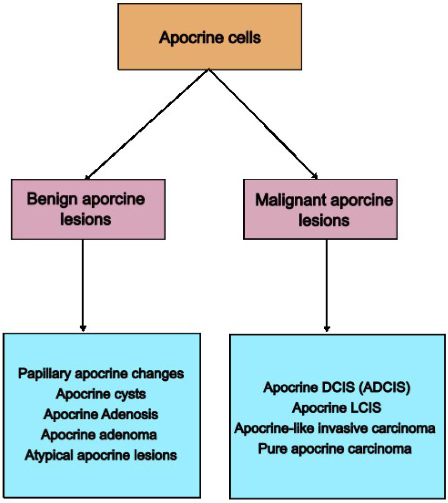
Figure 1:
Classification of apocrine lesions in the breast.
Table 1:
Immunohistology characteristics of ADCIS and pure apocrine carcinoma.
| IMMUNOHISTOLOGY MARKERS | PURE APOCRINE CARCINOMA | ADCIS | INVOLVING PATHWAY |
|---|---|---|---|
| ER, PR (−), AR (+) | 100% strictly match to AR (+,) ER(−), PR (−) criteria | >90% | AR pathway |
| HER-2 | 1/3 positive, 2/3 negative | 47.1% | HER2 signaling pathway |
| GCDFP-15 | 75% | 96.4% | AR pathway |
| AMACR | > 90% | > 90% | Oxidative degradation pathways |
| ER-A36 | 94.7% | 94.7% (classified with pure apocrine carcinoma) | MAPK/ERK signaling pathway EGFR/Src/ERK signaling pathway |
| PIK3CA | >80% | 100% | PI3K/mTOR pathway |
| P53 | 46–50% | 61.8% | PI3K/mTOR/p53 pathway |
| PD-L1 | 10–40% | N/A | PD-1/PD-L1 pathway |
| AR-V7 | 57% | N/A | AR pathway |
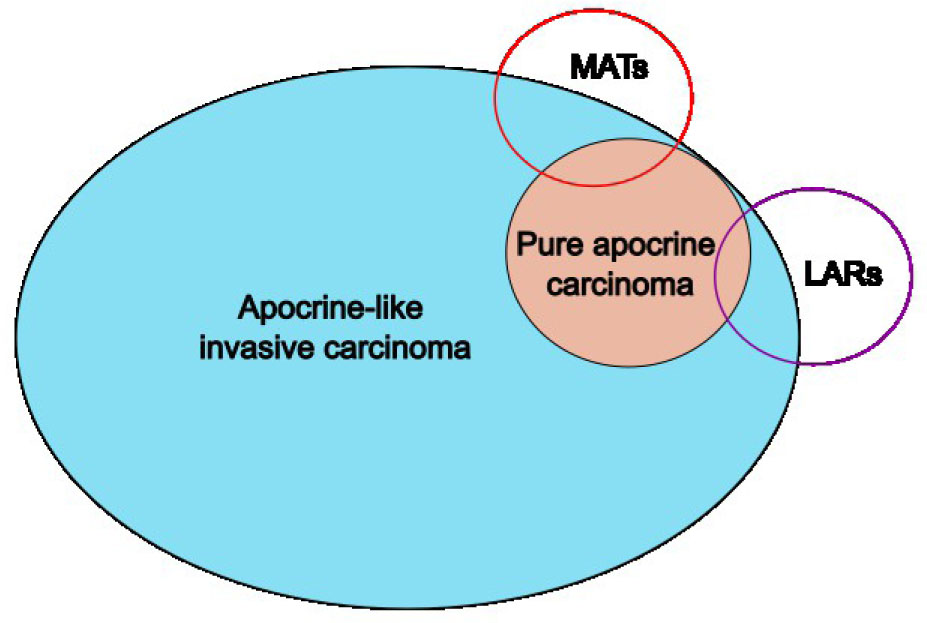
Figure 2:
Illustration shows the relationship between apocrine carcinoma definitions.
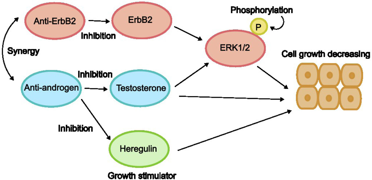
Figure 3:
The Cross-talk of AR and ErbB2 pathways In Naderi's study, the function of ERK1/2, testosterone, and Heregulin can be suppressed by both AR and ErbB2 pathways. Anti-ErB2 and anti-androgen therapy can inhibit the function of testosterone and ErbB2 and further cause the phosphorylation of ERK1/2, followed by negative regulation of cell growth.[52]
Table 2:
Treatment recommendation from the literature review.
| CLASSIFICATION | INTERVENTION |
|---|---|
| ATYPICAL APOCRINE LESIONS (AA) | No intervention +− Core needle biopsy |
| APOCRINE DCIS (ADCIS) | Mastectomy or conservative surgery +/− adjuvant radiation therapy |
| APOCRINE CARCINOMA | Neoadjuvant chemotherapy + surgery +/− radiation therapy + anti-androgen therapy (if AR+) + anti-HER2 antibodies (if HER2+) +/− PI3 kinase inhibition and CDK4/6 inhibitors (if biomarkers detected) |