
INTRODUCTION
Diabetes mellitus (DM) reprsents a chronic metabolic disease with a high prevalence and a substantial burden on health-care systems, with a global prevalence exceeding half a billion people older than 20 years, corresponding to roughly 10% of the adult population (1). The pathophysiological basis of DM involves the combined effects of chronic hyperglycemia, oxidative stress, activation of the polyol pathway with intracellular sorbitol accumulation, and non-enzymatic protein glycation, which can lead to microcirculatory disturbances, structural damage, and impaired homeostasis in various tissues, including the eye (2,3).
The impact of DM on the posterior segment and the development of diabetic retinopathy is well established (1,2). In recent decades, however, the effects of DM on the anterior segment have received increasing attention (1,3). On the corneal surface and in the epithelial layer, changes consistent with diabetic keratopathy are often observed, including thickening and multilamination of the epithelial basement membrane, reduced corneal sensitivity, and delayed epithelial healing (4,5). Chronic hyperglycemia activates the sorbitolaldose reductase pathway, and the accumulation of sorbitol and fructose disturbs Na+/K+-ATPase activity in the endothelial cell membrane, contributing to reduced barrier function (6). In DM, corneal endothelial cells may exhibit decreased endothelial cell density (ECD), increased polymegatism and pleomorphism, and a reduced percentage of hexagonal cells, indicating compromised morphological stability and a reduced functional reserve. Given that cataract in patients with DM occurs earlier and progresses more rapidly than in the general population, these endothelial alterations become particularly relevant (7). Multiple studies report lower ECD and increased central corneal thickness (CCT) in patients with type 2 DM (DM2) compared with healthy individuals, suggesting impaired endothelial stability (8). At the same time, cataract surgery is among the most frequently performed surgical procedures in medicine (6). Together, these factors may influence postoperative visual outcomes and satisfaction in patients with DM after cataract surgery.
Corneal endothelial status is particularly important in the perioperative period of phacoemulsification. Although phacoemulsification is the gold standard in cataract surgery, it exposes the corneal endothelium to multimodal stress, including irrigation fluids and ultrasound energy with acoustic cavitation, which may induce oxidative stress (10,11). Because the corneal endothelium in DM2 is morphologically weakened and has a lower functional reserve, the consequences of intraoperative stress may be more pronounced, with greater early postoperative endothelial cell loss (4,10). Patients with DM may also develop more pronounced postoperative corneal thickening (increased CCT), especially early after phacoemulsification, compared with non-diabetic patients (12). These findings suggest that DM may prolong corneal recovery and, consequently, delay visual recovery after phacoemulsification (9,10,11,12)
The primary aim of this study was to evaluate the effect of DM on central corneal thickness and corneal endothelial changes after phacoemulsification.
MATERIALS AND METHODS
Study design and participants
This retrospective cohort study was conducted from November 2023 to May 2024 and included 76 eyes: 36 eyes of patients with type 2 diabetes mellitus and 40 eyes of non-diabetic participants (control group). The eye was used as the unit of analysis. The follow-up time point was 1 month after cataract surgery.
Inclusion criteria were age >18 years, preoperative ECD ≥1500 cells/mm2, and the presence of a developed cataract. Exclusion criteria included previous intraocular trauma or surgical interventions, conditions with a potential major impact on the corneal endothelium (corneal dystrophies, uveitis, glaucoma), and high myopia or hyperopia greater than 5 diopters. Data on sex, age, comorbidities, and patient habits were collected from patient history and medical documentation.
Ethics and informed consent
The study was approved by the Ethics Committee of Specijalna bolnica Sveti Vid (Approval No. 19) and conducted in accordance with the Declaration of Helsinki. Written informed consent was obtained from all patients, and all data were analyzed in anonymized form.
Preoperative assessment
All patients underwent a complete preoperative ophthalmic examination, including best-corrected visual acuity (BCVA) assessment using Snellen charts, intraocular pressure (IOP) measurement by applanation tonometry (Haag-Streit), slit-lamp examination of the anterior segment, dilated fundus examination, intraocular lens (IOL) power calculation (optical biometry), and ocular ultrasonography. Corneal endothelial parameters were obtained by specular microscopy (Tomey EM-3000) (Figure 1). The analyzed parameters were ECD, percentage of hexagonal cells, coefficient of variation, and CCT.
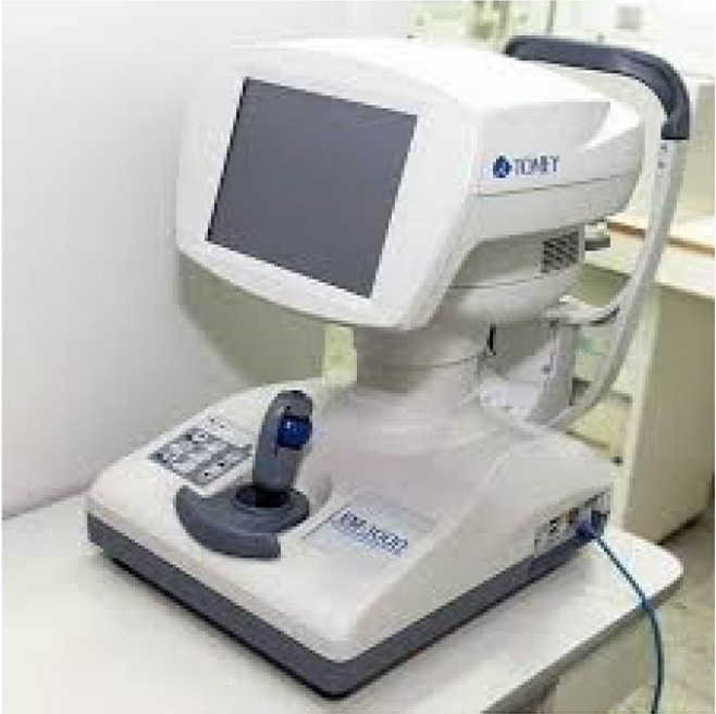
Figure 1.
Tomey EM-3000 specular microscope
Surgical procedure
All surgeries were performed by three experienced cataract surgeons under topical anesthesia (tetracaine). The phacoemulsification machine used in all procedures was Stellaris (Bausch & Lomb, Laval, Quebec, Canada). Maximal preoperative mydriasis was achieved with topical phenylephrine and homatropine. Two 1.5 mm paracenteses were created at 2 and 10 o’clock, and the anterior chamber was filled with a cohesive viscoelastic (ProVisc®, Alcon Laboratories, Inc., Fort Worth, TX, USA). A 2.2 mm clear corneal incision was made at 12 o’clock. Continuous curvilinear capsulorhexis, hydrodissection, and nucleus rotation were performed. The nucleus was fragmented using the divide-andconquer technique. Residual cortex was removed using bimanual irrigation and aspiration. After filling the capsular bag with cohesive viscoelastic, an IOL was implanted. Viscoelastic was aspirated, and intracameral cefuroxime (1 mg/0.1 mL in balanced salt solution) was injected. Corneal incisions were hydrated using balanced salt solution. At the conclusion of surgery, standard topical medication was administered according to the institutional protocol. Postoperatively, patients were prescribed topical dexamethasone/tobramycin five times daily and nepafenac three times daily, followed by gradual tapering according to the standard postoperative regimen.
Postoperative assessment
One month after phacoemulsification, all patients underwent a complete ophthalmic examination with repeated corneal measurements (ECD, percentage of hexagonal cells, coefficient of variation, and CCT).
Sample size estimation
The effect size used for sample size estimation was derived from the study by El-Saied et al. (2023) (20), which reported a statistically significant postoperative difference in endothelial cell density between diabetic and non-diabetic eyes at 1 month after phacoemulsification.
Statistical analysis
Statistical analysis was performed using IBM SPSS Statistics (version 27.0). Continuous variables were summarized as mean ± standard deviation (SD) for normally distributed data or median (interquartile range) for non-normally distributed data. Categorical variables were presented as frequencies and percentages. Normality of distribution was assessed using the Shapiro–Wilk test. Pre- and postoperative comparisons within groups were performed using the paired Student’s t-test for normally distributed variables and the Wilcoxon signed-rank test for non-normally distributed variables. Between-group comparisons (type 2 diabetes mellitus vs control group) were conducted using the independent-samples t-test or the Mann–Whitney U test, as appropriate. Effect sizes were calculated using Cohen’s d for parametric tests and r for non-parametric tests. A two-sided p value < 0.05 was considered statistically significant.
RESULTS
A total of 45 patients (76 eyes) were included. The diabetes group comprised 22 patients (36 eyes), and the control group comprised 23 patients (40 eyes). In the overall sample, there were 24 women (53.3%) and 21 men (46.7%), with no statistically significant difference in sex distribution between the groups. Mean age was 67.4 ± 10.1 years in the diabetes group and 65.8 ± 12.6 years in the control group, without a statistically significant difference (p > 0.05) (Table 1).
Table 1.
Age and sex distribution
| Variable | DM group (n = 36 eyes / 22 patients) | Control group (n = 40 eyes / 23 patients) |
|---|---|---|
| Age (years), mean ± SD | 67.4 ± 10.1 | 65.8 ± 12.6 |
| Sex (patients), n (%) | ||
| Male | 11 (50.0%) | 10 (43.5%) |
| Female | 11 (50.0%) | 13 (56.5%) |
Table 2.
Preoperative and postoperative corneal parameters in the diabetes and control groups
| Parameter | DM Pre-op (Mean ± SD) | DM 1 Month (Mean ± SD) | p (within DM) | Control Pre-op (Mean ± SD) | Control 1 Month (Mean ± SD) | p (within Control) | p (between groups) |
|---|---|---|---|---|---|---|---|
| ECD (cells/mm2) | 2577.25 ± 191.22 | 1899.19 ± 682.59 | <0.001 | 2460.35 ± 307.80 | 1786.18 ± 702.56 | < 0.001 | Pre-op: >0.05 Post-op: <0.05 |
| HEX (%) | 43.2 ± 5.4 | 39.4 ± 8.2 | >0.05 | 45.1 ± 7.8 | 42.2 ± 7.6 | >0.05 | >0.05 |
| CV (%) | 38.9 ± 5.5 | 40.7 ± 10.2 | >0.05 | 35.6 ± 3.3 | 37.5 ± 6.1 | >0.05 | >0.05 |
| CCT (μm) | 545.61 ± 32.45 | 556.32 ± 21.07 | >0.05 | 522.74 ± 36.49 | 536.69 ± 36.63 | >0.05 | Pre-op: <0.05 Post-op: <0.05 |
In the diabetes group, preoperative ECD was 2577.25 ± 191.22 cells/mm2 and decreased to 1899.19 ± 682.59 cells/mm2 at 1 month postoperatively, representing a highly significant change (p < 0.001). In the control group, ECD was 2460.35 ± 307.80 cells/mm2 preoperatively and 1786.18 ± 702.56 cells/mm2 postoperatively, representing a statistically significant reduction (p < 0.001). Intergroup comparison revealed no statistically significant difference in preoperative ECD (p > 0.05), but a significant postoperative difference was found (p < 0.05).
In both groups, cataract surgery did not result in a statistically significant change in the percentage of hexagonal cells (p > 0.05). In the diabetes group, the percentage of hexagonal cells decreased from 43.2 ± 5.4% preoperatively to 39.4 ± 8.2% postoperatively, while in the control group it decreased from 45.1 ± 7.8% to 42.2 ± 7.6%. Intergroup comparisons of hexagonality before and after surgery showed no statistically significant differences.
The coefficient of variation increased from 38.9 ± 5.5% preoperatively to 40.7 ± 10.2% postoperatively in the diabetes group, without a statistically significant difference (p > 0.05). In the control group, the coefficient of variation was 35.6 ± 3.3% before surgery and 37.5 ± 6.1% after surgery, also without a statistically significant difference (p > 0.05).
Central corneal thickness in the diabetes group was 545.61 ± 32.45 μm preoperatively and 556.32 ± 21.07 μm postoperatively, without a statistically significant within-group change (p > 0.05). In controls, CCT was 522.74 ± 36.49 μm preoperatively and 536.69 ± 36.63 μm postoperatively, also without a statistically significant within-group change. However, CCT differed significantly between the diabetes and control groups both before and after phacoemulsification (p < 0.05).
DISCUSSION
Diabetes mellitus is a systemic disease that affects multiple organs, and ocular involvement is common. In addition to diabetic retinopathy, patients may develop cataract, neovascular glaucoma, and other complications that can threaten vision. Corneal involvement in diabetes, including diabetic keratopathy and endothelial dysfunction, may reduce the functional reserve of the corneal endothelium, thereby increasing susceptibility to postoperative stromal edema, dry eye, decreased corneal sensitivity, and delayed visual recovery (1).
Phacoemulsification has become the standard technique for cataract surgery over the past several decades. The procedure is based on a phaco probe containing a piezoelectric crystal that vibrates at ultrasonic frequencies, generating cavitation within the crystalline lens and allowing its fragmentation and removal. This technique has significantly improved surgical precision, reduced incision size, and enhanced postoperative visual recovery. It has also lowered the incidence of many intraoperative and postoperative complications. Nevertheless, complications may still occur (13). Corneal edema remains one of the most common postoperative complications, primarily due to endothelial cell stress or loss during surgery. In patients with diabetes mellitus, chronic hyperglycemia further increases endothelial vulnerability.
Oxidative stress and metabolic imbalance impair endothelial cell function, predisposing these patients to a more pronounced postoperative response leading to dysfunction of corneal endothelial cells. Damage to these cells results in corneal edema, clinically manifested by decreased visual acuity.
In the present study, eyes of patients with type 2 diabetes mellitus showed a statistically significant reduction in ECD one month after phacoemulsification. A statistically significant endothelial cell loss was observed in both groups; however, however, postoperative endothelial parameters were less favorable in the diabetes group. In addition, CCT was higher in the diabetes group both preoperatively and postoperatively, and intergroup differences were significant at both time points. Earlier studies have suggested that increased CCT may represent an early manifestation of diabetes-related corneal involvement (15,16). By reducing Na⁺/K⁺-ATPase activity in the endothelial layer, diabetes mellitus promotes corneal hydration. In addition, hyperglycemia increases aldose reductase activity, an enzyme responsible for sorbitol synthesis. Accumulated sorbitol acts as an osmotic agent, drawing water into the cornea and contributing to increased thickness (16).
These findings support the recommendation that preoperative assessment of corneal endothelial status, including specular microscopy and CCT measurement, should be part of routine evaluation in diabetic patients scheduled for phacoemulsification (17). Endothelial-protective surgical strategies - appropriate viscoelastic use, minimizing ultrasound time and energy, maintaining anterior chamber stability, and gentle cortical manipulation - may help limit endothelial loss. Postoperatively, closer follow-up and adequate anti-inflammatory therapy may be warranted in patients with diabetes (6).
Although diabetes mellitus may increase the risk of corneal endothelial damage, with careful preoperative assessment and contemporary surgical technique, phacoemulsification can remain a safe and effective procedure in this population (19). Phacoemulsification remains the gold standard in cataract surgery; however, special caution is required in patients with diabetes mellitus due to existing structural and functional alterations of the corneal endothelium. Numerous clinical and experimental studies have demonstrated that diabetic patients exhibit reduced endothelial cell density, increased polymegatism and pleomorphism, as well as diminished endothelial functional reserve, making them more susceptible to postoperative complications (8).
Our results further indicate that diabetes mellitus had a significant impact on central corneal thickness. Phacoemulsification in diabetic patients was associated with a more pronounced endothelial cell loss and an increase in central corneal thickness at 1 month postoperatively compared to nondiabetic patients, particularly in cases of long-standing diabetes and poor glycemic control. Mechanical, thermal, and oxidative stress during surgery additionally burden the already compromised endothelium, thereby increasing the risk of postoperative corneal edema and prolonged visual recovery (19).
Our findings are consistent with several previously published studies that evaluated corneal endothelial changes in diabetic patients undergoing phacoemulsification. Chaurasia et al. (7) reported less favorable postoperative endothelial parameters in diabetic eyes compared with non-diabetic controls after cataract surgery, which is in line with our results. Similarly, a recent meta-analysis by Yang et al. (8) demonstrated that diabetes mellitus is associated with increased endothelial vulnerability and a higher susceptibility to postoperative endothelial damage. In addition, studies investigating central corneal thickness have shown that diabetic patients tend to exhibit thicker corneas both before and after surgery, likely reflecting impaired endothelial pump function (12,17). Our results support these observations, as we also observed significantly higher CCT values in the diabetes group at both time points. Taken together, these findings suggest that diabetes-related endothelial alterations may contribute to less favorable postoperative corneal status after phacoemulsification.
However, by applying modern surgical techniques, careful intraoperative management, adequate selection of viscoelastic agents, and reduction of ultrasound time and energy, the negative impact of phacoemulsification on the corneal endothelium in diabetic patients can be significantly minimized. Preoperative endothelial assessment using specular microscopy, together with appropriate systemic control of diabetes mellitus, remains essential for achieving optimal surgical outcomes.
Although phacoemulsification in patients with diabetes mellitus carries an increased risk of endothelial damage, with proper patient selection and an individualized surgical approach, this method remains safe and effective in the treatment of cataracts in this population.
CONCLUSIONS
Eyes of patients with type 2 diabetes mellitus showed lower postoperative endothelial cell density 1 month after phacoemulsification compared with non-diabetic controls. Central corneal thickness was higher in the diabetes group both before and after surgery, while changes in hexagonality and the coefficient of variation were not statistically significant. Routine preoperative endothelial evaluation and endothelial-protective surgical technique may be particularly important in diabetic patients undergoing phacoemulsification.
Notes
[1] STATEMENT OF HUMAN RIGHTS
This study was conducted in accordance with the ethical standards of the institutional research committee (Ethics Committee of Eye Hospital „Sveti Vid“, Approval No. 19) and with the 1964 Helsinki Declaration and its later amendments.
ACKNOWLEDGMENTS
The authors have no acknowledgments to declare.