
INTRODUCTION
Multiple sclerosis (MS) is a chronic demyelinating disease of the central nervous system (CNS), mediated by immune and inflammatory mechanisms (1). The immune response is involved in its pathogenesis, and an imbalance between pro- and anti-inflammatory cytokines has been demonstrated in numerous studies showing a correlation with changes in MS disease activity (2, 3, 4, 5, 6). All these perturbations could be useful for further improvement of clinical practice.
In addition to the various functions of cells of the immune system in the periphery, cells of the nervous system, microglia, and astrocytes are also important sources of cytokines (7). By acting on specific structures in the brain, proinflammatory cytokines can cause behavioural changes (8, 9). The main symptoms of this inflammation-induced behaviour are fatigue, anhedonia, anorexia, depression, and increased pain sensitivity (10, 11, 12). One of the proposed theories is that the behavioural changes represent a variant of “inflammation-induced sickness behaviour”, which is an adaptive defence mechanism against infection (13). The onset of sickness behaviour is associated with infiltration of inflammatory cells in the brain and increased mitochondrial ribonucleic acid (mRNA) expression of interleukin-1 beta (IL-1β), tumor necrosis factor-alpha (TNF-α), and prostaglandin E2 in brain tissue, whereas anti-inflammatory treatment with dexamethasone, interleukin-1 (IL-1) receptor antagonist (IL-1Ra), and indomethacin ameliorates behavioural symptoms (14, 15). The production of TNF-α in acute inflammation is always followed by the production of IL-1β (16).
The blockade of IL-1 in experimental autoimmune encephalomyelitis (EAE) has a protective effect, whereas interferon-beta (IFN-β) induces the expression of IL-1Ra in microglia, increasing the concentration of this mediator in serum (17). Serum levels of pro-inflammatory cytokines are certainly increased in relapsing MS (18). In this context, the relationship between fatigue, depression, and inflammatory cytokines has also been demonstrated in various autoimmune diseases (19, 20). Further, proinflammatory cytokines play an important role in the genesis of MS-related depression and fatigue (21, 22). An increase in the production of cytokines such as TNF-α, interferon-gamma (IFN-γ), and IL-1β has been demonstrated in depressed MS patients (23). Depression and fatigue are symptoms that are strongly correlated, and it has been shown that successful treatment of depression can in turn decrease fatigue (24).
In addition to the aforementioned cytokine IL-1β, cytokine family 1 also includes IL-18, whose active forms, as in the case of IL-1, are released by the action of caspase 1, which is activated by the nucleotide-binding oligomerization domain-, leucine-rich repeat, and pyrin domain-containing 3 (NLRP3) inflammasome (25). IL-18 is a pleiotropic pro-inflammatory cytokine involved in the regulation of innate and adaptive immune responses (26). It exists in an inactive precursor form (proIL-18) and converts to an active form in the presence of caspase-1 in response to inflammatory and infectious stimuli (27, 28). Formerly called interferon-gamma-inducing factor, it causes the production of IFN-γ in T cells and natural killer (NK) cells but can also be produced by various cell types, including monocytes/macrophages, dendritic cells, B cells, and other antigen-presenting cells, as well as astrocytes and microglia (29, 30, 31). Dysregulation of IL-18 production was observed in various autoimmune diseases such as bowel disease, rheumatoid arthritis, systemic juvenile idiopathic arthritis, and lupus erythematosus, suggesting its involvement in inflammatory cascades (32, 33, 34, 35).
Recently, a strong correlation between serum levels of IL-18 and C-reactive protein (CRP) was found in patients with severe Coronavirus infection (36). Further, uric acid (UA) has been extensively studied as an alarmin in MS, and it is known to activate the NLRP3 inflammasome and promote inflammation through maturation and release of the pro-inflammatory cytokines IL-1β and IL-18 (37, 38). Decreased concentrations of UA have been recorded in patients with MS compared to healthy individuals, as well as in relapses compared to concentrations in the remission phase of MS (39). All this suggests that these cascades involving the effects of UA and CRP on IL-18 secretion should be further explored in MS.
The similarities between the IL-1 and IL-18 systems suggest the possibility that, like IL-1β, IL-18 may also be a mediator of the sickness behaviour symptomatology (12, 40). An increased concentration of IL-18 has been confirmed in the serum and cerebrospinal fluid (CSF) of patients with MS, but there are no data on the relationship between this cytokine and fatigue and depressive symptomatology (41).
In this study, we aimed to measure and compare serum levels of IL-18 in patients with MS at different stages, in patients with a stable remitting form, in acute relapses before and after 2 months of treatment with pulse corticosteroid therapy, and in healthy subjects who served as controls. We sought to explore more complex interactions and the presence of a correlation between the levels of IL-18 and CRP, as a peripheral biomarker of inflammation, and UA, as an alarmin, and also, for the first time in this context, its association with clinical scores representing the severity of fatigue and depressive symptomatology.
PATIENTS AND METHODS
Subjects
The study included patients with clinically confirmed MS diagnosis according to the revised McDonald’s criteria and matched healthy controls recruited during blood donation at the Blood and Blood Products Service of the University Clinical Centre Kragujevac (42). In addition, the MS patients were divided into two groups: MS patients in the acute relapse phase (MS relapse group) and MS patients in the remitting phase (MS remitting group). An exacerbation of MS or relapse was defined as the appearance of new symptoms that lasted longer than 24 hours and occurred in the absence of fever and infection, or an exacerbation of existing symptoms after 30 days of a stable phase of the disease (43).
Patients in the MS relapse group were treatment-naïve regarding immunomodulatory therapy. Patients in the relapse phase were referred to the neurology unit because of the current exacerbation of the disease and indicated treatment with pulse corticosteroid therapy. In this group of patients, laboratory measurements and clinical assessments were performed before the initiation of treatment for acute relapse and eight weeks after the use of pulse corticosteroid therapy in the stable phase of the disease. In the remitting MS patients, these procedures were performed at scheduled three-month visits. All patients in the remitting group were treated with IFN-β-1a administered subcutaneously three times weekly.
Exclusion criteria were acute or chronic medical conditions, acute infections, recent surgical procedures, alcohol or drug abuse, any disease that is immune- or redox-mediated, allergies, or autoimmune disorders. The study was conducted in accordance with the ethical standards of the committee responsible for human experimentation and the presented protocol of this study was approved by the Ethics Committee of the University Clinical Centre Kragujevac. Voluntary written informed consent was obtained from each participant prior to enrolment in the study.
Laboratory measurements
Blood samples for the measurement of serum levels of CRP and UA and for the determination of serum cytokine concentrations were obtained in the morning and additionally before the use of corticosteroid therapy in MS recurrent patients. Serum cytokine levels were measured in patients and healthy volunteers using conventional enzyme-linked immunosorbent assay (ELISA) kits (R & D Systems Minneapolis, MN for IL-18 according to the manufacturer’s instructions). Immunological measurements were performed at the Center for Molecular Medicine and Stem Cell Research, Faculty of Medical Sciences, University of Kragujevac.
Clinical assessment
Auto- and heteroanamneses were performed to obtain sociodemographic and clinical data. Depressive symptomatology was assessed with the Beck Depression Inventory (BDI), which is recommended by the American Academy of Neurology for screening depressive symptoms in MS patients (44). The 21-item BDI includes somatic and cognitive-affective symptoms, and each item is graded on a 0 to 4 scale reflecting the patient’s feelings over the past two weeks. The degree of severity of depression symptoms was quantified as follows: normal (range 0–10), mild mood disturbance (range 11–16), borderline clinical depression (range 17–20), moderate depression (range 21–30), severe depression (range 31–40), and extreme depression (over 40).
The occurrence of fatigue was assessed with a self-report questionnaire - Fatigue Severity Scale (FSS) (45). The scale consists of nine items on a seven-point scale. The presence of mild fatigue is indicated by an average score greater than 3 points, and as disabling fatigue by an average FSS score greater than 4.
The Expanded Disability Status Scale (EDSS) was used to assess the degree of neurological disability (46). The EDSS consists of eight functional domains: visual (optic), brainstem, pyramidal, cerebellar, sensory, bowel and bladder, cerebral function, and ambulation. The EDSS score ranges from 0 (normal neurologic examination) to 10 (death due to MS), in half-point increments, to indicate a mild, moderate or severe disability.
Statistical analysis
Statistical analyses were performed using SPSS 20.0 software (IBM Corp. Armonk, NY, USA). Numerical values are expressed as mean and standard deviation (SD). Normal distribution of the data was tested with the Shapiro-Wilk test and the Kolmogorov-Smirnov test. A paired-sample test and the Wilcoxon test were used to compare the clinical parameters in the same group before and after therapy. For nonparametric variables, the Mann-Whitney test was used to detect mean differences between two groups of patients. A Kruskal-Wallis test was used to assess differences in IL-18 serum levels between three groups (MS relapse, MS remitting, and HC group). We examined the correlations between IL-18 serum levels and CRP and UA serum levels and the correlations between IL-18 serum levels and EDSS, BDI, and FSS scores using Pearson’s correlation for parametric or Spearman’s correlation for nonparametric data. To determine the best prediction of explanatory variables such as sex, age, and serum levels of UA and IL-18 on illness onset, a binary logistic regression analysis was performed. A p-value of 0.05 was considered statistically significant.
RESULTS
Sociodemographic data are shown in Table 1. The study included 133 participants, MS patients (n = 98) and healthy controls (n = 35). Of the MS population, 48 patients or 48.9% were in an acute relapse (MS relapse group) and 50 patients or 51.1% were in a stable phase of the disease and receiving immunomodulatory treatment (MS remitting group). No differences were found between the groups in terms of gender and age. The mean disease duration was significantly longer in the MS remitting group (6.19 ± 7.11 vs. 8.64 ± 4.46; p = 0.01), while the relapse rate was higher in the MS relapse group (2.42 ± 1.22 vs. 1.24 ± 1.39; p = 0.05).
Table 1.
Sociodemographic characteristics of the study population
| variables | MS relapse group (n =48) | MS remitting group (n = 50) | control group (n = 35) | p - value |
|---|---|---|---|---|
| age (years) | 41.7 ± 9.13 | 38.6 ± 7.88 | 38.6 ± 8.73 | 0.14* |
| sex (female / male) | 30 / 18 | 35 / 15 | 22 / 13 | 0.20* |
| mean duration of illness (years) | 6.19 (1–34) | 8.64 (2–18) | NA | 0.01** |
| relapse rate | 2.42 (0–5) | 1.24 (0–6) | NA | 0.05** |
We analysed serum concentrations of IL-18 and observed statistically significant differences between the MS group and healthy controls (468.96 ± 840.77 vs. 405.20 ± 146.16 pg/ml, p = 0.017). Further, the MS group was divided into the MS relapse and MS remitting groups and the data were compared between all three groups. We found a statistically significant difference in the mean values of IL-18 concentration between MS relapse, MS remitting and the control group (578.41 ± 1160.70 vs. 357.195 ± 206.75 vs. 405.20 ± 146.16 pg/ml, p = 0.040) (presented in Figure 1).
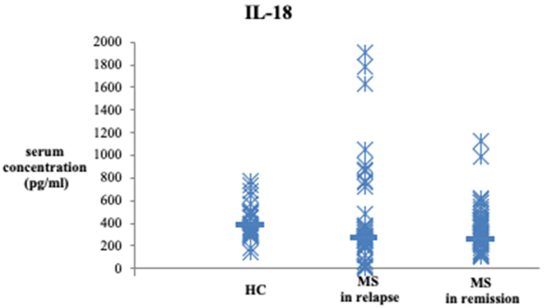
Figure 1.
Serum levels of IL-18.
Serum levels of IL-18 were determined by ELISA in healthy control subjects (n = 35), MS relapse patients (n = 48) and MS remitting patients (n = 50). Data are presented as mean ± SD. Statistical significance was tested using the Kruskal-Wallis test. Serum levels of IL-18 were significantly higher in MS relapse and MS remitting patients compared to the group of healthy control subjects (578.41 ± 1160.70 vs. 357.195 ± 206.75 vs. 405.20 ± 146.16 pg/ml, p = 0.040).
In addition, a comparison of serum concentrations of IL- 18 in the MS group in relapse and remission revealed no statistically significant differences between these patient groups (578.41 ± 1160.70 vs. 357.192 ± 206.75 pg/ml, p = 0.597). We also analysed serum levels of IL-18 in the 19 patients in the MS relapse group before and after treatment with pulse corticosteroid therapy and no statistically significant differences were found (p = 0.658).
We found a significant, moderate positive correlation between IL-18 and serum levels of CRP in relapse (p = 0.012, r = 0.305) (Figure 2A). In remission, a significantly strong negative correlation was observed between IL-18 and serum concentrations of UA (p = 0.014, r = −0.551) (Figure 2B). Clinical characteristics (EDSS, FSS and BDI scores) of the sample were previously presented in Vesic et al. (2020) (39). Regarding behavioural changes, we found a statistically significant, strong positive correlation between serum concentrations of IL-18 and BDI score (p = 0.018, r = 0.535) (Figure 2C) and a moderate positive correlation with EDSS score (p = 0.047, r = 0.462) (Figure 2D) in the stabilization of MS. We found no significant correlation between IL-18 and FSS score.
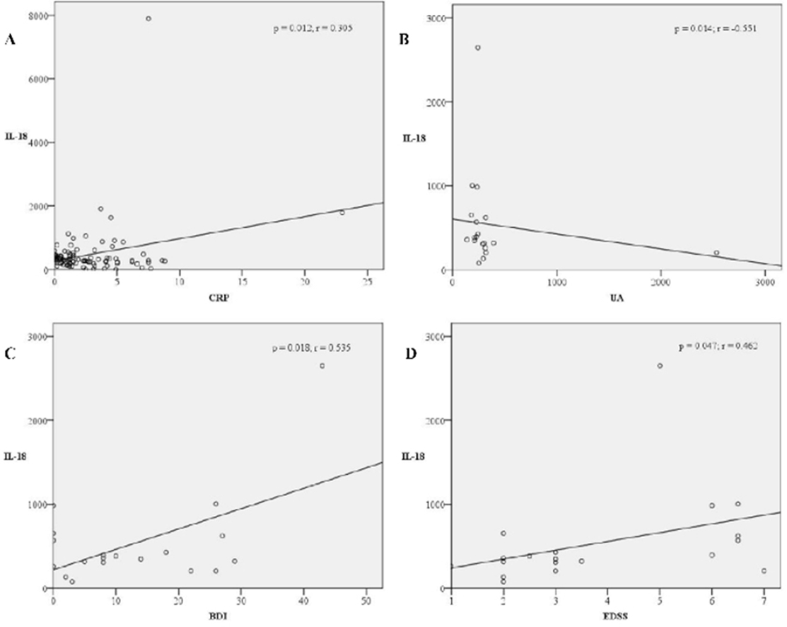
Figure 2
Legend. Correlations between serum levels of IL-18 and CRP and UA, and BDI and EDSS clinical scores. A significant, moderate positive correlation was observed between IL-18 and serum levels of CRP (p = 0.012, r = 0.305) in acute relapse (A). In stabilization, after eight weeks of corticosteroid therapy, a significant strong negative correlation was observed between IL-18 and serum concentrations of UA (p = 0.014, r = −0.551) (B), a strong positive correlation was observed between IL-18 and BDI score (p = 0.018, r = 0.535) (C), and a moderate positive correlation was observed with EDSS score (p = 0.047, r = 0.462) (D).
Abbreviations: IL - interleukin; CRP - C-reactive protein; EDSS - Expanded Disability Status Scale; UA - uric acid, BDI - Beck’s Depression Inventory.
Complementary binary logistic regression analysis was performed using available data on sex, age, serum IL-18 and UA levels to determine the best predictor of illness onset (Table 2). The results showed that only serum levels of UA influenced illness onset (p = 0.000), while sex (p = 0.950), age (p = 0.663), and serum levels of IL-18 (p = 0.884) were not significantly associated with illness onset.
Table 2.
Results of binary logistic regression analysis with illness onset as the dependent variable, and sex, age, serum UA and IL-18 levels as explanatory variables.
| category | explanatory variables | Wald | df | p - value | Odds ratio | Lower and upper 95% CI |
|---|---|---|---|---|---|---|
| illness onset | sex | 0.004 | 1 | 0.950 | 0.371 | 2.878 |
| age | 0.190 | 1 | 0.663 | 0.960 | 1.066 | |
| UA | 15.692 | 1 | 0.000 | 0.979 | 0.993 | |
| IL-18 | 0.021 | 1 | 0.884 | 0.999 | 1.001 |
DISCUSSION
The results of our study confirmed that the levels of IL-18 were significantly higher in patients with MS compared to the healthy control subjects. There were no significant differences in serum levels of this cytokine in MS relapse compared to the MS remitting group of patients. In addition, a slight but nonsignificant decrease in serum levels was observed after corticosteroid therapy. All these data could imply that IL-18 cannot be considered as a state marker but rather as a trait marker in MS. In the periphery, a positive correlation was found between CRP and IL-18 in the relapse phase and a negative one between UA and IL-18 in a remission phase after acute relapse. These results may suggest that IL-18 is involved in inflammation in the acute phase and regresses in the stabilization phase, which is associated with increased behavioural symptoms such as fatigue and depression.
Evidence is accumulating that IL-18 is involved in the pathogenesis of MS and other autoimmune diseases (47). In 1998, Jander and Stoll observed increased levels of IL-18 mRNA in the spinal cord of rats suffering from the acute phase of EAE (48). In EAE, IL18-/- mice have been shown not to develop the disease (49). In recent years, several studies have presented conflicting results regarding serum levels of IL-18 in MS patients. In the search for genetic factors associated with predisposition to MS, it has been suggested that the IL-18 gene polymorphism increases susceptibility to MS (42, 50). Previously, increased expression of IL-18 was found in demyelinating brain lesions of MS patients, and antibodies against IL-18 were shown to prevent autoimmune encephalomyelitis (51, 52). Similar to our results, increased serum levels of IL-18 were observed in MS patients compared to the control group (53, 54). In addition, Huang et al. reported that the expression of IL-18 in serum and the level of IL-18 mRNA in peripheral blood mononuclear cells were significantly increased in patients with MS (55).
We also analysed the serum levels of IL-18 at different stages of MS and there were no significant differences in serum levels of this cytokine in MS relapse compared to the MS remitting group of patients. The highest levels of IL-18 were detected in the MS relapse patient group. The results of Losy and colleagues emphasized significantly higher concentrations of IL-18 in patients with gadolinium-enhancing lesions on magnetic resonance imaging compared with patients without active lesions. This fact suggests the involvement of IL-18 in the acute exacerbation and active stage of MS (56). In contrast, Trenova et al. examined serum levels of IL-18 in MS-drug-naïve patients and in the stable phase of the disease as well as in healthy control subjects, and they found no significant differences of this cytokine between the groups, indicating that special attention should be paid to the applied therapy (57). Thus, we observed the lowest serum levels of IL-18 in MS patients with long stable phase of MS treated with IFN-β-1a compared to healthy control subjects. Previously, it was reported that the levels of IL-18 decreased significantly during therapy with glatiramer acetate in MS patients (56). In addition, we analysed sera IL-18 levels before, and after corticosteroids treatment in MS relapse patients. Corticosteroids regulate NLRP3‐inflammasome activation at both the transcriptional level via blocking NF-κB signal pathway and processing level via suppressing the reactive oxidative species generation and IL-18 levels (58, 59). In our study, a slight but non-significant decrease in serum levels was observed after two months of corticosteroid therapy. A possible reason is a small sample size that only 19 patients had a follow-up after treatment of corticosteroids. In current literature, a limited number of studies have been discussed about IL-18 sera levels in different stages of MS. The relationship of the IL-18, MS phenotype and corticosteroids treatments need to be investigated in the future on a bigger sample size.
To further explore the inflammatory milieu, IL-18 was placed in the context of interactions with CRP and UA. In a previous study, our research group observed significantly higher CRP levels as an inflammatory marker of acute exacerbation compared between MS remitting and relapsing patients and significantly lower UA levels in the serum of MS relapsing patients compared to MS remitting patient groups and healthy control subjects (39). The additional results presented from the same sample showed a significant, moderate positive correlation between CRP and IL-18 serum levels in acute relapse and a significant strong negative correlation between UA and IL-18 levels after treatment of acute relapse with pulse corticosteroid therapy during stabilization of the illness. IL-18 seems to interfere with CRP potentiating inflammation while downregulating UA levels in the acute phase of the illness. Later, after treatment with corticosteroids, it plays a lesser role in the phase of attenuated inflammation (lower CRP and higher UA serum levels), but may be more important in regulating behavioural symptoms such as fatigue and depression.
In present study, we revealed a moderate positive correlation between sera IL-18 levels and EDSS score in stable phase of disease. The findings of Soares et al. emphasized that a constitutive activation of NLRP3 inflammasome, and the consequent increased IL-1β and IL-18 production, represents a risk factor for both, the development of MS and the progression to higher EDSS score and severe forms of the disease. On the other hand, low IL-18 production and/or NLRC4 activation resulted beneficial for MS patients (60).
IL-18-deficient mice exhibited hippocampal abnormalities which resulted in impaired learning and memory, and depressive-like behaviour, but also stress-induced hyperactivity mediated by neuroinflammation and attenuated neurogenesis (61, 62). In addition, administration of IL-18 to the amygdala, but not to the hippocampus, resulted in severe depression-like behaviours (63). Haastrup et al. indicated that the genetic tendency toward higher IL-18 production may increase susceptibility to depression in response to stressful life events (64). Several clinical studies reported increased serum levels of IL-18 in patients with depression compared to healthy controls and demonstrated impaired brain activity (65, 66, 67, 68). Our results showed a statistically significant, strong positive correlation between serum concentrations of IL-18 and BDI score in the remission phase. Previous studies observed relationship between depressive symptoms and IL-18 in other inflammatory diseases. Zao et al. showed that IL-18 levels positive correlated with BDI scores in depressed patients with chronic obstructive pulmonary disease (69). Recent studies indicated that increased IL-18 in the brain causes depression-like behaviors by promoting the IL-18 receptor/NKCC1 signaling pathway and that this cytokine is a biomarker for post-stroke depression. The blockage of endogenous IL-18 by IL-18 binding protein, a specific antagonist of IL-18, repressed depressive phenotypes in mice (63). Potential mechanisms through cytokines mediate depression like behavior are various. Proinflammatory cytokines directly stimulate the hypothalamic-pituitary-adrenal axis and can mitigate serotonin synthesis by inducing the enzyme indoleamine 2, 3 dioxygenase, which catabolizes tryptophan into kynurenine (70, 71). In homeostatic concentrations, IL-18 support neurons and enhance long-term potentiation, but elevated IL-18 and other proinflammatory cytokines may promote the dysregulation of several neurotransmitter pathways, including the dopaminergic, serotoninergic, noradrenergic, and glutamatergic systems (71, 72).
This was clinically confirmed by the fact that the nonsteroidal anti-inflammatory drug ketoprofen decreased IL-18 levels in patients with depression when used as adjunctive treatment and also improved clinical outcomes (73). Future strategies must consider that higher serum UA levels may be responsible for chronic activation of the NLRP3 inflammasome, persistent activation of NF-κB, and elevated levels of pro-inflammatory cytokines, including IL-18, but also in MS related depression (38).
These conflicting results across studies might be a consequence of sample heterogeneity and measurement in different phases of illness. This study was limited in terms of comparative resources (lacked laboratory data from control subjects). In addition, not all patients had follow-up after treatment with corticosteroids (n = 19). Data on metabolic parameters and other additional clinical and sociodemographic variables must also be considered in the future.
CONCLUSION
The results of the present study show that the levels of the proinflammatory cytokine IL-18 are increased in MS patients compared with healthy subjects. The positive correlation of IL-18 with serum CRP in the relapse phase and the negative correlation with serum levels of UA in the remission phase suggests its inflammatory role in the active phase of the disease and a regressive one in the stabilization phase of the disease course. The positive correlation of IL-18 with depression and disability scale scores confirms its role as a contributor to depressiveness and disability in MS patients. MSrelated depression and fatigue are common and challenging symptoms of MS, which have a significant impact on patients’ quality of life. The cytokine pattern in MS patients varies according to disease stage, necessitating an expansion of research on cytokines as potential biomarkers of disease onset, progression, assessment of treatment efficacy and response to treatment.
Notes
[6] ETHIC APPROVAL
The study was conducted in accordance with the ethical standards of the committee responsible for human experimentation (institutional) and the Helsinki Declaration of 1975, as revised in 2013. Voluntary written and informed consent was obtained from each participant prior to enrolment in the study. The protocol of the study was approved by the local ethic committee.
ACKNOWLEDGMENTS
Prof. Miodrag L. Lukic and Prof. Nebojsa N. Arsenijevic contributed significantly to initiating this research project and shedding light on possible aspects of the IL-18 special role. Prof. Marija Milovanovic contributed significantly in the realization of the project and immunological measurement. Prof Milica Borovcanin contributed significantly to the concept and manuscript structure and gave some new insights through her specific competencies We thank Bojana Mircetic for language editing.