
Table 1.
Summary of products used in study.
| Sample name | Product description | Manufacturer |
|---|---|---|
| CP1 | Dry Pouch Filler 1 | Helix Innovations, LLC |
| CP2 | Wet Pouch Filler 1 | Helix Innovations, LLC |
| CP3 | Dry Pouch Filler 2 | Swedish Match |
| CP4 | Wet Pouch Filler 2 | Swedish Match |
Table 2.
Screen size of sieve stack fractions used by Labs 1–4 for particle size analysis by sieve. The bold values indicate fractions used by all laboratories, with the summary shown in the composite sieve stack used for graphical analysis.
| Sieve screen particle size (μm) | ||||
|---|---|---|---|---|
| Lab 1 | Lab 2 | Lab 3 | Lab 4 | Composite stack |
| 900 | 710 | 500 | 500 | 500 |
| 500 | 500 | 420 | 250 | 250 |
| 400 | 355 | 355 | 200 | 125 |
| 300 | 250 | 250 | 125 | Catch (<125) |
| 250 | 180 | 180 | 100 | |
| 200 | 125 | 125 | 75 | |
| 150 | 90 | Catch (<125) | 63 | |
| 125 | 63 | 45 | ||
| 100 | Catch (<60) | Catch (<45) | ||
| 90 | ||||
| Catch (<90) | ||||
Table 3.
Sieving particle size results for dry nicotine pouch filler. The values represent the average of intercepts at 10%, 50%, and 90% of three replicates (n = 3) and the error represents one standard deviation (1 σ).
| Particle size (μm) | ||||
|---|---|---|---|---|
| Value | Lab 1 | Lab 2 | Lab 3 | Lab 4 |
| CP1 – Dry filler 1 | ||||
| Dx[10] | 129.7 ± 7.0 | 143.1 ± 3.4 | 139.1 ± 10.6 | 131.7 ± 3.7 |
| Dx[50] | 315.3 ± 14.2 | 325.4 ± 2.0 | 314.3 ± 24.0 | 315.2 ± 12.9 |
| Dx[90] | 652.3 ± 29.9 | 623.2 ± 3.3 | 500.0 ± 0.0 | 534.8 ± 6.4 |
| CP3 – Dry filler 2 | ||||
| Dx[10] | 165.0 ± 8.7 | 151.2 ± 0.9 | 161.9 ± 10.1 | 156.4 ± 1.1 |
| Dx[50] | 288.7 ± 8.1 | 278.3 ± 2.0 | 277.7 ± 4.4 | 299.2 ± 4.8 |
| Dx[90] | 438.0 ± 10.4 | 434.7 ± 2.5 | 416.0 ± 0.7 | 469.2 ± 5.1 |
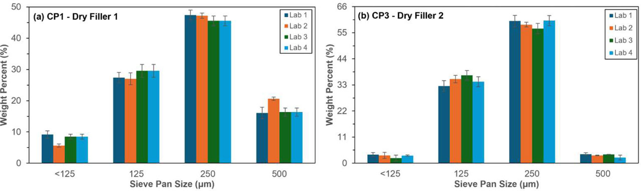
Figure 1.
Sieving particle size distributions using a consolidated sieve stack for dry nicotine pouch fillers (a) CP1 and (b) CP3. Each bar graph is the average of three replicates (n = 3) and the error bars represent one standard deviation (1 σ).
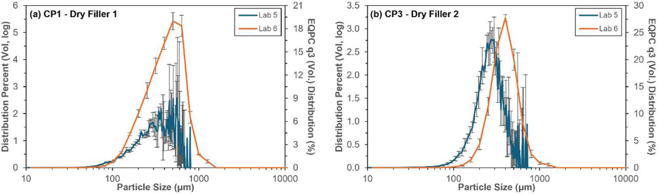
Figure 2.
Dynamic image analysis particle size distributions (PSD) plots for dry nicotine pouch fillers (a) CP1 and (b) CP3. For Lab 5, distributions are calculated as the Feret minimum diameter with the distribution percent shown and each bar is the average of three replicates (n =3). For Lab 6, distributions are calculated as the equal projection of a circle (EQPC) with the volume-weighted (q3) distribution of six replicates (n = 6) shown. The error bars represent one standard deviation (1 σ).
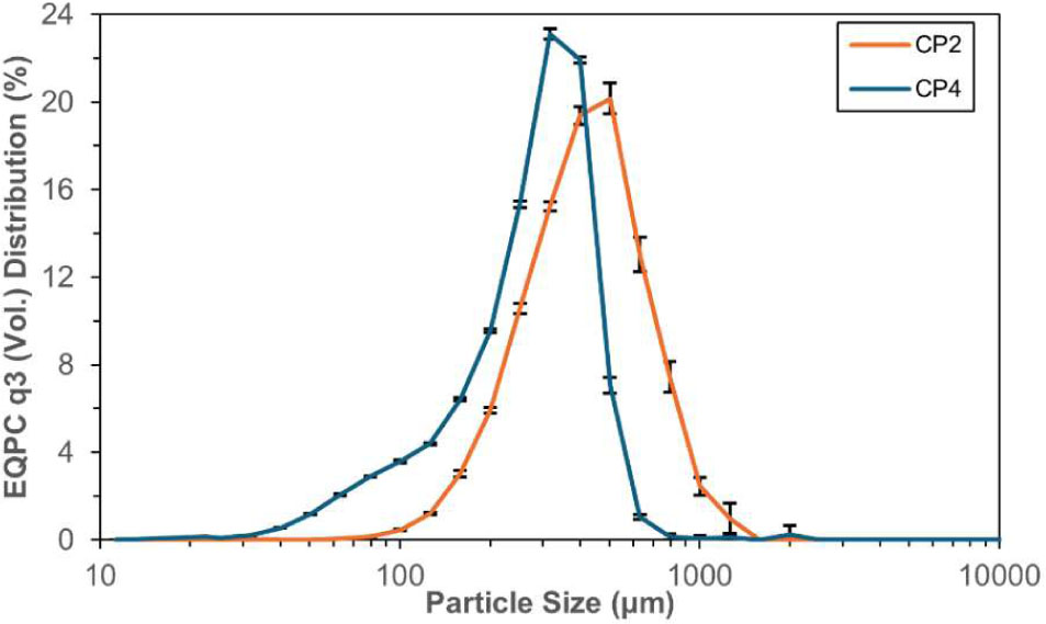
Figure 3.
Dynamic image analysis particle size distribution (PSD) plots for wet nicotine pouch products (CP2 and CP4). Distributions are calculated as the equal projection of a circle (EQPC) with the volume-weighted (q3) distributions of six replicates (n = 6) shown. The error bars represent one standard deviation (1 σ). Results performed by Lab 6.
Table 4.
Dynamic image analysis particle size results for nicotine pouch fillers. The values represent the average of intercepts at 10%, 50%, and 90% of three replicates (n = 3) for Lab 5 and six replicates (n = 6) for Lab 6. The error represents one standard deviation (1 σ). Note that CP2 and CP4 were not tested by Lab 5.
| Particle size (μm) | ||||
|---|---|---|---|---|
| Value | CP1 | CP2 | CP3 | CP4 |
| Lab 5 | ||||
| Dx[10] | 143.97 ± 4.18 | – | 156.76 ± 9.73 | – |
| Dx[50] | 327.66 ± 14.79 | – | 262.25 ± 13.55 | – |
| Dx[90] | 560.68 ± 39.43 | – | 420.29 ± 42.63 | – |
| Lab 6 | ||||
| Dx[10] | 174.65 ± 0.93 | 194.41 ± 2.54 | 216.76 ± 10.49 | 96.50 ± 0.95 |
| Dx[50] | 381.14 ± 15.44 | 373.68 ± 2.40 | 346.38 ± 11.31 | 262.20 ± 0.80 |
| Dx[90] | 625.96 ± 8.33 | 650.36 ± 25.22 | 535.26 ± 24.91 | 394.05 ± 0.55 |
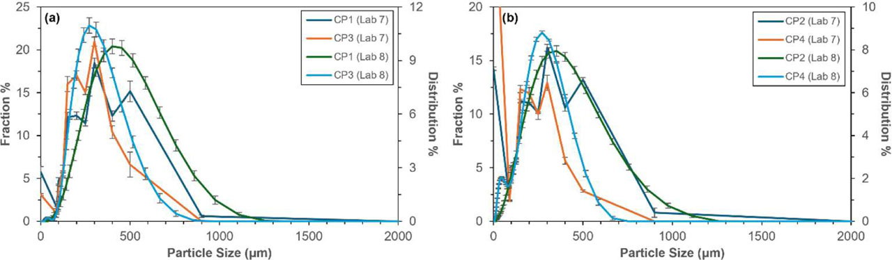
Figure 4.
Laser diffraction particle size distribution (PSD) plots for nicotine pouch fillers where (a) summarizes dry pouch fillers (CP1 and CP3) and (b) summarizes wet pouch fillers (CP2 and CP4). Samples tested by Lab 7 are measured as fraction % and samples tested by Lab 8 are represented as distribution %. Distributions are the average of six replicates (n = 6) shown and the error bars represent one standard deviation (1 σ).
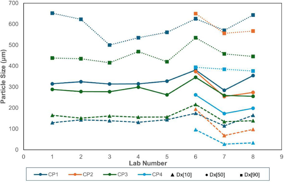
Figure 5.
Graph representing the distribution intercepts at 10%, 50%, and 90% (Dx[10], Dx[50], and Dx[90]) for the four nicotine pouch fillers tested by Labs 1–8. The Dx[10] results are represented as triangles and dashed lines. The Dx[50] results are represented as circles and solid lines. The Dx[90] results are represented as squares and dotted lines. Labs 1–4 performed sieving analysis, Labs 5 and 6 performed dynamic image analysis, and Labs 7 and 8 performed laser diffraction analysis.
Table 5.
Laser diffraction particle size results for nicotine pouch fillers. The values represent the average of intercepts at 10%, 50%, and 90% of six replicates (n = 6) and the error bars represent one standard deviation (1 σ).
| Particle size (μm) | ||||
|---|---|---|---|---|
| Value | CP1 | CP2 | CP3 | CP4 |
| Lab 7 | ||||
| Dx[10] | 114.7 ± 5.2 | 68.2 ± 0.7 | 134.7 ± 3.2 | 27.7 ± 2.6 |
| Dx[50] | 284.2 ± 9.7 | 254.3 ± 2.3 | 259.5 ± 8.0 | 173.5 ± 8.9 |
| Dx[90] | 570.7 ± 13.2 | 556.3 ± 9.6 | 458.3 ± 18.7 | 384.8 ± 4.7 |
| Lab 8 | ||||
| Dx[10] | 165.3 ± 12.2 | 97.8 ± 5.3 | 139.2 ± 5.8 | 33.6 ± 0.2 |
| Dx[50] | 354.8 ± 10.1 | 273.8 ± 4.9 | 255.8 ± 4.8 | 198.2 ± 1.3 |
| Dx[90] | 643.3 ± 14.6 | 567.2 ± 8.9 | 445.5 ± 9.3 | 376.0 ± 4.4 |
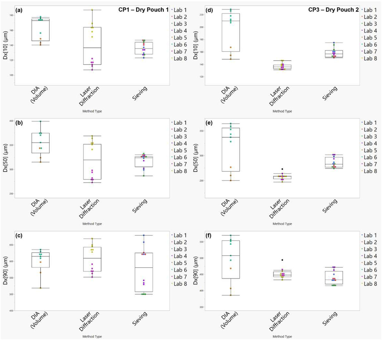
Figure 6.
Box and whisker plots used for particle size method comparison of dry pouch products. Left: CP1 (Dry Pouch Filler 1) (a-c) and right: CP3 (Dry Pouch Filler 2) (d-f) where (a) and (d) show Dx[10] results, (b) and (e) show Dx[50] results, and (c) and (f) show Dx[90] results.
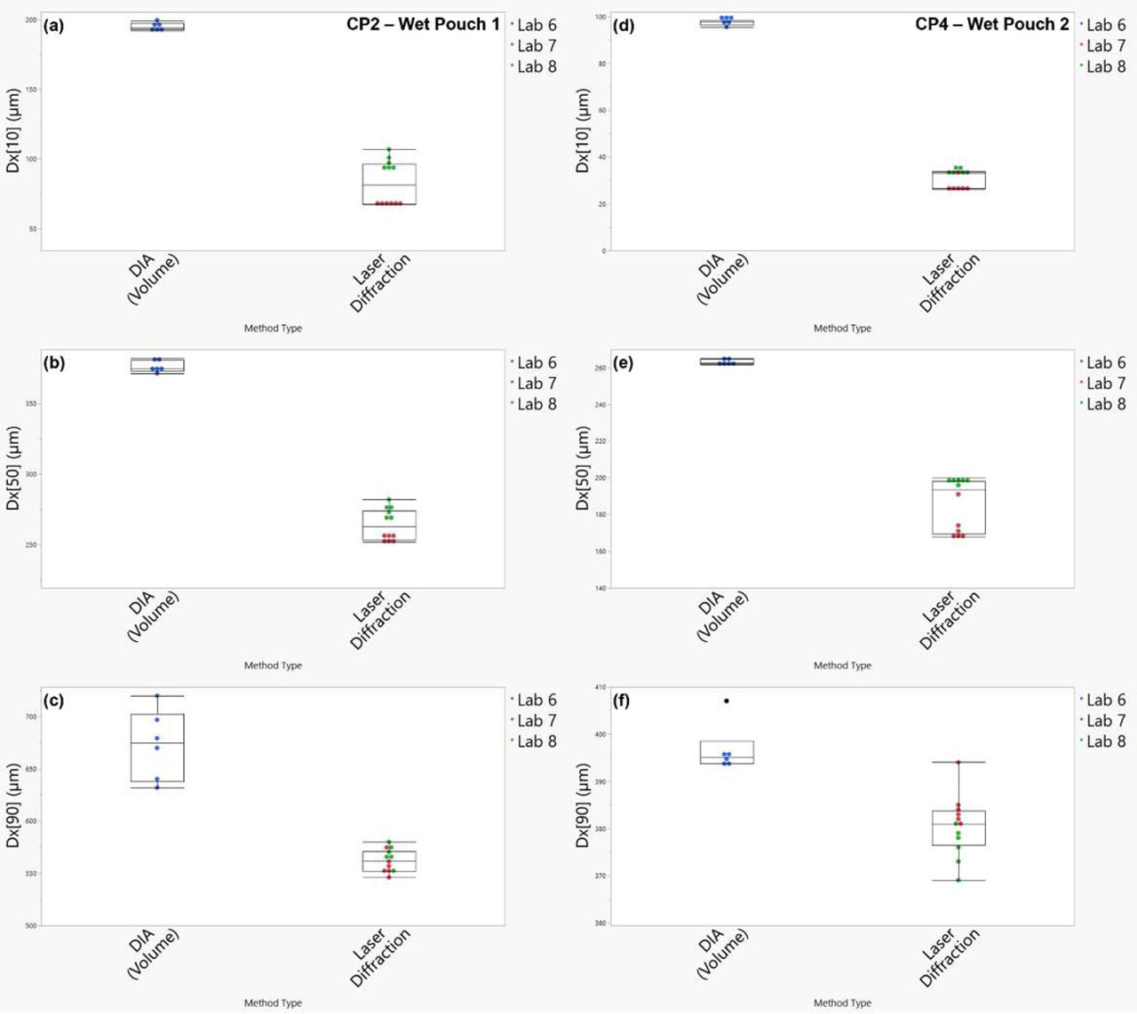
Figure 7.
Box and whisker plots used for particle size method comparison of wet pouch products. Left: CP2 (Wet Pouch Filler 1) (a-c) and right: CP4 (Wet Pouch Filler 2) (d-f) where (a) and (d) show Dx[10] results, (b) and (e) show Dx[50] results, and (c) and (f) show Dx[90] results.
Table 6.
Summary of the results of Tukey means comparison for each distribution intercept to compare differences between particle size methodologies. Levels not connected by the same letter (in different group) are significantly different.
| Mean particle size (μm) | ||||||
|---|---|---|---|---|---|---|
| Dx[10] | Dx[50] | Dx[90] | ||||
| Methodology | Mean | Group | Mean | Group | Mean | Group |
| CP1 – Dry filler 1 | ||||||
| DIA | 162.43222 | A | 355.49111 | A | 607.00000 | A |
| Laser diffraction | 140.00000 | B | 319.50000 | B | 601.49556 | A |
| Sieving | 135.87500 | B | 317.56667 | B | 577.56667 | A |
| CP2 – Wet filler 1 | ||||||
| DIA | 195.03833 | A | 376.17667 | A | 672.88167 | A |
| Laser diffraction | 82.99167 | B | 264.08333 | B | 561.75000 | B |
| CP3 – Dry filler 2 | ||||||
| DIA | 196.88000 | A | 319.03556 | A | 497.84778 | A |
| Laser diffraction | 158.64167 | B | 285.95000 | B | 451.91667 | B |
| Sieving | 136.91667 | C | 257.66667 | C | 439.46667 | B |
| CP4 – Wet filler 2 | ||||||
| DIA | 97.396667 | A | 263.04500 | A | 396.70333 | A |
| Laser diffraction | 30.633333 | B | 185.83333 | B | 380.41667 | B |