
INTRODUCTION
Induction and formation of gums (gummosis) are found through the plant kingdom. Generally gums are induced by biotic and abiotic environmental factors such as bacterial and fungal infection, insect attack, mechanical and chemical injury, water stress, and other environmental stressors (Boothby, 1983). All these factors have been shown to promote also ethylene synthesis in plant tissues. It was reported that ethylene or ethylene-releasing compounds (i.e. ethephon; 2-chloroethylphosphonic acid) stimulate gum formation in stone-fruit trees and their fruits of the Rosaceae, apricot, cherry, peach, plum, almond (Boothby, 1983; Saniewski et al., 2006), but also in tulip (Tulipa sp.) bulbs and other bulbous ornamental plants (Kamerbeek, de Munk, 1976).
Tulip bulbs infected by Fusarium oxysporum f. sp. tulipae have been shown to produce considerable quantities of ethylene, causing gummosis in diseased and healthy bulbs stored in the same conditions (Figure 1) (Kamerbeek, de Munk, 1976; Saniewska et al., 2004; Saniewski et al., 2007). These results suggest that ethylene is a common factor involved in the induction of gummosis (Boothby, 1983).
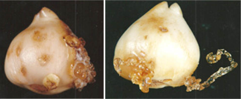
Figure 1.
Gummosis in tulip bulbs cv. Apeldoorn induced by Fusarium oxysporum f. sp. tulipae (on left) and by ethephon (2-chloroethylphosphonic acid) – ethylene releasing compounds (on right). Author of photographs: Justyna Goraj-Koniarska.
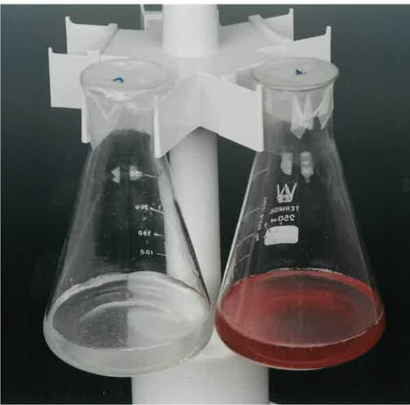
Figure 2.
Experiment with Fusarium oxysporum f. sp. tulipae. In the control treatment (left), no mycelial growth was observed. In the treatment supplemented with gums, the mycelium develops and produces pigments.
On the other hand it has been shown that jasmonates have a promoting effect on the induction and/or production of gums in tulip bulbs and in stone-fruit trees and their fruits of Rosaceae family, such as cherries, plums, peaches, apricots (Saniewski, Puchalski, 1988).
This was reported that a rapid increase in endogenous levels of jasmonates, mainly Jasmonic acid (JA), occurs in plants or their organs under stress conditions, e.g. after mechanical wounding, under osmotic stress conditions and after pathogens infection or insect attack (Saniewski, 1997). These facts strongly suggest that jasmonates are important key compounds involved in induction and/or production of gums in plants together with or without endogenous ethylene. Jasmonates have also been well known to control ethylene production in plant tissues (Saniewski, 1997).
Gums are complexes of different substances, but most important constituents are polysaccharides of highly individual structure (Boothby, 1983). The composition of tulip gum polysaccharides has been determined and shown to be consisted of glucuronoarabinoxylan with an average molecular weight of ca 700 kDa (Skrzypek et al., 2005). It is well known that different kinds of polysaccharides function in plants as molecular signals (elicitors) that regulate growth, development and survival in the environment, through elicitation of various physiological and biochemical processes (Aldington et al., 1991; Darvill et al., 1992; Côte, Hahn, 1994; Creelman, Mullet, 1997; Ebel, Mithöfer, 1998; Shibuya, Minami, 2001; Radman et al., 2003; Delattre et al., 2005; Courtois, 2009; Zhong et al., 2016). It is possible that a polysaccharides of tulip gums act as an elicitor, which regulates some processes connected with or responsible for mycelial growth and other physiological processes of Fusarium oxysporum f. sp. tulipae. Saniewska (2002b) studied the effect of gums induced by Fusarium oxysporum f. sp. tulipae in tulip bulbs on the mycelium growth and development of the pathogen in vitro.
It was previously demonstrated that incubation of Fusarium oxysporum f. sp. tulipae spores in the water suspension of tulip gums stimulated spore germination and greatly induced the appearance of red-coloured hyphae and medium. It seems that the polysaccharide of tulip gums is the main agent responsible for the elicitation of the secondary metabolite(s) by the hyphae of the pathogen (Saniewski et al., 2011). The aim of the present study was the identification of chemical structure of the red coloured pigment occurring in Fusarium oxysporum f. sp. tulipae elicited by tulip gums.
MATERIALS AND METHODS
Isolate Fusarium oxysporum f. sp. tulipae was obtained from infected tulip bulbs of the “Apeldoorn” variety from a bulb-producing farm. The spores of isolates of Fusarium oxysporum f. sp. tulipae were produced from 10 day old cultures grown on PDA medium, at 25 °C in the darkness. After that period the mycelium was washed with 20 cm3 of sterilized water. Then the mycelium was smoothed by glass stirrer for liberation of spores and after 30 minutes spores were separated from mycelium using filter paper. The spore density in 1 cm3 of filtered suspension estimated under a microscope using a Bürker camera was 1.5 × 10⁶.
Gums that formed on tulip bulbs after infection with Fusarium oxysporum f. sp. tulipae were used in these studies. Tulip gums, were dissolved in distilled and sterilized water. To a water solution of gum with a volume of 19 cm3, 1 cm3 of solution with F. oxysporum f. sp. tulipae spores was added. The density of the pathogen spores was then checked again and determined to be 1.2 × 10⁶. The total concentration of tulip gums was 1.0% (w/w). As a control distilled water with F. oxysporum f. sp. tulipae spores was used. The solution of gums and pathogen was kept at room temperature for 15 days with occasional shaking. The mycelium was sampled after 7 and 15 days. The experiment was performed in triplicate.
The mycelium samples were freeze-dried for 48 h, during which the temperature of the condenser was 218 K (−55 °C) and the final pressure was 600 pA (0.06 mBar). This process was carried out in laboratory freeze-dryer (Labconco, Kansas City, MO, USA).
The lyophilized coloured mycelium was extracted with 70% MeOH. The extract was evaporated in vacuo, dissolved in MeOH and used for analysis. Thermo Finnigan ICQ Advantage MAX mass spectrometer was used for quantitation and identification of compounds. An LC system consisting of Finnigan Surveyor pump equipped with a gradient controller, an automatic sample injector and Photodiode Array Detector (PDA) was used. The separation was performed on a 250 × 4 mm i.d., 5 µm Eurospher 100 C18 column (Knauer, Germany). A mobile phase consisted of 0.05% acetic acid in water (B) and 0.05% acetic acid in acetonitrile (A) was used for the separation. The flow-rate was kept constant at 0,5 mL/min. for a total run time of 60 min. The system was run with a gradient program: 18% A to 36% A in 55 min., 36% A to 100% A in 20 min. The sample injection volume was 25 µL. Quantitation was based on external standardization by employing calibration curves for commercial standard in the range 0.0362 – 6.94 µg/mL. The standard was prepared in methanol prior to analysis. Structural analyses were performed with Thermo Finnigan LCQ Advantage Max ion-trap mass spectrometer with an electrospray ion source coupled to the HPLC system described above. The spray voltage was set to 4.2 kV and a capillary offset voltage –60 V. All spectra were acquired at a capillary temperature of 220 °C. The calibration of the mass range (200–2000 Da) was performed in negative ion mode. Nitrogen was used as sheath gas and the flow rate was 0.9 L/min. The structure of compounds was confirmed on the basis of pseudomolecular ion formation.
RESULTS
Incubation of Fusarium oxysporum f. sp. tulipae in water solution of tulip gums resulted in the appearance of red color product in mycelium. Lyophilized mycelium extract was analysed with liquid chromatography, which revealed two dominant peaks (1: Rt = 44.02 min. and 2: Rt = 47.15 min.) in the chromatogram (Figure 3). Based on the peak area these two peaks made over 90% of total. Their chemical structures were confirmed by comparing UV and MS data with those previously reported (Lebeau et al., 2019).
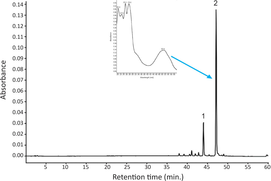
Figure 3.
The UV chromatogram of the extract from mycelium of Fusarium oxysporum f. sp. tulipae grown in tulip gum solution.
The UV spectrum taken from PDA detector of dominant compound 2 exhibited λmax at 201.8, 227.6, 251.2, 274.9, 336sh and 507,8 nm (Figure 3), and m/z value of 381 in negative mode [M-H]-. These values were similar to those obtained previously for bikaverin (6,11-dihydroxy-3-methoxy-1-methylbenzo[b]xantene-7,10,12-trione) the naphthoquinone pigment found in the wild Fusarium oxysporum LCP531 strain (Lebeau et al., 2019).
Compound 1 exhibited λmax at 201.5, 227.5, 251.4, 275.2, 336sh and 508.2 nm and m/z value of 367 in negative mode [M-H]-. These values were quite similar to compound 2 and were identical to those obtained previously for norbikaverin (6,10,11-trihydroxy-3-methoxy-1-methylbenzo[b] xantene-7,8,12-trione] (Lebeau et al., 2019).
The other minor peaks, the peak area of which made up less than 10% of total peak area were based on absorption spectra estimated as bikaverin related compounds, but their structures were not confirmed in this study. Presumably these were yellow naphthoquinone pigments (absorption maxima at 441 nm) reported previously (Lebeau et al., 2019).
Both bikaverin and norbikaverin content in the mycelium of Fusarium oxysporum f. sp. tulipae were determined after 7 and 15 days of incubation (Table 1). After 7 days of incubation the concentration of bikaverin was 2.94 mg/100 mg mycelial dry matter and was nearly 4.5 higher than that of norbikaverin. Incubation of the solution for the next 8 days resulted in nearly doubled concentration of both naphthoquinones.
Table 1.
Bikaverin and norbikaverin content (mg/100 mg dry matter) in Fusarium oxysporum f. sp. tulipae mycelium grown with tulip gum solution.
| Sample | Norbikaverin (Rt = 44.02) | Bikaverin (Rt = 47.15) | Total |
|---|---|---|---|
| Mycelium after 7 days | 0.64 ± 0.06 | 2.94 ± 0.26 | 3.58 ± 0.32 |
| Mycelium after 15 days | 1.02 ± 0.11 | 4.62 ± 0.55 | 5.64 ± 0.62 |
DISCUSSION
Fungal secondary metabolites display diverse biological activities. They may act as phytotoxins causing damages to plants, have important roles in other interactions with other plants or may exhibit antimicrobial activities and be involved in competition for survival with other microbes. Bikaverin, the reddish pigment, is produced by different fungal species, especially those belonging to the genus Fusarium. This compound shows antibiotic activity against certain fungi and protozoa (Balan et al., 1970; Son et al., 2008), nematocidal activity (Kwon et al., 2007) and anti-cancer activity (Fuska et al., 1975; Henderson et al., 1977; Limon et al., 2010), but its biological function in Fusarium remains unknown (Limon et al., 2010; Chełkowski et al., 1992).
It was reported that the synthesis of bikaverin is regulated by the culture conditions, such as carbon and nitrogen sources, calcium content, pH and many other factors (Giordano et al., 1999; Wiemann et al., 2009; Rodriguez-Ortiz et al., 2010). For example, in Fusarium fujikuroi fungus the synthesis of bikaverin was induced by nitrogen starvation and acidic pH, and it was favoured by aeration, and sulphate and phosphate starvation (Limon et al., 2010).
Species of genus Fusarium produced also other red coloured naphthoquinone metabolites (Lebeau et al., 2019; Medentsev et al., 2006; Brimble et al., 1999).
Poly- or oligosaccharides are the most well studied signal molecules in elicitors signal transduction. Many elicitors, such as chitin, chitosan, xyloglucans, β-glucans and their fragments, oligogalacturonides, laminarin, exhibit elicitor activity across different plant species and highly induce phytoalexins in plants, suggesting that different plants possess some common receptors to sense these signals (Zhao et al., 2005). One oligosaccharide elicitor can be recognized by several plants through their receptors with similar binding properties (Okada et al., 2002). Dass and Ramawat (2009) showed that gum arabic obtained from Acacia senegal (arabinogalactan-proteins) and mesquite gum obtained from Prosopis cineraria (arabinogalactan-proteins) evidently increased guggulsterone production in cell cultures of Commiphora weightii. Water-extracted mycelial polysaccharide prepared from the endophytic fungus Fusarium oxysporum Dzf17 isolated from the rhizomes of Discorea zingiberensis evidently increased diosgenin accumulation in cell suspension culture of D. zingiberensis (Li et al., 2011). Chitosan, pectin and alginate enhanced production of anthocyanin in Vitis vinifera cell suspension cultures (Cai et al., 2012). In Cocos nucifera cell culture treatment with chitosan enhanced production of different secondary metabolites, e.g., p-hydroxbenzoic acid, pcoumaric acid and ferulic acid (Chakraborty et al., 2009). Chitosan/chitin and pectin were effective in inducing of anthraquinone synthesis in cultured cells of Morinda citrifolia (Doernenburg, Knorr, 1994). Gum arabic significantly promoted accumulation of phenolic acids, particularly 3-0-glucosyl-resveratrol, in Vitis vinifera cultures, as well in the culture medium (Cai et al., 2012). Letarte et al. (2006) showed that arabinogalactan-protein from gum arabic increased microspore viability and induced embryogenesis in the microspore culture of wheat (Triticum aestivum), and finally obtained regenerated green plants. Arabinogalactan-protein from cashew nut (Anacardium occidentale) gum greatly stimulated somatic embryogenesis in carrot culture and enhanced of the conversation rate of somatic embryos into plantlets (Pereira-Netto et al., 2007).
The induction of red colour pigment in mycelium of Fusarium oxysporum f. sp. tulipae by tulip gums was previously reported, but its chemical structure was not confirmed (Saniewski et al., 2011). In present study the red colour pigment was identified to be a mixture of bikaverin and norbikaverin. Their concentration in mycelium was quite high, which may suggest that this process can be applied in pigment production for industrial purposes. However, it is interesting to mention that tulip gums induced pigment production exclusively by Fusarium oxysporum pathogenic for tulip bulbs. This gum did not elicit the red pigment formation through mycelium of F. oxysporum f. sp. callistephi, F. oxysporum f. sp. dianthi and F. oxysporum f. sp. narcissi (Saniewski et al., 2011). These pathogens are not pathogenic for tulip bulbs and tulip gums did not stimulate linear mycelium growth of these pathogens on Czapek-Dox-Agar (CzDA) medium (Saniewska, 2002a).
CONCLUSIONS
Infection of tulip bulbs by Fusarium oxysporum f. sp. tulipae induced gum formation, and the main constituent of these gums was previously identified as glucuronoarabinoxylan. Tulip gums elicited red pigments formation in Fusarium oxysporum f. sp. tulipae, identified as bikaverin and norbikaverin. Further studies are needed to confirm positive effect of glucuronoarabinoxylan isolated from tulip gums on the bikaverin and norbikaverin formation.