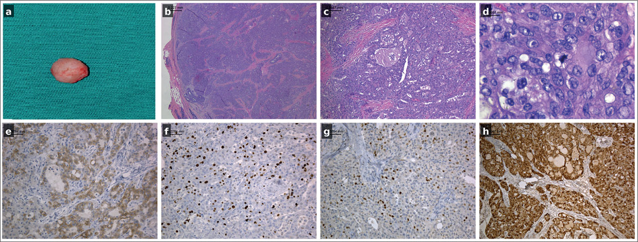
Figure 1.
Gross, histopathological, and immunohistochemical features of secretory breast carcinoma in a six-year-old girl: (a) Gross specimen of the excised tumor, approximately 10 × 10 mm, well-circumscribed and lobulated; (b) Lobular tumor architecture with a clearly visible pseudocapsule (HE, ×25); (c) Microcystic, cribriform, and tubular growth patterns with intraluminal secretory material (HE, ×100); (d) High mitotic activity, reflecting the proliferative potential of the tumor (HE, ×1000); (e) Focal membranous/cytoplasmic CD117 positivity (×200); (f) Ki-67 nuclear positivity in approximately 20% of tumor cells (×200); (g) Rare scattered p63-positive nuclei (<5% of tumor cells) (×200); (h) Strong and diffuse nuclear/cytoplasmic S-100 immunopositivity (×100).
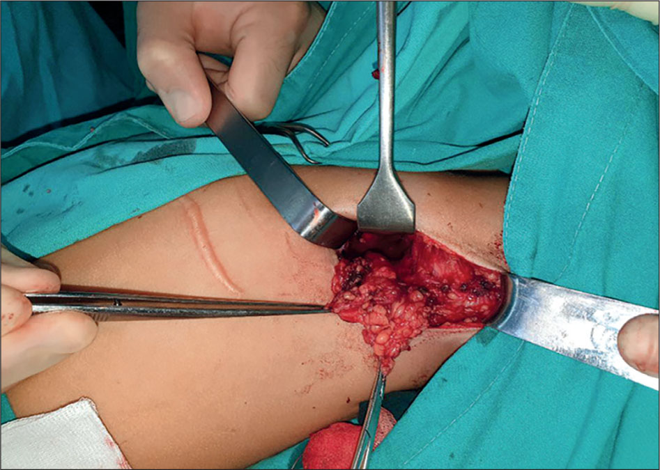
Figure 2.
Intraoperative photograph during axillary lymph node dissection performed at six-month follow-up due to two ultrasonographically suspicious nodes (22 × 18 mm and 6 × 2 mm), which were subsequently confirmed to be histologically benign.
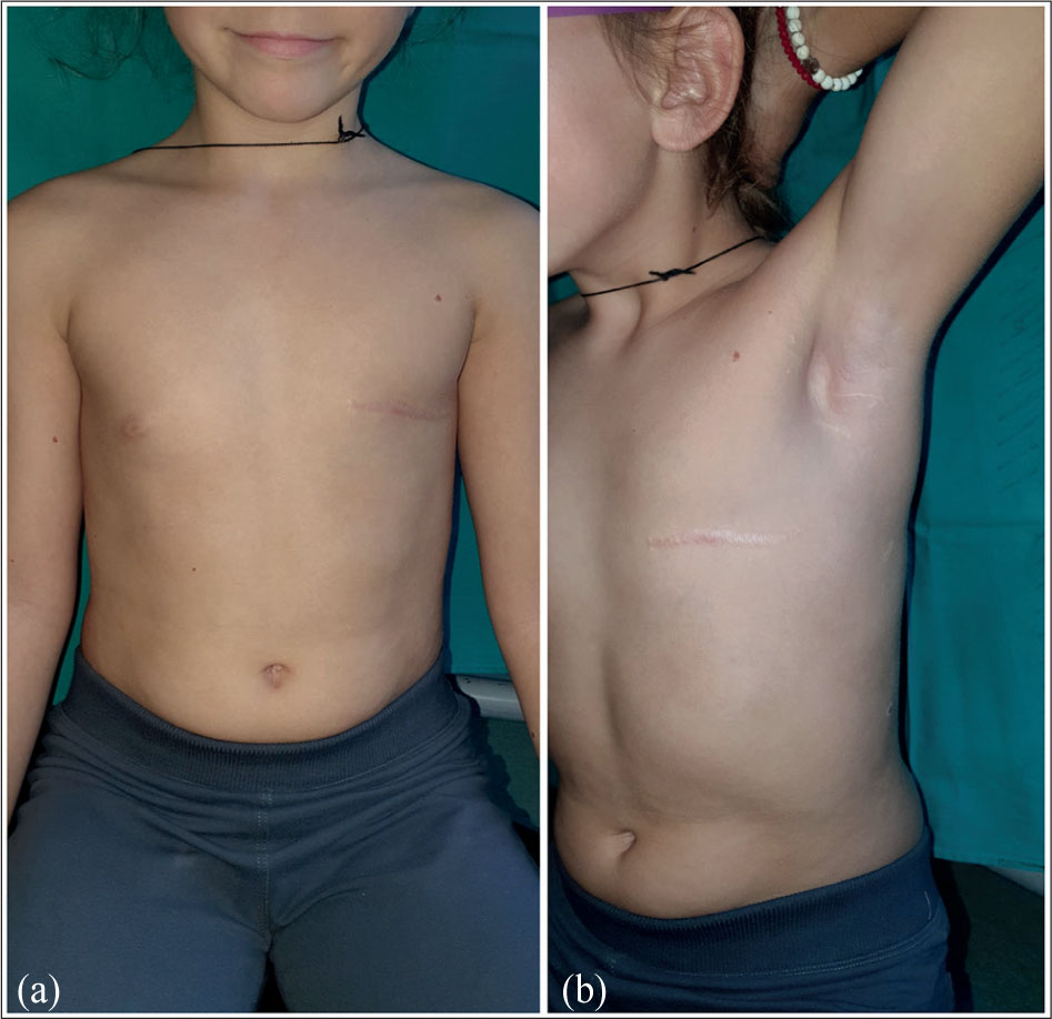
Figure 3.
Long-term postoperative follow-up of the patient: (a) Frontal view demonstrating good cosmetic outcome after mastectomy; (b) Slightly angled frontal view, confirming a well-healed scar and symmetrical chest contour.