
1. Introduction
Resistance training improves musculoskeletal strength, movement efficiency, and injury resilience, supporting both clinical rehabilitation and athletic performance (Pedersen et al., 2013; Winett & Carpinelli, 2001). Upper-body pulling exercises are widely prescribed to develop the posterior shoulder and upper-back musculature, making an understanding of exercise variation practically relevant.
Pulling exercises differ by movement plane. Horizontal rowing involves pulling resistance toward the torso in the transverse plane, whereas vertical pulling (e.g., lat pull-down, LPD) occurs in the frontal plane (Lusk et al., 2010). Although both recruit the latissimus dorsi (LD), trapezius, rhomboids, deltoid, erector spinae, and elbow flexors, relative muscle contributions may vary with joint orientation, grip width, and forearm position (Hewit et al., 2018). Consequently, findings from vertical pulling exercises may not generalize to horizontal rowing.
Surface electromyography (sEMG) provides an indirect estimate of neuromuscular activity through changes in signal amplitude. Most EMG investigations of grip width and forearm orientation have focused on the LPD, with inconsistent effects on sEMG amplitude and little influence on maximal strength (Lusk et al., 2010; Signorile et al., 2002; Sperandei et al., 2009). Fewer studies have examined horizontal rowing, and those that have often used submaximal or isometric conditions (Fenwick et al., 2009; García-Jaén et al., 2021; Vasconcelos et al., 2023). EMG data collected during dynamic rowing at loads exceeding 75% of one-repetition maximum (1RM) remain limited, constraining translation to strength-oriented training contexts (Schoenfeld et al., 2014).
The purpose of this study is to examine the effects of grip width and forearm orientation on maximal strength and sEMG amplitude during the barbell incline prone row (IPR) performed at a high relative load. Because horizontal rowing exercises are frequently prescribed in both rehabilitation and strength training, understanding whether simple grip modifications alter maximal load capacity and sEMG amplitude has practical implications for exercise prescription. Despite their widespread use, these variables remain poorly characterized during heavy dynamic rowing. The present findings extend prior EMG research by evaluating these variables under conditions more representative of maximal strength training, while also highlighting the constraints inherent to such analyses. It was hypothesized that grip width and forearm orientation would independently influence sEMG amplitude patterns, and that estimated 1RM would differ between grip-width conditions.
2. Material and methods
Participants: An a priori sample size estimate was calculated using G*Power (version 3.1.9.7) for a 2 × 2 repeated-measures ANOVA design. Based on a moderate effect size (f = 0.25), an alpha level of 0.05, and desired statistical power of 0.80, a total sample size of 24 participants were estimated. Twenty-three resistance-trained individuals (training experience: 5.7 ± 2.9 years; age: 23.3 ± 4.3 years; height: 176.3 ± 8.9 cm; body mass: 81.6 ± 18.3 kg; biacromial distance: 39.3 ± 5.0 cm; resting heart rate: 74.6 ± 15.3 bpm) ultimately completed repetition maximum testing. Participants were eligible if they were between 18 and 45 years of age and reported a minimum of 1 year of consistent resistance training experience, defined as an average of at least two training sessions per week. Exclusion criteria included a significant upper-extremity musculoskeletal injury within the previous 6 months requiring cessation of training, known neurological disorders, diabetes, or any medical condition deemed unsafe for maximal resistance exercise based on American College of Sports Medicine guidelines and PAR-Q screening. All participants provided written informed consent prior to participation. The study protocol was approved by the University of Findlay Institutional Review Board (Approval #1853) and conducted in accordance with the Declaration of Helsinki. To minimize acute fatigue and variability between sessions, participants were instructed to abstain from vigorous physical activity, including upper-body resistance training, for at least 48 h prior to each testing session (Sperandei et al., 2009). At each visit, participants completed a brief subjective readiness questionnaire addressing sleep quality, hydration, nutrition, and recent training. Participants were also instructed not to practice any of the IPR variations outside of the laboratory sessions.
3. Experimental design
A within-subject, repeated-measures design was employed to examine the effects of grip width and forearm orientation on estimated 1RM and sEMG activity during the barbell IPR. Signals were sampled at 2,048 Hz. Four exercise variations were tested: close-supinated (CS), close-pronated (CP), wide-supinated (WS), wide-pronated (WP). Grip width was individualized using each participant’s biacromial distance (BAD), measured as the linear distance between the most lateral aspects of the acromion processes. Close and wide grip widths were defined as 1.33 × and 1.66 × BAD, respectively, consistent with prior literature and modified for barbell-based pulling movements (Andersen et al., 2014; Haff & Triplett, 2016; Sperandei et al., 2009). Grip positions were clearly marked on the barbell using colored tape to ensure consistency across sessions. Participants completed two experimental sessions separated by 2–7 days. Session one was dedicated to determining estimated 1RM for each grip variation. Session two involved sEMG data collection while performing each variation at 85% of its respective 1RM. The order of grip conditions was randomized for each participant and maintained across sessions to reduce learning and sequencing effects.
3.1. Session one
Following completion of consent and readiness screening, baseline anthropometric and physiological measurements were collected, including height and body mass (Health O Meter, Illinois, USA), resting heart rate (Mibest Pulse Pro, Beijing, China), and blood pressure (Omeron Healthcare, Kyoto, Japan). BAD was measured by a single investigator, who palpated and marked the acromion process for each shoulder before measuring the distance using a tape measure (Parul et al., 2019). This investigator remained consistent across all participants and sessions to reduce measurement variability. Participants then completed a standardized education and familiarization period. Proper IPR technique was demonstrated, and standardized verbal instructions were provided: participants were instructed to “pull the bar toward the chest and return to full elbow extension while maintaining contact with the bench and matching the metronome tempo.” Participants practiced each variation for 2–5 min until proper range of motion, scapular control, and tempo compliance were visually confirmed. The warm-up protocol consisted of 8 min of arm ergometry at 60–80 RPM (rate of perceived exertion 3–5 on the modified Borg scale), followed by a one-minute rest and ten repetitions of each IPR variation using an unloaded barbell. Upon successful completion of the warm-up without adverse symptoms, participants progressed to strength testing. Variation order was determined via random number generation to reduce learning effect impacts. Estimated 1RM testing was conducted and calculated from a 5RM using the National Strength and Conditioning Association Training Load Chart (Haff & Triplett, 2016). Each condition was completed within three to five attempts, with 2–3 min of rest between sets. A metronome set at 60 beats per minute enforced a standardized tempo (1 s raising, 1 s lowering). Testing was terminated upon technical failure, defined as deviation from prescribed tempo, loss of bench contact, or incomplete range of motion. Estimated 1RM values were calculated based on Haff and Triplett’s (2016) 1RM estimation table (Weight divided by 0.87) and used to determine 85% 1RM loads for session two.
3.2. Session two
Participants returned for sEMG testing after maintaining at least 48 h without upper-body resistance training. Upon arrival, readiness screening was repeated to ensure consistency in recovery status between sessions. Skin preparation included shaving (if necessary), cleansing with isopropyl alcohol, and light abrasion until mild erythema was observed to reduce skin impedance. Disposable Noraxon Ag/AgCl electrodes (4 × 2.2 cm, 2 cm inter-electrode spacing; Noraxon, Scottsdale, USA) were placed on the dominant limb. Electrode placement was based on SENIAM guidelines (Hermens et al., 2000). Ten muscles were assessed: upper trapezius (UT), middle trapezius (MT), lower trapezius (LT), LD, pectoralis major (PM), middle deltoid (MD), posterior deltoid (PD), biceps brachii (BB), brachioradialis (BR), and triceps brachii (TB). The infraspinatus was excluded due to known limitations in surface EMG validity for this muscle (Johnson et al., 2011). Electrode impedance and baseline signal quality were visually inspected using Noraxon MR 4.0 software (Noraxon, Scottsdale, USA) prior to data collection. Trials were monitored in real time to identify excessive noise or signal dropout. Despite these precautions, occasional electrode detachment occurred during high-load trials, resulting in reduced sample sizes for select muscles. Only trials with clear, uninterrupted signals were retained for analysis.
3.3. Maximal voluntary isometric contractions (MVIC)
MVIC were collected for normalization purposes following established recommendations (Konrad, 2005). Two 5 s MVIC trials were recorded for each muscle with 30 s of rest between efforts. Resistance was applied using straps, cables, or manual stabilization depending on muscle group. Strong verbal encouragement was provided during each trial to ensure maximal effort. Participants were also coached extensively to perform maximally. While some inter-individual variability in MVIC performance is acknowledged, normalization procedures were applied consistently across all conditions, and the repeated-measures design minimized the influence of between-subject variability on within-condition comparisons.
3.4. sEMG trials
Following a warm-up identical to session one, participants performed each IPR variation for two sets of three repetitions at 85% of the previously determined 1RM. Order of variations was randomized via random number generation. A metronome maintained movement tempo, and technique was continuously monitored. Rest periods of 2.5 min were provided between sets to minimize fatigue accumulation. Raw sEMG signals were band-pass filtered (10–500 Hz), full-wave rectified, and smoothed using a root mean square algorithm. Signals were normalized to %MVIC for each muscle. To reduce start-up and fatigue artifacts, the first and last repetitions of each set were excluded from analysis (Jakobsen et al., 2012). The mean amplitude of the middle repetition from each set was averaged to represent sEMG amplitude for each condition.
3.5. Statistical analysis
Estimated 1RM values and normalized sEMG amplitudes (%MVIC) were analyzed using separate 2 × 2 factorial repeated-measures ANOVAs with grip width (close vs wide) and forearm orientation (supinated vs pronated) as within-subject factors. Due to signal loss and quality control exclusions, sample size varied across muscles for sEMG analyses. Only participants with complete data for a given muscle across all four conditions were included in that muscle’s analysis. Descriptive statistics are presented as mean value ± standard deviation. All analyses were conducted using JASP software (version 0.19.3; JASP Team, 2024). Statistical significance was set a priori at p < 0.05. Effect sizes were reported as partial eta squared (η²ₚ) for ANOVA outcomes, interpreted according to conventional thresholds.
4. Results
4.1. 1RM
Grip width significantly influenced estimated 1RM (p < 0.001, η²ₚ = 0.655), with higher loads achieved in close-grip conditions. Forearm orientation and interaction terms were not significant (Table 1).
4.2 sEMG
4.2.1. Trunk muscles
MT sEMG amplitude was influenced by grip width (p = 0.044, η²ₚ = 0.218) and forearm orientation (p = 0.002, η²ₚ = 0.449), with greater amplitude during wide and pronated grip positions. LD amplitude was influenced by forearm orientation (p = 0.040, η²ₚ = 0.238), greater in supinated grips. UT and LT showed no significant effects. Trunk sEMG amplitude results are presented in Table 2 and Figure 1.
Table 2
2 × 2 factorial repeated measures Anova – Trunk muscles sEMG amplitude
| Muscle | Effect | DF | F value | p value | Partial Eta Squared |
|---|---|---|---|---|---|
| UT | Width | 1.15 | 0.218 | 0.287 | 0.075 |
| UT | Orientation | 1.15 | 0.875 | 0.365 | 0.055 |
| UT | Width:Orientation | 1.15 | 3.487 | 0.082 | 0.189 |
| MT | Width | 1.17 | 4.738 | 0.044 | 0.218 |
| MT | Orientation | 1.17 | 13.863 | 0.002 | 0.449 |
| MT | Width:Orientation | 1.17 | 2.233 | 0.153 | 0.116 |
| LT | Width | 1.15 | 0.822 | 0.379 | 0.052 |
| LT | Orientation | 1.15 | 3.921 | 0.066 | 0.207 |
| LT | Width:Orientation | 1.15 | 2.321 | 0.148 | 0.134 |
| LD | Width | 1.16 | 0.896 | 0.358 | 0.053 |
| LD | Orientation | 1.16 | 4.986 | 0.040 | 0.238 |
| Ld | Width:Orientation | 1.16 | 0.865 | 0.366 | 0.051 |
UT = Upper Trapezius, MT = Middle Trapezius, LT = Lower Trapezius, LD = Latissimus Dorsi. N = 16(UT), 18(MT), 16(LT), 17(LD).
Source: Authors’ contribution.
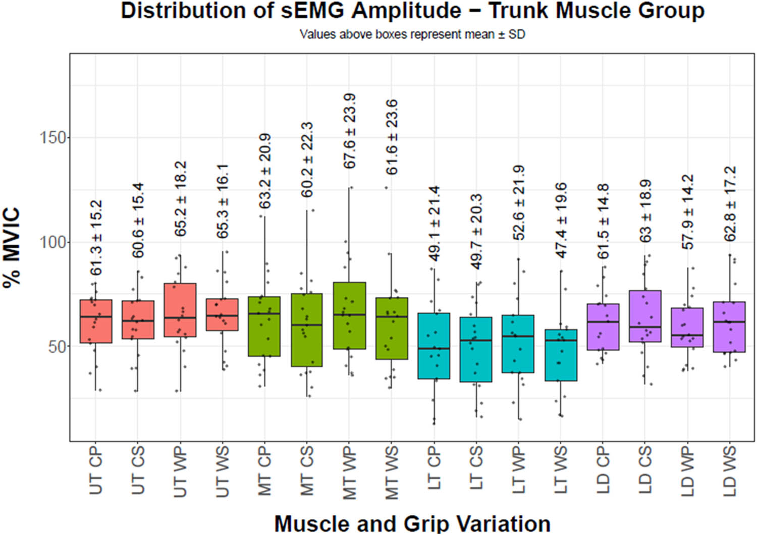
Figure 1
sEMG amplitudes of trunk muscles. Boxes represent the interquartile range; center line indicates the median; whiskers represent the range; points represent individual participants. Values above each box indicate mean value ± SD. The vertical axis reports the percentage of MVIC, while the horizontal axis reports the must group (UT = Upper Trapezius, MT = Middle Trapezius, LT = Lower Trapezius, LD = Latissimus Dorsi) along with grip position (CP = Close pronated, CS = Close supinated, WP = Wide pronated, WS = Wide supinated)
Source: Authors’ contribution.
4.2.2. Shoulder muscles
Forearm orientation influenced MD (p = 0.027, η²ₚ = 0.287) and PD (p = 0.049, η²ₚ = 0.198), with greater amplitude during pronated conditions. PM showed no significant effects. Shoulder sEMG amplitude results are presented in Table 3 and Figure 2.
Table 3
2 × 2 factorial repeated measures ANOVA – shoulder muscles sEMG amplitude
| Muscle | Effect | DF | F value | p value | Partial Eta Squared |
|---|---|---|---|---|---|
| MD | Width | 1.15 | 0.541 | 0.474 | 0.035 |
| MD | Orientation | 1.15 | 6.049 | 0.027 | 0.287 |
| MD | Width:Orientation | 1.15 | 3.357 | 0.087 | 0.183 |
| PD | Width | 1.18 | 1.893 | 0.186 | 0.095 |
| PD | Orientation | 1.18 | 4.436 | 0.049 | 0.198 |
| PD | Width:Orientation | 1.18 | 1.420 | 0.249 | 0.073 |
| PM | Width | 1.13 | 0.230 | 0.640 | 0.017 |
| PM | Orientation | 1.13 | 4.321 | 0.058 | 0.249 |
| PM | Width:Orientation | 1.13 | 0.057 | 0.815 | 0.004 |
MD = Middle Deltoid, PD = Posterior Deltoid, PM = Pectoralis Major. N = 16 (MD), 19 (PD), 14 (PM).
Source: Authors’ contribution.
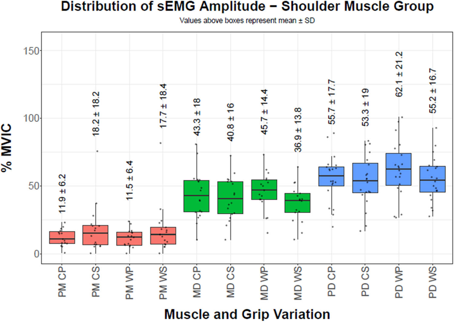
Figure 2
sEMG amplitudes of shoulder muscles. Boxes represent the interquartile range; center line indicates the median; whiskers represent the range; points represent individual participants. Values above each box indicate mean value ± SD. The vertical axis reports the percentage of MVIC, while the horizontal axis reports the muscle group (MD = Middle Deltoid, PD = Posterior Deltoid, PM = Pectoralis Major) along with grip position (CP = Close pronated, CS = Close supinated, WP = Wide pronated, WS = Wide supinated)
Source: Authors’ contribution.
4.2.3. Elbow muscles
BB amplitude was influenced by grip width (p = 0.001, η²ₚ = 0.487) and forearm orientation (p = 0.006, η²ₚ = 0.386), greater in wide and supinated grips. BR (p = 0.040, η²ₚ = 0.237) and TB (p = 0.024, η²ₚ = 0.295) were influenced by forearm orientation, greater in pronated grips. Elbow sEMG amplitude results are shown in Table 4 and Figure 3.
Table 4
2 × 2 factorial repeated measures ANOVA – elbow muscles sEMG amplitude
| Muscle | Effect | DF | F value | p value | Partial Eta Squared |
|---|---|---|---|---|---|
| BB | Width | 1.16 | 15.217 | 0.001 | 0.487 |
| BB | Orientation | 1.16 | 10.057 | 0.006 | 0.386 |
| BB | Width:Orientation | 1.16 | 0.646 | 0.433 | 0.039 |
| BR | Width | 1.16 | 3.326 | 0.087 | 0.172 |
| BR | Orientation | 1.16 | 4.970 | 0.040 | 0.237 |
| BR | Width:Orientation | 1.16 | 2.161 | 0.161 | 0.119 |
| TB | Width | 1.15 | 0.004 | 0.952 | 0.000 |
| TB | Orientation | 1.15 | 6.265 | 0.024 | 0.295 |
| TB | Width:Orientation | 1.15 | 1.506 | 0.239 | 0.091 |
BB = Biceps Brachii, BR = Brachioradialis, TB = Triceps Brachii, N = 17 (BB), 17 (BR), 16 (TB).
Source: Authors’ contribution.
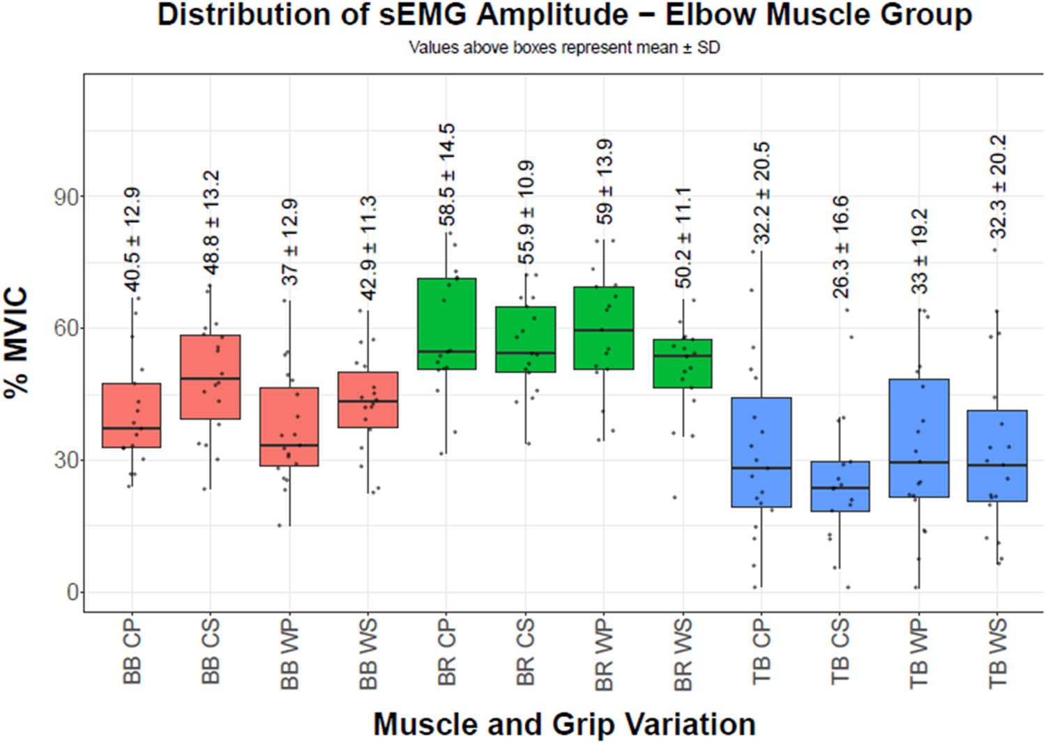
Figure 3
sEMG amplitudes of elbow muscles. Boxes represent the interquartile range; center line indicates the median; whiskers represent the range; points represent individual participants. Values above each box indicate mean value ± SD. The vertical axis reports the percentage of MVIC, while the horizontal axis reports the muscle group (BB = Biceps Brachii, BR = Brachioradialis, TB = Triceps Brachii) along with grip position (CP = Close pronated, CS = Close supinated, WP = Wide pronated, WS = Wide supinated)
Source: Authors’ contribution.
5. Discussion
The primary strength-related finding was a significant main effect of grip width on estimated 1RM, with narrower grips allowing greater loads to be lifted compared to wider grips. Although the absolute differences in estimated 1RM across grip conditions were modest, the effect size associated with grip width was large. In contrast, forearm orientation did not significantly alter estimated 1RM, and no interaction between grip width and forearm orientation was observed. From a biomechanical perspective, grip width and forearm orientation may alter the length tension relationship of the tested trunk, shoulder, and elbow musculature. These findings suggest the importance of accounting for grip-specific load capacity rather than assuming that all horizontal rowing variations have equivalent load capacities.
Consistent with the study hypotheses, forearm orientation exerted a broader influence on sEMG amplitude than grip width. Pronated grips were associated with greater sEMG amplitude of the MT, MD, PD, and BR, whereas supinated grips resulted in greater amplitude of the LD and BB. Grip width influenced amplitude primarily in the MT and BB. The absence of significant interaction effects suggests that grip width and forearm orientation independently influence sEMG amplitude rather than interacting synergistically. These findings partially align with prior EMG investigations of vertical pulling exercises while also diverging in meaningful ways. Studies examining the LPD have reported inconsistent effects of grip width and forearm orientation on sEMG amplitude, often without corresponding differences in maximal strength (Lusk et al., 2010; Signorile et al., 2002; Sperandei et al., 2009). Theoretically, changes in forearm orientation may alter the length tension relationship of these muscles. Increased sEMG amplitude of the MT and PD during pronated grips is consistent with previous studies comparing rowing exercises performed at different shoulder abduction angles (García-Jaén et al., 2021; Vasconcelos et al., 2023). Similarly, greater BB amplitude with supination aligns with established elbow flexion biomechanics (Kleiber et al., 2015).
Limitations in this study should be acknowledged. Although a a priori power analysis was conducted, the final sample size fell slightly below the estimated requirement, and sEMG analyses were further limited by occasional electrode detachment during high-load trials. Given the number of ANOVAs performed, findings near the threshold for statistical significance should be interpreted cautiously. Effect sizes are therefore reported to aid interpretation. While standardized electrode placement, normalization procedures, and a repeated-measures design were employed to mitigate variability, measurement noise remains an inherent limitation of surface EMG, particularly during high-intensity free-weight movements.
The practical implications of this study should be considered within the scope of its methodological constraints. The findings suggest that grip width influences maximal load capacity during the IPR and may be considered when prescribing load-based training variables. Forearm orientation appears to influence sEMG amplitude patterns under high load, which may be relevant when programming horizontal pulling exercises to emphasize specific muscle groups. Future research should prioritize larger sample sizes, improved EMG signal stability, and the integration of biomechanical measures such as joint angles and moment arms. Longitudinal studies examining training adaptations to specific grip configurations, as well as investigations involving clinical or novice populations, would further enhance the applicability of horizontal pulling research.
In conclusion, grip width influences maximal strength capacity during the barbell IPR, with narrower grips allowing greater load expression. Forearm orientation independently affects sEMG amplitude patterns across multiple upper-body pulling muscles when exercises are performed at high relative loads. While these effects are modest, they appear systematic and may have implications for load prescription and exercise selection in strength-oriented contexts.
Funding information
Authors state that no funding information.
Author contributions
AF: Conceptualization, Methodology, Investigation, Data Curation, Formal Analysis, Project Administration, Supervision, Writing – Original Draft, Writing – Review & Editing. HD: Methodology, Investigation, Data Curation, Formal Analysis, Writing – Review & Editing. SM: Conceptualization, Methodology, Formal Analysis, Writing – Review & Editing. DV: Conceptualization, Methodology. SB: Conceptualization, Methodology, Investigation, Data Curation, Supervision, Writing – Original Draft, Writing – Review & Editing.
Conflict of interest statement
Authors state no conflicts of interest.