
1. Introduction
In infants at risk of atypical development, neurodevelopmental therapy should be initiated as early as possible to normalize disturbed postural and movement patterns. Early intervention is initiated for a variety of reasons, including immaturity or damage to the central nervous system (CNS), preterm birth, atypical muscle tone, postural asymmetry, delayed movement patterns, and developmental defects (Zanon et al., 2019). Therapeutic interventions rely mainly on early and accurate diagnosis as well as established and validated therapeutic methods that offer the potential for reducing developmental deficits (Bartík et al., 2025). The most popular methods include Vojta therapy and the Bobath concept (neurodevelopmental treatment), supplemented by sensorimotor integration (Hohendahl et al., 2022; Pathak et al., 2021). The duration of therapy is determined individually for each patient. Therapy is continued until age-appropriate movement and postural patterns are achieved. Vojta therapy enables children with neurodevelopmental deficits to achieve spontaneous motor activity (SMA) patterns that are as close as possible to ideal movement patterns (IMPs) (De-La-Barrera-Aranda et al., 2021). This occurs as a result of brain neuroplasticity, which, through compensatory processes, enables the formation of new functional connections within the CNS, allowing for the partial or complete restoration of lost functions and skills, as well as the acquisition of previously unattained movement patterns (Ungureanu et al., 2022). Vojta therapy promotes the activation of neurons in the brain and induces changes in neuron structure and functions by forming new connections, thereby enabling the child to acquire age-appropriate locomotor patterns (Parau et al., 2024).
Numerous studies have shown that the therapeutic approach developed by Václav Vojta significantly contributes to improvements in motor skills and postural control in infants (Zafeiriou, 2004; Torró-Ferrero et al., 2022; Igual Blasco et al., 2023; Trafalska & Paprocka-Borowicz, 2025). Vojta therapy is based on eliciting global locomotor patterns that are activated through appropriate positioning and stimulation of specific areas on the patient’s body, referred to as trigger zones in this method (Vojta & Schweizer, 2009). Global to locomotor patterns, such as reflex creeping and reflex rolling, consist of partial movement patterns that can be elicited even in infants with a damaged or highly immature nervous system (Sánchez-González et al., 2024). Healthy infants use these movement patterns during SMA as part of the genetically programmed process of postural and motor ontogeny. Vojta therapy enables improvement of automatic body posture control through the activation of the deep muscles of the spine and trunk, promoting synergistic action of all muscle groups, including atrophied muscles, around the key joints of the limbs (Vojta, 1989; Parau et al., 2023). This approach facilitates access to three important components of movement generation, including automatic postural control, phasic movement, and the ability to overcome gravity, thus enabling the child to reduce motor deficits and achieve successive milestones in psychomotor development (Vojta, 1973; Sanz-Mengíbar et al., 2021). By stimulating the nervous system, the therapist may positively influence speech development, tongue motor function, and autonomic functions (Falahati Nezhad et al., 2023). In the Vojta method, daily therapeutic exercises are performed primarily by the parent under the guidance of a certified therapist. This task may be challenging for many parents due to the close emotional bond with their child (Qian et al., 2025). However, parent-led therapy reduces costs, eliminates the need for travel to a therapy center, and enables parents to increase the intensity of exercises without incurring additional costs (San-Martin-Gomez et al., 2026). In children, therapeutic progress and skill development are influenced by many factors. The importance of the type and course of delivery, as well as neonatal adaptation following different types of delivery, has been emphasized by many researchers (Yoshida et al., 2023). Some studies have suggested that delivery by cesarean section may be associated with delayed neuromotor development in infants (Polidano et al., 2017; Huang et al., 2023; Fritz et al., 2024) and may even be considered a potential predisposing factor for autism spectrum disorder (Curran et al., 2015). The prevalence of cesarean section deliveries is on the rise, including elective procedures without clear medical indications, which warrants further research into the psychomotor development of infants following various types of delivery (Boerma et al., 2018).
Therefore, the aim of this study is to assess the effects of Vojta therapy on central coordination disorders (CCDs), SMA, attainment of IMPs, reflexes, and postural asymmetry in infants, with respect to gender and the type of delivery.
2. Materials and methods
2.1. Participants
The study was conducted at the Center for Rehabilitation and Education in Szczytno (Poland), and it involved 197 infants who were referred for Vojta therapy by a rehabilitation physician. The study had a prospective design, and the data collected over the entire course of therapy were analyzed.
The study group consisted of 107 male subjects aged 4.15 ± 2.18 months and 90 female subjects aged 3.84 ± 2.12 months who underwent baseline assessments. A total of 149 infants were included in the therapy, and 139 of them completed the therapy (77 males and 62 females). The parents of 10 children withdrew from therapy during the course of treatment (Figure 1).
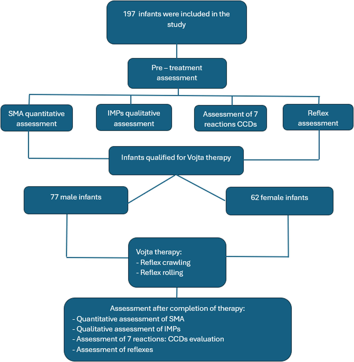
Figure 1
Flow diagram of the research protocol
Source: Authors’ contribution.
The inclusion criteria were: history of complications during pregnancy and delivery, preterm birth, low Apgar score at 1 min of life, low birth weight, postural asymmetry (at 7 weeks of age), atypical muscle tone, delayed psychomotor development, and non-age-appropriate involuntary reflexes. All the subjects were healthy and without any signs of infection during the study. Children with genetic disorders were excluded from the study.
The characteristics of the study group are presented in Table 1. According to the information provided by the parents, the studied infants were born between the 24th and 42nd weeks of pregnancy. The mean gestational age was 37.53 ± 3.47 weeks in males and 38.4 ± 2.73 weeks in females. The difference favoring girls approached statistical significance (p = 0.055). The remaining parameters did not differ significantly between the genders. Birth weight was 3118.51 ± 859.2 g in males and 3078.40 ± 783.2 g in females. Apgar scores at 1 min were high, averaging 8.9 ± 2.0 in males and 9.3 ± 1.4 in females (Table 1).
Table 1
Characteristics of the studied male and female infants
| Variable | Male N = 107 | Female N = 90 | t | p | ||||||
|---|---|---|---|---|---|---|---|---|---|---|
| Mean value | SD | Min | Max | Mean value | SD | Min | Max | |||
| Gestational age (weeks) | 37.53 | 3.5 | 24 | 42 | 38.4 | 2.7 | 26 | 42 | −1.92 | 0.055 |
| Birth weight (g) | 3118.5 | 859.2 | 666 | 4900 | 3078.4 | 783.2 | 660 | 4450 | 0.34 | 0.734 |
| Apgar score* (points) | 8.86 | 2.0 | 1 | 10 | 9.29 | 1.43 | 2 | 10 | −1.70 | 0.091 |
| Age (months) | 4.15 | 2.2 | 1 | 16 | 3.84 | 2.1 | 1 | 11 | 0.99 | 0.324 |
Notes: *- at 1 min of life.
Source: Authors’ contribution.
2.2. Ethical statement
The study was conducted in accordance with the guidelines and policies of the Health Science Council and the Declaration of Helsinki. It was approved by the Ethics Committee of the Medical University of Poznań (179/2012). Each child’s parent received detailed information about the purpose of the study, potential risks, the study protocol, and the procedures performed during the study. All parents gave voluntary informed consent to participate in the study.
2.3. Procedures, data collection, and equipment
Before therapy initiation, parents were interviewed to obtain information on pregnancy and delivery course, maternal diseases during pregnancy, birth weight, and Apgar score. Subsequently, infants underwent a detailed assessment including SMA, IMPs, and seven postural reactions used to determine the severity of CCDs.
The SMA assessment was performed in supine and prone positions and focused on postural control, motor coordination, antigravity abilities, developmental milestones, body symmetry, segmental alignment, head and girdle positioning, limb activity, and movement fluency and variability. Quantitative assessment enabled determination of motor developmental age and identification of postural asymmetries.
The quality of IMPs was evaluated with emphasis on movement execution rather than mere occurrence. Qualitative assessment included movement organization, fluency, variability, complexity, precision, and age-appropriate motor performance, enabling early detection of subtle neurological dysfunctions.
Seven postural reactions were assessed according to the Václav Vojta method to classify the severity of CCDs. The examination followed Vojta’s standardized protocol and included: the Prechtl traction response (Vojta modification), Landau reaction, axillary suspension reaction, Vojta lateral tilting reaction, Collis horizontal suspension reaction (Vojta modification), Peiper–Isbert vertical suspension reaction, and Collis vertical suspension reaction (Vojta modification) (Vojta, 1973). Infants with abnormal responses were classified according to Vojta’s four-grade CCD scale.
Reflex development was assessed according to chronological or corrected age (in preterm infants), including palmar reflex and the Moro reflex, with consideration of age-appropriate occurrence, stereotypical responses, and reflex inhibition. Following assessment, Vojta therapy was initiated according to each infant’s individual clinical needs.
Vojta therapy was conducted by a therapist two to three times per week and by the parents four times daily at home. Various forms of reflex locomotion were used during therapy, including reflex creeping and reflex rolling (phases I–IV), performed in the supine and side-lying positions, as well as the first therapeutic position. Stimulation zones on the trunk and limbs were used during the exercises. All therapeutic components, including the sequence of exercises, positioning, and selection of stimulation zones, were adapted to the child’s individual needs and diagnosed problems. Depending on the therapeutic outcomes and the infant’s avoidance behaviors, the rehabilitation program was modified and progressed seamlessly to subsequent stages, enabling the acquisition of age-appropriate motor skills. The duration of therapy ranged from 1 to 33 months, depending on the child’s progress and normalization of postural and locomotor patterns. After completion of the therapeutic program, a follow-up assessment of psychomotor development was performed in the supine and prone positions in infants grouped by gender. The severity of CCDs and reflex development were evaluated. The results were compared to determine the overall effectiveness of Vojta therapy, its effects on postural reaction tests (CCD score), presented motor patterns, achievement of IMPs, and reflex development in infants.
2.4. Statistical analysis
The significance of differences between mean values was determined by Student’s t-test for dependent variables. A two-proportion test was used to determine differences in the prevalence of a given feature between groups. The results were processed statistically in Statistica PL v. 13.0 at a significance level of p < 0.05.
3. Results
The changes in CCD, SMA, and IMP scores after Vojta therapy are presented in Table 2. Vojta therapy led to a significant decrease in the CCD scores of both male (from 4.8 to 2.2 points) and female (from 4.7 to 1.8 points) subjects. With increasing age and duration of therapy, significant improvements were observed in both genders (p < 0.001 in all cases): SMA scores increased from 3.2 to 6.6 months in males and from 2.6 to 6.5 months in females, whereas IMP scores increased from 1.6 to 5.6 months in males and from 1.2 to 5.7 months in females, indicating progressive approximation to age-appropriate motor patterns.
Table 2
Changes in CCD, SMA, and IMP scores in infants who completed Vojta therapy
| Score | Male (n = 77) | Female (n = 62) | ||||||||||
|---|---|---|---|---|---|---|---|---|---|---|---|---|
| Before | After | Difference | Before | After | Difference | |||||||
| Mean value | SD | Mean value | SD | t | p | Mean value | SD | Mean value | SD | t | p | |
| CCD (points) | 4.78 | 2.09 | 2.23 | 2.39 | 7.04 | <0.001 | 4.66 | 2.22 | 1.77 | 2.31 | 7.09 | <0.001 |
| SMA (months) | 3.16 | 1.81 | 6.55 | 2.22 | 10.40 | <0.001 | 2.60 | 2.01 | 6.48 | 1.92 | 11.01 | <0.001 |
| IMPs (months) | 1.64 | 1.93 | 5.58 | 2.56 | 10.81 | <0.001 | 1.23 | 1.89 | 5.72 | 2.27 | 11.96 | <0.001 |
Notes: CCDs - central coordination disorders; SMA - spontaneous motor activity; IMPs - ideal movement patterns.
Source: Authors’ contribution.
The effects of Vojta therapy on postural reactions denoting CCDs in infants are presented in Table 3. Significant (p < 0.001 in the vast majority of cases) improvements in all seven postural reactions (increasing frequencies) were observed in both male and female subjects.
Table 3
Frequency of postural reactions before and after Vojta therapy in infants who completed therapy
| Test | Male (n = 77) | Female (n = 62) | Total (n = 139) | |
|---|---|---|---|---|
| Pull-to-sit (traction response) | Before | 22.1 (17) | 32.3 (20) | 26.6 (37) |
| After | 64.9 (50) | 80.6 (50) | 71.9 (100) | |
| p (difference) | <0.001 | <0.001 | <0.001 | |
| Landau reaction | Before | 24.7 (19) | 22.6 (14) | 23.7 (33) |
| After | 72.7 (56) | 79.0 (49) | 75.5 (105) | |
| p (difference) | <0.001 | <0.001 | <0.001 | |
| Axillary suspension test | Before | 29.9 (23) | 30.6 (19) | 30.2 (42) |
| After | 58.4 (45) | 61.3 (38) | 59.7 (83) | |
| p (difference) | <0.001 | 0.001 | <0.001 | |
| Vojta reaction | Before | 29.9 (23) | 38.7 (24) | 33.8 (47) |
| After | 74.0 (57) | 72.6 (45) | 73.4 (102) | |
| p (difference) | <0.001 | <0.001 | <0.001 | |
| Collis horizontal suspension test | Before | 50.6 (39) | 37.1 (23) | 44.6 (62) |
| After | 71.4 (55) | 74.2 (46) | 72.7 (101) | |
| p (difference) | 0.012 | <0.001 | <0.001 | |
| Peiper–Isbert suspension test | Before | 27.3 (21) | 35.5 (22) | 30.9 (43) |
| After | 62.3 (48) | 75.8 (47) | 68.3 (95) | |
| p (difference) | <0.001 | <0.001 | <0.001 | |
| Collis vertical suspension test | Before | 37.7 (29) | 37.1 (23) | 37.4 (52) |
| After | 72.7 (56) | 79.0 (49) | 75.5 (105) | |
| p (difference) | <0.001 | <0.001 | <0.001 | |
Notes: values are presented as percentages; numbers of infants are given in parentheses.
Source: Authors’ contribution.
The development of spontaneous motor skills and the attainment of IMPs in male infants born by vaginal delivery and cesarean section are presented in Table 4. A significant (p < 0.001) improvement in SMA scores was noted in both delivery groups; however, the final mean scores were slightly higher in males born by vaginal delivery (vaginal delivery: 7.4; cesarean section: 6.3), which may suggest more rapid development of locomotor functions. A significant (p < 0.001) improvement in IMPs was also observed in both groups after Vojta therapy, but males born by vaginal delivery exhibited slightly more mature movement patterns in the final assessment (vaginal delivery: 5.9; cesarean section: 5.4).
Table 4
Changes in SMA and IMP scores after Vojta therapy in male infants who completed therapy, according to the type of delivery
| Score | Vaginal delivery (n = 16) | Cesarean section (n = 24) | ||||||||||
|---|---|---|---|---|---|---|---|---|---|---|---|---|
| Before | After | Difference | Before | After | Difference | |||||||
| Mean value | SD | Mean value | SD | t | p | Mean value | SD | Mean value | SD | t | p | |
| SMA (months) | 3.84 | 2.23 | 7.38 | 2.00 | 4.72 | <0.001 | 2.84 | 1.88 | 6.25 | 2.45 | 5.25 | <0.001 |
| IMPs (months) | 2.41 | 1.89 | 5.91 | 2.19 | 4.84 | <0.001 | 1.42 | 1.89 | 5.40 | 2.61 | 6.06 | <0.001 |
Notes: SMA - spontaneous motor activity; IMPs - ideal movement patterns.
Source: Authors’ contribution.
The development of spontaneous motor skills and the achievement of IMPs in female infants born by vaginal delivery and cesarean section is presented in Table 5. A significant (p < 0.001) improvement in SMA and IMP scores was noted in both delivery groups. Similar to the studied males, the final mean scores were higher in females born by vaginal delivery (vaginal delivery: 7.2; cesarean section: 6.1). A significant improvement in IMP scores was also noted, and the mean score was higher in females born by vaginal delivery (6.5) than by cesarean section (4.9).
Table 5
Changes in SMA and IMP scores after Vojta therapy in female infants who completed therapy, according to the type of delivery
| Score | Vaginal delivery (n = 13) | Cesarean section (n = 17) | ||||||||||
|---|---|---|---|---|---|---|---|---|---|---|---|---|
| Before | After | Difference | Before | After | Difference | |||||||
| Mean value | SD | Mean value | SD | t | p | Mean value | SD | Mean value | SD | t | p | |
| SMA (months) | 2.73 | 2.15 | 7.15 | 2.00 | 5.44 | <0.001 | 2.65 | 2.12 | 6.06 | 2.22 | 4.58 | <0.001 |
| IMPs (months) | 1.38 | 1.89 | 6.46 | 1.98 | 6.67 | <0.001 | 1.24 | 1.95 | 4.88 | 2.69 | 4.52 | <0.001 |
Notes: SMA - spontaneous motor activity; IMPs - ideal movement patterns.
Source: Authors’ contribution.
The influence of the type of delivery on CCDs was also evaluated in infants who completed Vojta therapy. The CCD scores of male and female infants born by vaginal delivery and cesarean section are presented in Table 6. In the group of children born by vaginal delivery, the mean CCD score was 2.0 in males and 1.8 in females. No significant (p > 0.05) differences in CCD scores were observed between the genders.
Table 6
CCD scores in male and female infants who completed Vojta therapy, according to the type of delivery
| Type of delivery | Male | Female | Differences | |||||
|---|---|---|---|---|---|---|---|---|
| n | Mean value | SD | n | Mean value | SD | t | p | |
| Vaginal | 16 | 2.01 | 2.34 | 13 | 1.77 | 2.17 | 0.27 | ns |
| Cesarean section | 24 | 2.38 | 2.50 | 17 | 2.35 | 2.91 | 0.03 | ns |
| t (p) | ns | ns | ||||||
Source: Authors’ contribution.
Similar observations (p > 0.05) were made in the group of infants born by cesarean section. In this group, the mean CCD score was 2.38 for males and 2.35 for females, without significant (p > 0.05) differences between the genders. No significant (p > 0.05) differences in CCD scores were observed between infants categorized by the type of delivery. These results suggest that the type of delivery (vaginal vs cesarean section) had no significant influence on CCD scores after completion of Vojta therapy. In other words, the effectiveness of Vojta therapy in normalizing movement patterns was comparable regardless of the type of delivery.
The prevalence of three features – palmar grasp reflex, Moro reflex, and postural asymmetry – before and after completion of Vojta therapy is compared in Table 7. Before therapy, the palmar grasp reflex was observed in 48.2% of the infants (46.8% of males and 50.0% of females). After completion of therapy, the prevalence of the palmar grasp reflex decreased significantly (p < 0.001) and was observed in only 4.3% of the infants (6.5% of males and 1.6% of females). The Moro reflex was observed in 18.7% of the subjects before therapy and in none after therapy (0.0%), indicating a significant (p < 0.001) decrease. Before therapy, postural asymmetry was observed in 85.6% of subjects, more frequently in males (87.0% of males and 83.9% of females). The prevalence of postural asymmetry decreased significantly (p < 0.001) after therapy, to 27.3% (32.5% of males and 21.0% of females), indicating an improvement in postural symmetry, which is one of the primary goals of Vojta therapy.
Table 7
Prevalence of the palmar grasp reflex, Moro reflex, and postural asymmetry in infants before and after completion of Vojta therapy
| Feature | Male (n = 77) | Female (n = 62) | Total (n = 139) | |
|---|---|---|---|---|
| Palmar grasp reflex | Before | 46.8 (36) | 50.0 (31) | 48.2 (67) |
| After | 6.5 (5) | 1.6 (1) | 4.3 (6) | |
| p (difference) | <0.001 | <0.001 | <0.001 | |
| Moro reflex | Before | 18.2 (14) | 19.4 (12) | 18.7 (26) |
| After | 0.0 (0) | 0.0 (0) | 0.0 (0) | |
| p (difference) | <0.001 | <0.001 | <0.001 | |
| Postural asymmetry | Before | 87.0 (67) | 83.9 (52) | 85.6 (119) |
| After | 32.5 (25) | 21.0 (13) | 27.3 (38) | |
| p (difference) | <0.001 | <0.001 | <0.001 | |
Notes: values are presented as percentages; numbers of infants are given in parentheses.
Source: Authors’ contribution.
4. Discussion
The postural patterns and motor skills exhibited by infants in each developmental stage reflect the maturity and functional status of the CNS. Therefore, when a child fails to achieve age-appropriate motor development milestones or presents them in a manner that considerably deviates from typical patterns, a detailed neurodevelopmental assessment should be carried out to identify CCDs (Vojta & Peters, 2007; Podstawski et al., 2025). The assessment enables early identification of the risk of abnormal psychomotor development and timely initiation of the appropriate therapy (Hamer & Hadders-Algra, 2016; Merino-Andres et al., 2022). In the study population, 149 infants required developmental therapy. Therefore, the study aimed to examine the relationships between SMA, attainment of IMPs, and the prevalence of CCDs before and after completion of Vojta therapy vs the type of delivery, the presence of primitive reflexes, and postural asymmetry. Following Vojta therapy, the number of abnormal postural reactions indicative of CCDs decreased markedly, and infants’ SMA and IMP scores improved. The statistical analysis revealed a high level of statistical significance, supporting the conclusion that the Vojta method is an effective tool for improving motor development in infants with motor developmental deficits. In preterm infants, Vojta therapy led to significant improvements in all seven postural reactions, particularly in Vojta reaction, traction response, Peiper–Isbert suspension test, and Collis suspension tests. The greatest improvement was observed in children born after 34 weeks of gestation (Trafalska & Paprocka-Borowicz, 2025). In the present study, all seven postural reactions improved significantly after completion of therapy, indicating that the Vojta method can be an effective tool supporting motor development in infants with motor developmental deficits.
Ha and Sung (2022) reported that Vojta therapy significantly improved trunk control, abdominal muscle thickness, and Gross Motor Function Measure-88 in children with central hypotonia. They examined 20 children with central hypotonia, where Vojta therapy was applied to the experimental group (n = 10) and general physical therapy to the control group (n = 10). In the experimental group, the thickness of internal oblique and transversus abdominis muscles increased significantly (p < 0.05), which was accompanied by significant improvements in trunk control. The authors concluded that Vojta therapy can be recommended as an effective intervention method for improving trunk control and gross motor function in children with central hypotonia (Ha & Sung, 2022). In the current study, conducted in a much larger population sample, Vojta therapy also led to significant improvements in trunk control and postural symmetry. The results (Table 7) indicate that Vojta therapy clearly contributed to the attainment of symmetrical postural patterns and the reduction in postural asymmetry.
In a review of studies on the use of the Vojta method across different age groups, Sánchez-González et al. (2024) noted that the continued lack of randomized controlled trials limits the ability to draw strong causal conclusions. In their review, 55 studies were included in the qualitative analysis, and 18 were included in the meta-analysis. The cited authors concluded that although current evidence supporting Vojta therapy is limited in quality, there are indications suggesting its potential usefulness for the treatment of respiratory, neurological, and orthopedic pathologies (Sánchez-González et al., 2024).
The effectiveness of the Vojta method, observed in this study, can be attributed to the stimulation of specific body zones, combined with the activation of global locomotor patterns, such as reflex crawling and reflex rolling, and the activation of the deep muscles of the spine and trunk. The improvement in SMA and IMP scores, together with the reduction in CCDs and postural asymmetry, may reflect the structural and functional activation of the postural system. This interpretation is consistent with the principles of the Vojta concept, which posits that neuroplasticity enables compensation and the formation of new neural connections, ultimately leading to improved motor patterns (Vojta, 1988). According to the literature on pediatric physiotherapy and developmental neuro-kinesiology, infant motor development is strongly linked to sensorimotor experiences from birth (Deoni et al., 2019; Zhang et al., 2021). During vaginal delivery, the infant actively passes through the birth canal, which stimulates the proprioceptive and vestibular systems, improves neuromotor coordination, promotes the activation of postural muscles, and facilitates appropriate regulation of muscle tone (Menéndez-Pardiñas et al., 2023). In turn, cesarean section limits these mechanical and sensory stimuli. Infants born by cesarean section often exhibit movement patterns that deviate from ideal patterns in the first months of life, a higher risk of postural asymmetry, and delayed attainment of motor developmental milestones (Patra et al., 2017; Sandall et al., 2018). The results of the present study (Tables 4–6) validate these observations. Although motor development improved significantly (p < 0.001) in both groups, male and female infants born by vaginal delivery achieved higher final scores, suggesting that vaginal delivery has a more beneficial influence on early motor development. The absence of significant differences between delivery groups may indicate that perinatal factors, such as the type of delivery, do not permanently determine the occurrence or persistence of CCDs when early targeted neurodevelopmental intervention is implemented. The present results indicate that the type of delivery (vaginal vs cesarean) had no significant impact on CCDs after Vojta therapy. This finding is noteworthy and may provide a basis for further research in this area. The analysis incorporating the type of delivery, gender, and birth parameters (gestational age, birth weight, and Apgar score), conducted in this study, provides a broad and comprehensive overview of the infant population. The statistical analysis revealed no differences in final CCD scores after Vojta therapy, which could indicate that therapeutic outcomes were comparable regardless of the type of delivery, but it could also suggest that the effect of delivery was minimized or was not fully captured in the analysis.
Menéndez-Pardiñas et al. (2023) suggested that Vojta therapy may be highly effective in integrating the palmar grasp reflex. In the current study, the percentage of children in whom the palmar grasp reflex persisted beyond its physiological period of occurrence decreased significantly after therapy (from 48.2% before therapy to 4.3% after therapy). In addition, complete integration of the Moro reflex within the CNS was observed, as the reflex was no longer present in any of the infants after therapy (18.7% before therapy and 0% after therapy).
The study demonstrated that Vojta therapy is associated with significant benefits, including improvements in SMA, attainment of IMPs, reductions in CCDs, the integration of primitive reflexes, and decreased postural asymmetry, which aligns with the findings reported in the literature.
5. Limitations
Several limitations of this study should be acknowledged. First, the study group was not stratified according to the age at therapy initiation or the presence of specific risk factors (preterm birth, low birth weight, low Apgar score). Second, the absence of control groups that did not receive Vojta therapy or received alternative therapeutic approaches limits comparative interpretation of the findings. Although the duration of therapy ranged from 1 to 33 months, no long-term follow-up data were available, which restricts conclusions regarding the persistence of therapeutic effects.
6. Conclusion
Vojta therapy contributes to significant improvements in SMA, attainment of IMPs, as well as reductions in CCDs. The present findings suggest that Vojta therapy may mitigate differences relating to the course of delivery, and the mode of birth itself did not significantly influence the outcomes of neurodevelopmental therapy. Infants born by vaginal delivery tended to exhibit somewhat more advanced skills and more mature motor patterns after therapy. Vojta therapy was effective in facilitating the integration of primitive reflexes and reducing postural asymmetry. Overall, the results of this study corroborate the efficacy of the Vojta method in stimulating healthy motor development in infants.
Acknowledgments
The authors would like to express their gratitude to the parents whose children participated in the study.
Funding information
This research received no external funding.
Authors’ contribution
Conceptualization: R.P. and K.B.-J.; Data curation: K.B.-J.; Formal analysis: K.B.; Funding acquisition: R.P.; Investigation: K.B.-J. and R.P.; Methodology: R.P. and A.S.; Validation: K.B. and R.P.; Project administration: R.P.; Software: K.B.; Supervision: R.P.; Writing – original draft: R.P., K.B., K.B.-J., and A.S.; Writing – review and editing: R.P., K.B.-J., K.B., and A.S.; Visualization: R.P.; and Resources: R.P. All authors have read and agreed to the published version of the manuscript.
Conflict of interest statement
The authors declare no conflicts of interest.
Ethics approval and consent to participate
The research was conducted in accordance with the guidelines and policies of the Health Science Council and the Declaration of Helsinki, and it was approved by the Ethics Committee of the Medical University of Poznań (179/2012). Each child’s parent received detailed information about the purpose of the study, potential risks, the study protocol, and the procedures performed during the study. All parents gave voluntary informed consent to participate in the study.
Consent for publication
Consent was obtained from each parent for the publication of survey data.
Data availability statement
The access to Excel data generated during this study has been restricted by the Ethics Committee of the Medical University of Poznań to protect the participants’ privacy. Researchers who meet the criteria for access to confidential data can submit a data request by email to podstawskirobert@gmail.com.