
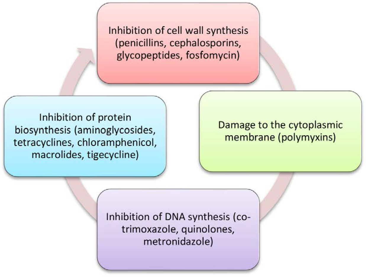
Figure 1.
Mechanisms of Antibiotic Action.
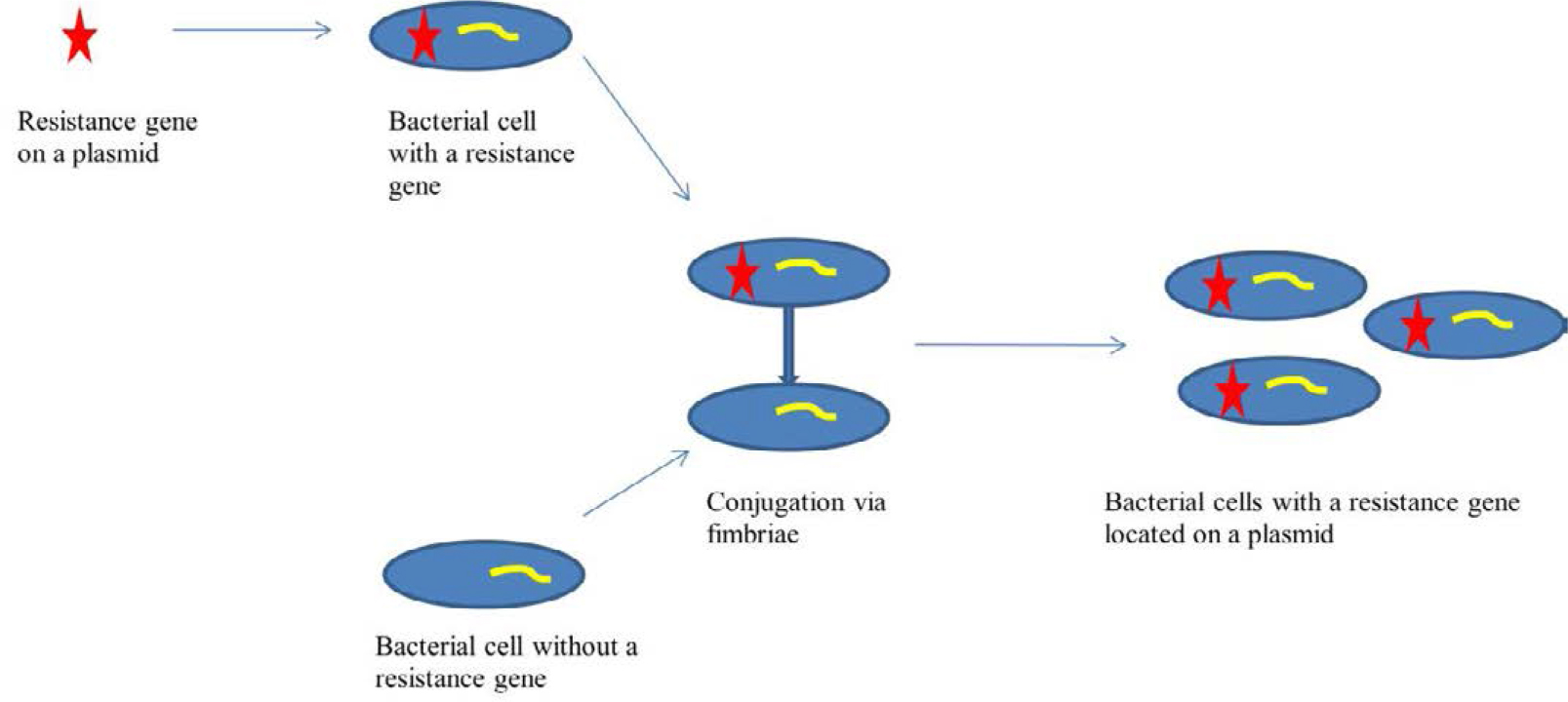
Figure 2.
Gene transfer of resistance in the conjugation process involving fimbriae.
Table 1.
Classification of β-lactamases according to Ambler.
| Class | Active site | Type of β-lactamases | Substrates |
|---|---|---|---|
| A | Serine | Penicillinases | Penicillins, first-generation cephalosporins |
| ESBL | Penicillins, first, second, third, and fourth-generation cephalosporins, aztreonam | ||
| Carbapenemases | Penicillins, first, second, third, and fourth-generation cephalosporins, carbapenems, monobactams | ||
| B | Zn++ | Metallo-β-lactamases | β-lactams except monobactams |
| C | Serine | AmpC cephalosporinases | Penicillins, cephalosporins (except for fourth-generation cephalosporins and carbapenems) |
| D | Serine | Oxacillinases | Penicillins (including isoxazolyl penicillins), first and second-generation cephalosporins |
| Carbapenemases | Carbapenems |
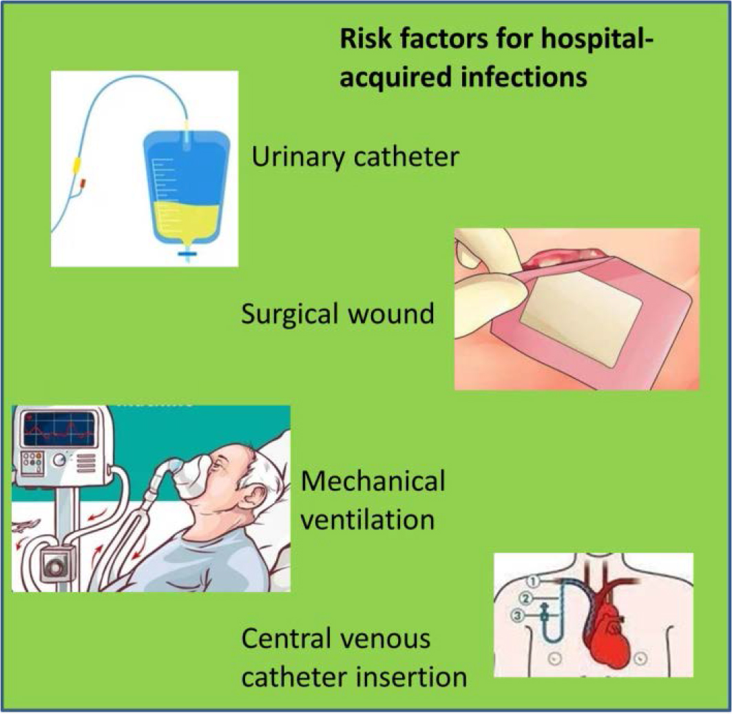
Figure 3.
Risk Factors for the Development of Hospital Infections
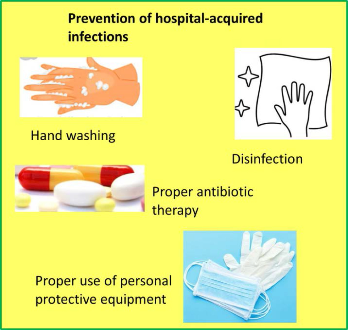
Figure 4.
Prevention Methods Effective in Preventing Hospital Infections
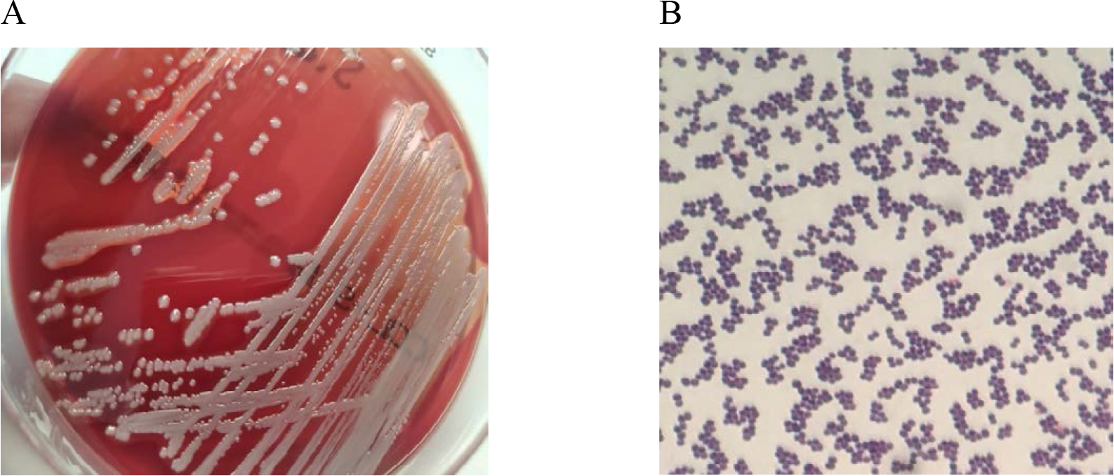
Figure 5.
In vitro cultures of Staphylococcus aureus on Columbia Agar with 5% sheep blood (A) and a Gram-stained preparation of Staphylococcus aureus viewed under a light microscope (magnification x1000). Source: own elaboration.
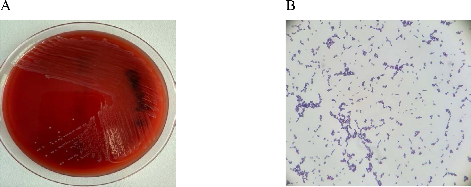
Figure 6.
In vitro cultures of Enterococcus faecalis on Columbia Agar with 5% sheep blood (A) and a Gram-stained preparation of Enterococcus faecalis viewed under a light microscope (magnification x1000). Source: own elaboration.
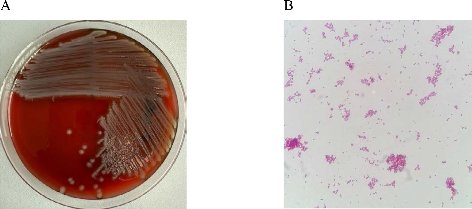
Figure 7.
In vitro cultures of Escherichia coli on Columbia Agar with 5% sheep blood (A) and a Gramstained preparation of Escherichia coli viewed under a light microscope (magnification x1000). Source: own elaboration.
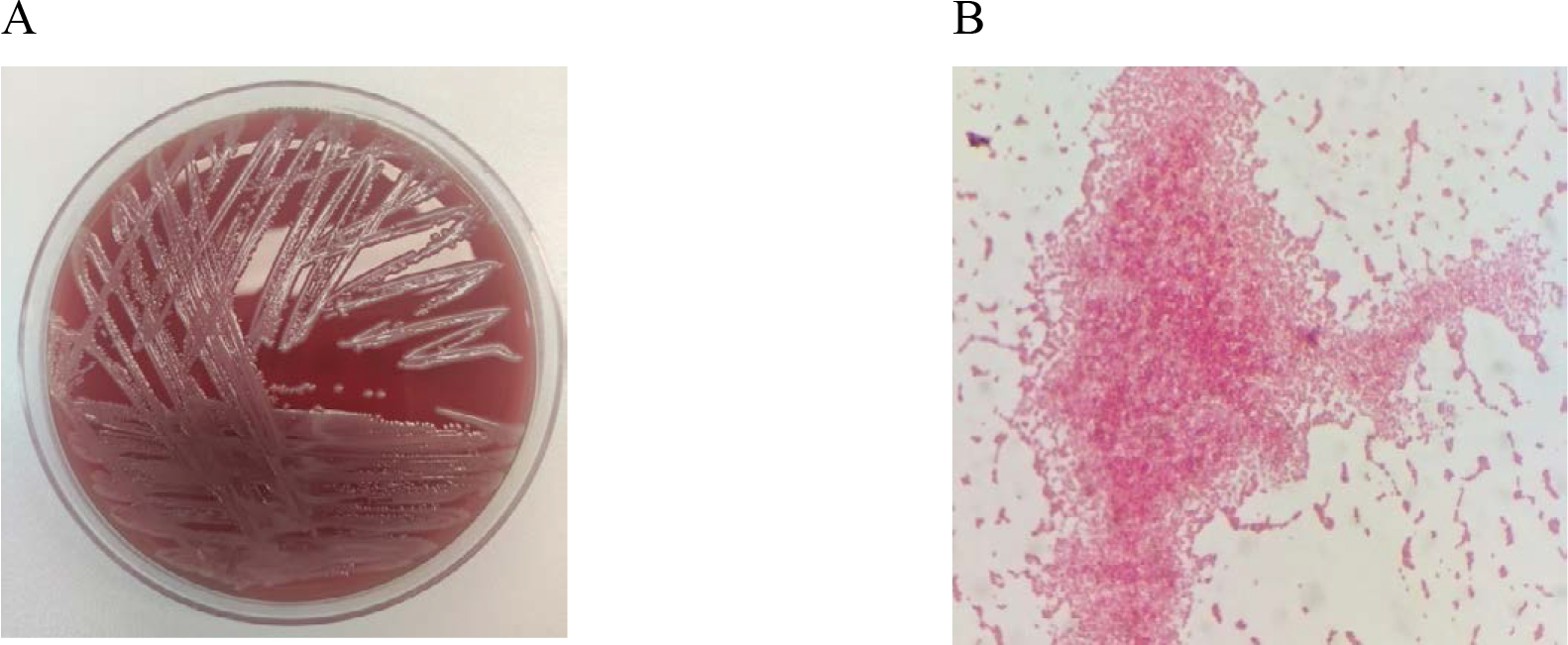
Figure 8.
In vitro cultures of Acinetobacter baumannii on Columbia Agar with 5% sheep blood (A) and a Gram-stained preparation of Acinetobacter baumannii viewed under a light microscope (magnification x1000). Source: own elaboration.
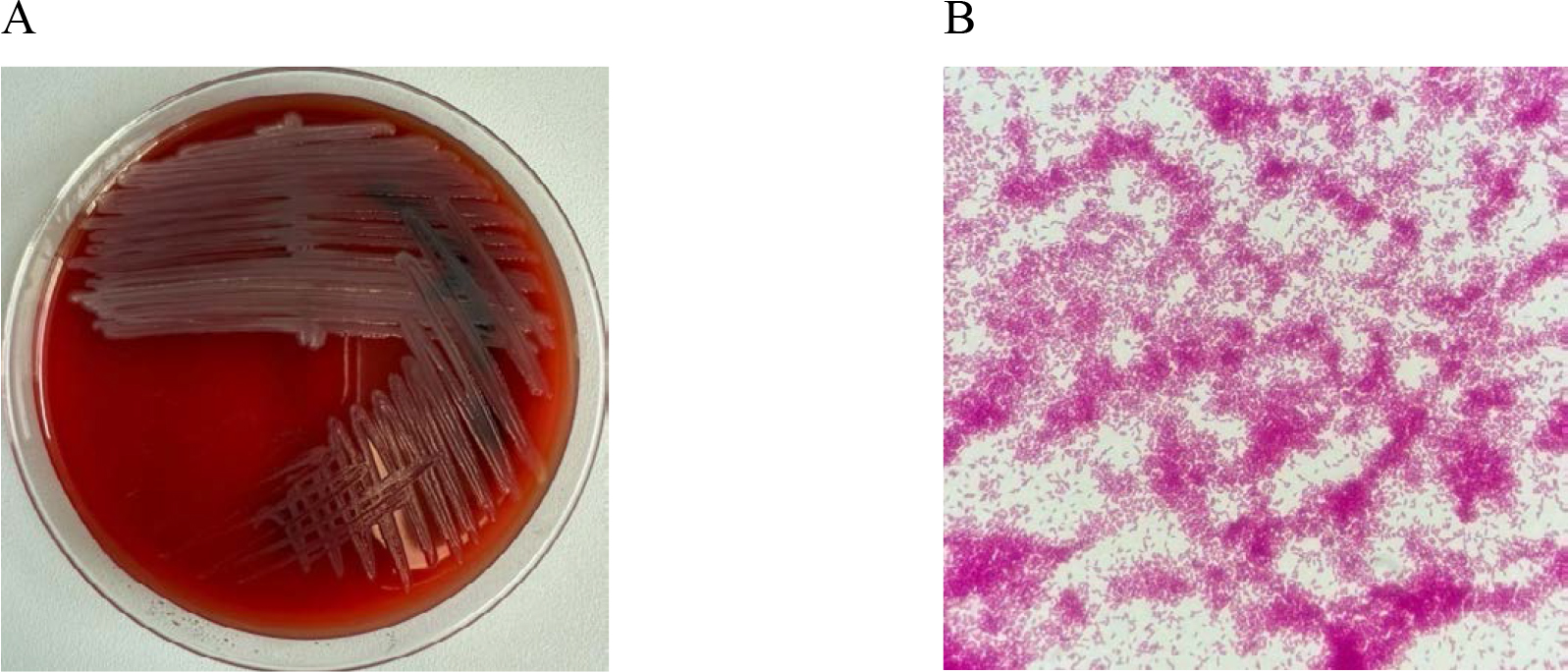
Figure 9.
In vitro cultures of Pseudomonas aeruginosa on Columbia Agar with 5% sheep blood (A) and a Gram-stained preparation of Pseudomonas aeruginosa viewed under a light microscope (magnification x1000). Source: own elaboration.