
Introduction
Ankylosis of the maxillary incisors is a common sequela of dental–alveolar trauma and presents significant challenges in orthodontic management. Fusion between the root surface and alveolar bone prevents physiologic tooth movement, rendering conventional mechanics ineffective and limiting treatment choices. 1 Current management approaches include: (1) extraction followed by orthodontic space closure, (2) extraction with prosthetic or implant rehabilitation, and (3) segmental osteotomy to reposition the ankylosed tooth–bone complex.2,3,4 Each option carries distinct advantages and drawbacks regarding esthetics, alveolar bone preservation, periodontal health, and long-term stability.
Techniques specifically described for ankylosed maxillary incisors fall into three major categories: (1) Single-tooth dento-osseous osteotomy, primarily used for vertical repositioning, with reported periodontal stability over 12–18 months;5 (2) Segmental osteotomy with or without grafting, enabling single-stage correction of infraoccluded or labially displaced incisors;3 and (3) Block osteotomy with distraction osteogenesis, used for staged vertical augmentation before consolidation.5,6,7,8,9 Although these techniques demonstrate feasibility, most evidence derives from case reports or small series, with heterogeneous protocols and limited follow-up.
The present case differs from previously reported examples in three clinically meaningful aspects. First, rather than simple vertical correction, this patient required combined retraction and impaction of flared ankylosed incisors within a skeletal class II relationship, creating a more complex biomechanical scenario. Second, the procedure was performed under local anesthesia through a single vestibular incision, with deliberate preservation of facial and palatal pedicles—consistent with the tissue-sparing principles of piezoelectric surgery. Third, despite achieving substantial improvements in overjet, overbite, and skeletal relationships, the overall treatment duration was longer, reflecting the biologically cautious, staged approach necessary for this case.
Taken together, this report illustrates the feasibility of minimally invasive, piezoelectric-assisted segmental osteotomy for three-dimensional correction of ankylosed maxillary incisors while preserving alveolar and soft-tissue form. As with all single-case evidence, interpretation should remain cautious, and longer follow-up is needed to evaluate long-term periodontal and orthodontic stability.
Diagnosis and aetiology
A 21-year-old female presented with concerns about protrusive lips and misaligned anterior teeth following previous dental–alveolar trauma. Clinical examination revealed a skeletal class II relationship (ANB 10°) with an Angle class II division 1 malocclusion, 8.0 mm overjet, 6.5 mm overbite, flared maxillary central incisors, and midline discrepancies of 1.5 mm (upper to the right, lower to the left) (Figures 1, 2). No Bolton discrepancy was detected.
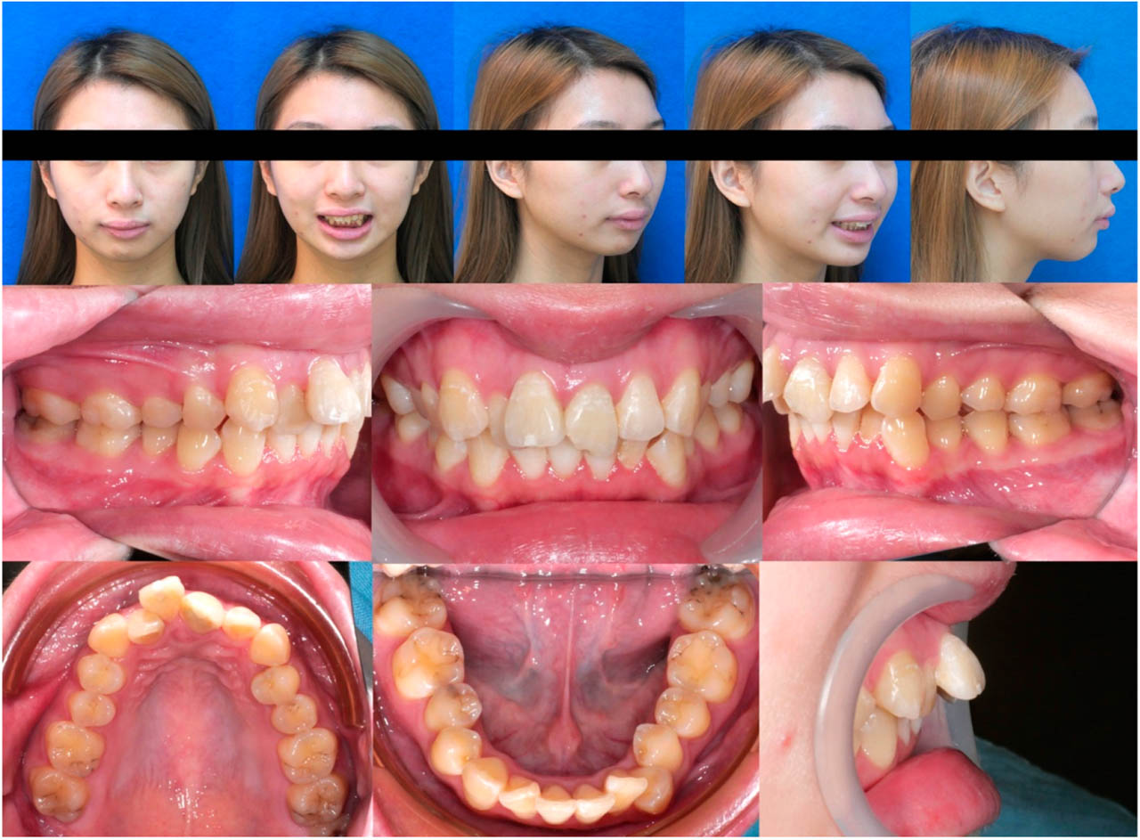
Figure 1.
Initial extraoral and intraoral photographs before comprehensive orthodontic treatment.
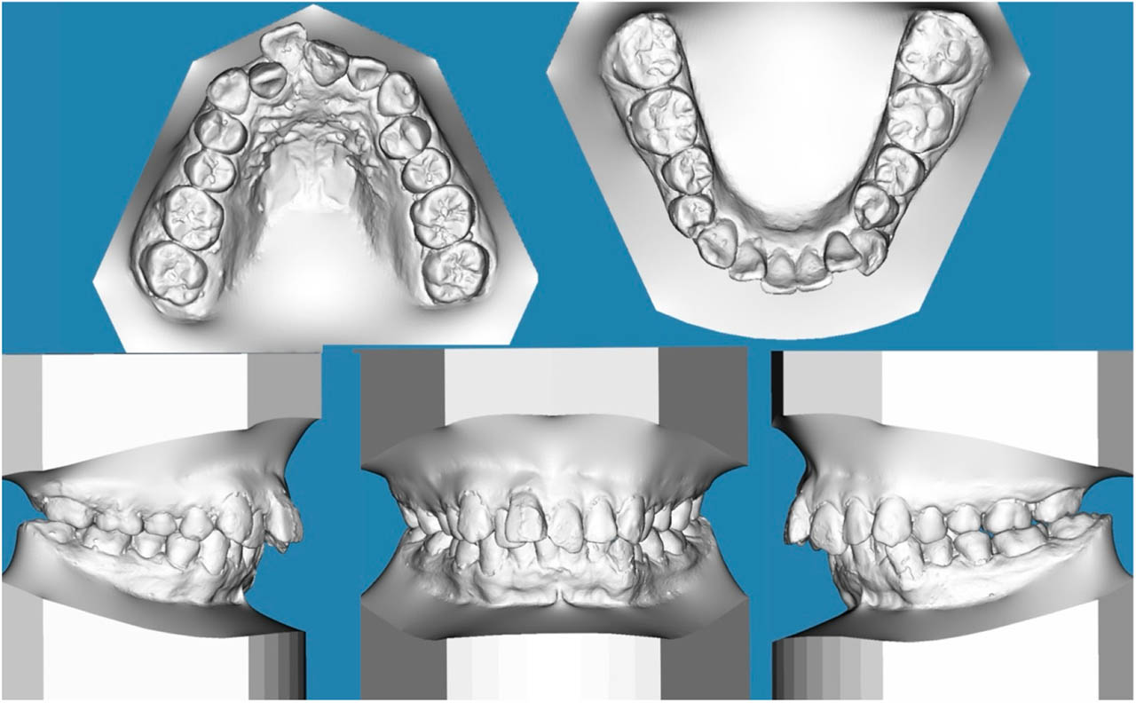
Figure 2.
Initial digital study model.
Pretreatment periapical radiographs showed that tooth 11 had been avulsed and replanted, while tooth 21 displayed an uncomplicated crown fracture (Figure 3). During initial orthodontic alignment, tooth 11 remained completely immobile, and follow-up radiographs confirmed absence of a periodontal ligament space, establishing the diagnosis of ankylosis. Concurrently, tooth 21 exhibited progressive external root resorption (Figure 4). Intraoral photographs corroborated anterior protrusion and dental misalignment (Figure 1). Cephalometric evaluation demonstrated a retrusive mandible (SNB 72.5°) and proclined maxillary incisors, exacerbating the sagittal discrepancy (Figure 5, Table I).
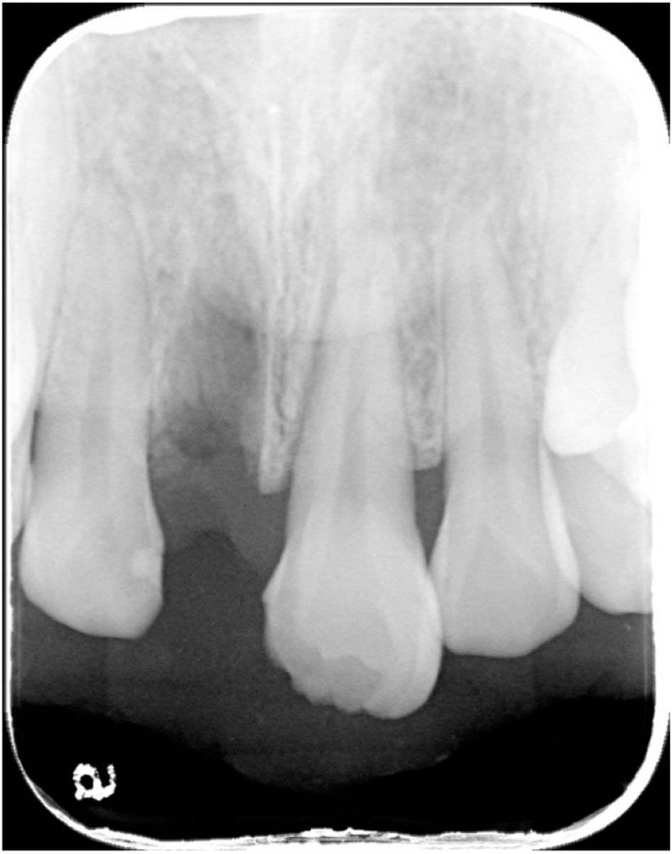
Figure 3.
Periapical radiograph showing tooth 11 avulsion before repositioning and an uncomplicated crown fracture of tooth 21.
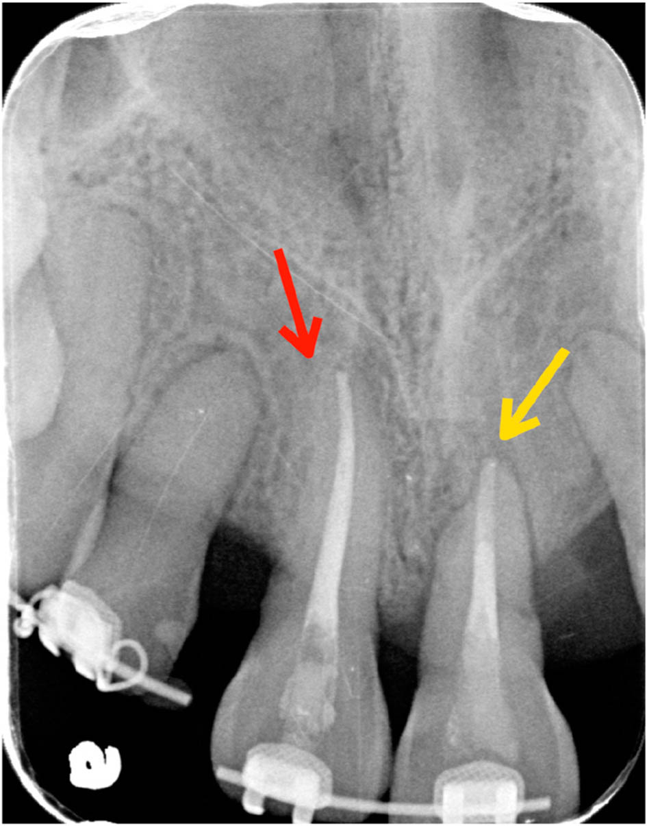
Figure 4.
Preoperative periapical radiograph showing tooth 11 with absence of a periodontal ligament space (red arrow) and external root resorption of tooth 21 (yellow arrow).
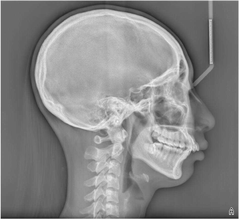
Figure 5.
Initial cephalometric radiograph.
TABLE I.
Initial and posttreatment cephalometric measurements
| 15–30 y/o, female | Taiwanese norm | Initial | Final | Change |
|---|---|---|---|---|
| Skeletal | ||||
| SNA (°) | 79.7 ~ 86.1 | 82.5 | 80.0 | −2.5 |
| SNB (°) | 77.6 ~ 83.7 | 72.5 | 74.5 | +2.0 |
| ANB (°) | 0.7 ~ 3.8 | 10.0 | 5.5 | −4.5 |
| SN-MP (°) (Gn-Go) | 25.5 ~ 34.5 | 39.7 | 36.1 | −3.6 |
| FMA (°) | 26.0 ~ 34.1 | 31.0 | 26.7 | −4.3 |
| Dental | ||||
| U1 - SN (°) | 102.2 ~ 115.1 | 107.0 | 102.5 | −4.5 |
| U1 - NA (mm) | 3.2 ~ 7.3 | 5.2 | 1.4 | −3.8 |
| L1 - MP (°) (Gn-Go) | 90.5 ~ 103.1 | 104.0 | 110.0 | +6.0 |
| L1 - NB (mm) | 4.0 ~ 8.4 | 11.1 | 9.1 | −2.0 |
| Overjet (mm) | 2.0 ~ 3.0 | 8.0 | 2.0 | −6.0 |
| Overbite (mm) | 2.0 ~ 4.0 | 6.5 | 2.0 | −4.5 |
| Soft tissue | ||||
| E - line upper lip (mm) | −2.9 ~ 0.4 | 1.5 | −0.5 | −2.0 |
| E - line lower lip (mm) | −1.9 ~ 2.1 | 2.5 | −0.5 | −3.0 |
| Nasolabial angle (°) (Cm-Sn-Ls) | 103.0 ~ 119.8 | 98.5 | 106.1 | +7.6 |
| Facial convexity (°) (G′-Sn′-Pg′) | 165.0 ~ 174.8 | 154.8 | 162.8 | +8.0 |
The etiology was attributed to the patient's history of dental trauma, a well-documented cause of both ankylosis and external root resorption.10 No systemic conditions, hereditary factors, or periodontal pathology were present. These findings guided treatment planning toward improving the skeletal Class II relationship while simultaneously addressing the ankylosed incisor segment.
Treatment objectives and treatment alternatives
The treatment aimed to correct the skeletal and dental Class II relationship, achieve a stable Class I occlusion, and improve facial esthetics. Key objectives included repositioning the ankylosed maxillary central incisors through controlled retraction and impaction to reduce overjet, establishing functional transverse alignment, decreasing the deep overbite, and preserving the anterior alveolar bone and soft-tissue architecture. Ensuring adequate vascular supply to the dento-osseous segment was essential to avoid ischemia or necrosis during healing.
Several treatment alternatives were considered. Extraction of teeth 11 and 21 with subsequent prosthetic replacement was rejected due to anticipated alveolar bone loss and esthetic concerns in this young patient. A non-surgical camouflage plan with four first premolar extractions was initiated but abandoned when ankylosis of tooth 11 prevented canine retraction, confirming the need for surgical intervention. Conventional segmental osteotomy using rotary instruments was an option but carried greater risks of soft-tissue trauma and compromised vascularity due to the need for multiple incisions. The final choice—piezoelectric-assisted segmental osteotomy through a single vestibular incision— offered precise bone removal, preservation of facial and palatal pedicles, and reduced surgical morbidity, providing the most favorable balance of safety, predictability, and esthetic outcomes.
Treatment progress
Treatment was initiated with a non-surgical camouflage approach involving extraction of all four first premolars to address the skeletal Class II pattern. The patient presented for orthodontic treatment approximately 8 months after the traumatic injury and the replantation of tooth 11. Fixed 0.022- inch slot appliances were placed, and alignment progressed with a 0.014-inch nickel–titanium wire followed by a 0.016 × 0.022-inch stainless steel wire for canine retraction. Two temporary anchorage devices (TADs) were inserted bilaterally in the IZC7 region to reinforce vertical and sagittal anchorage. Despite these efforts, tooth 11 remained immobile, and radiographs confirmed ankylosis, while tooth 21 demonstrated progressive external root resorption, prompting a shift to a surgical approach.
Prior to surgery, teeth 11 and 21 were bonded together to enhance stability and maintain vascular continuity within the dento-osseous segment (Figure 6). Under local anesthesia, a single vestibular incision placed 5 mm above the mucogingival junction provided broad horizontal visualization of the surgical field and access for segmental osteotomy (Figure 7). Using a piezoelectric unit, a vertical and occlusally diverging osteotomy was performed, with apical and interdental bone removed while preserving the facial and palatal pedicles to maintain blood supply (Figures 8, 9). The segmental osteotomy was performed approximately 44 months after the initiation of orthodontic treatment.
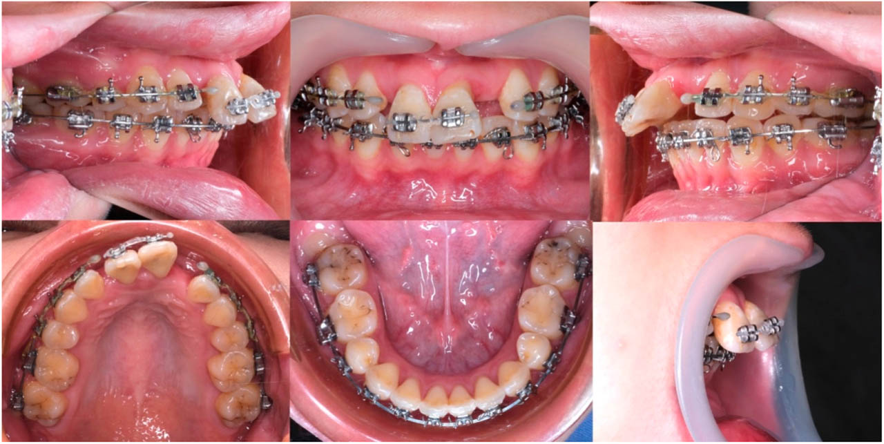
Figure 6.
Intraoral photographs before surgery.
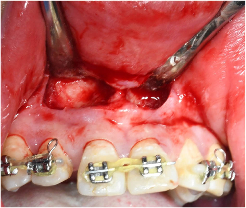
Figure 7.
A vestibular incision was made approximately 5 mm above the mucogingival junction using a scalpel blade. This incision and flap design provided excellent horizontal visibility, allowing precise osteotomy placement 5 mm apical to teeth 11 and 21. It also facilitated clear access for removing interdental and apical bone while preserving palatal-side blood supply to the dento-osseous segment.
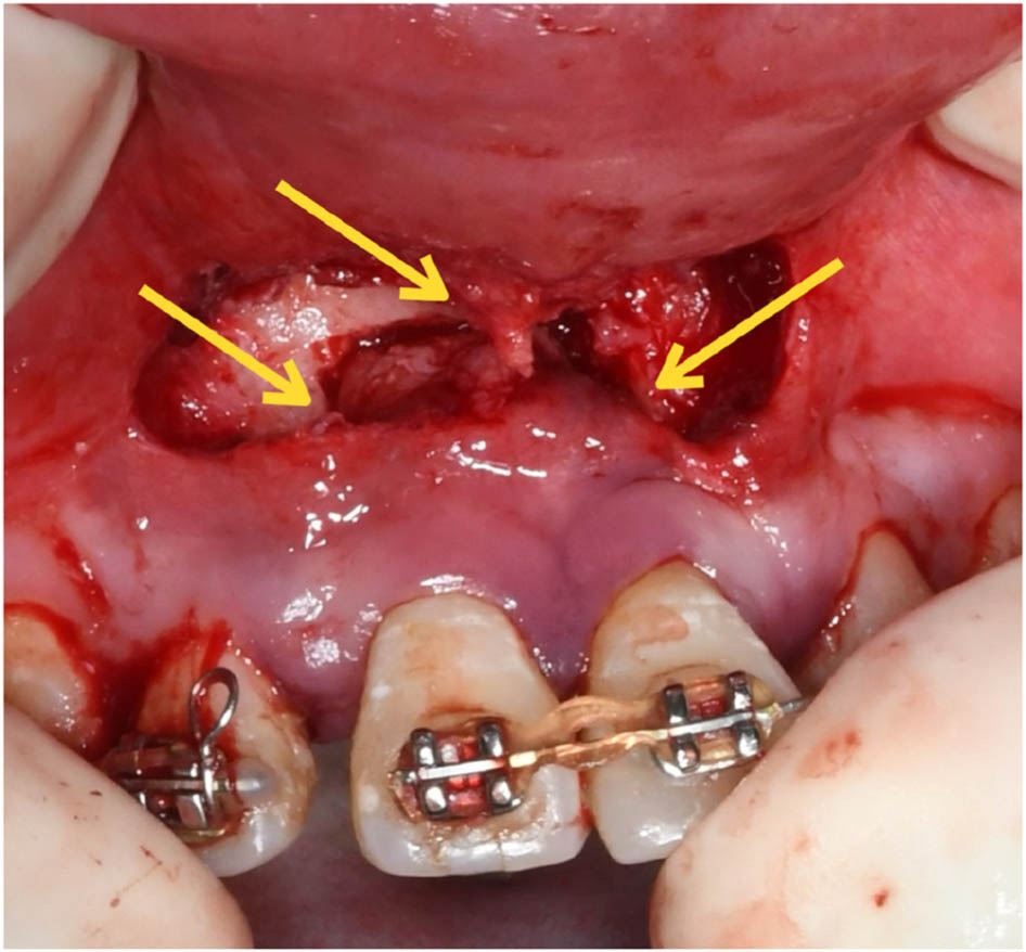
Figure 8.
A vertical and occlusally diverging osteotomy was performed, after which bony interferences were removed and the dento-osseous segment was repositioned. The yellow arrow indicates the apical and interdental bone that was removed, while both the facial and palatal pedicles were preserved.
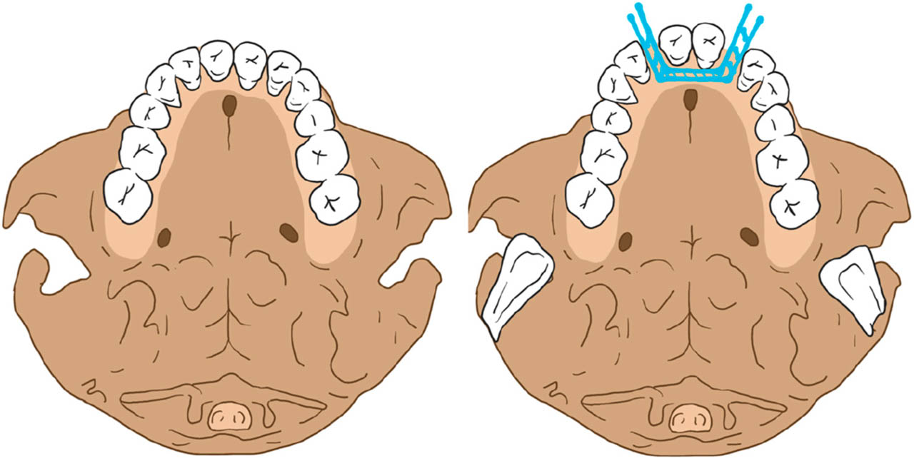
Figure 9.
The surgical diagram illustrates the osteotomy lines (blue line) in axial view.
Immediate soft-tissue tension prevented ideal positioning of the segment; therefore, gradual repositioning was carried out postoperatively using light continuous forces delivered by a power chain (Figures 10, 11). This allowed controlled retraction and impaction of the ankylosed segment over the following weeks. Radiographic follow-up at four months demonstrated progressive bone healing and consolidation of the osteotomy site (Figures 12, 13). Comprehensive orthodontic finishing was completed, and treatment concluded after 48 months with the placement of clear retainers.
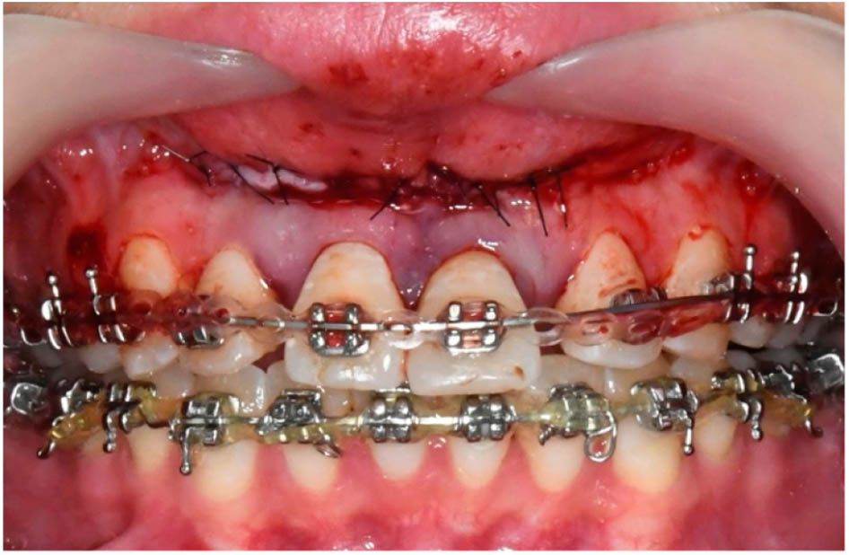
Figure 10.
Teeth 11 and 21 were fixed with the main wire with the help of power chain.
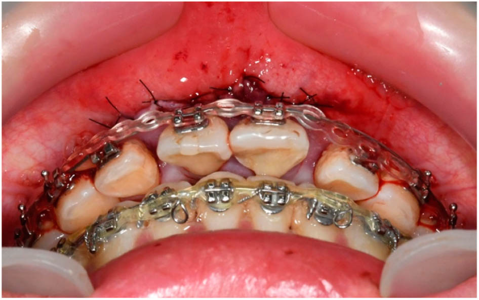
Figure 11.
Due to the non-ideal position, the main wire could not fit in the brackets of teeth 12 and 22. The force of power chain can retract the teeth-bone segment continuously.
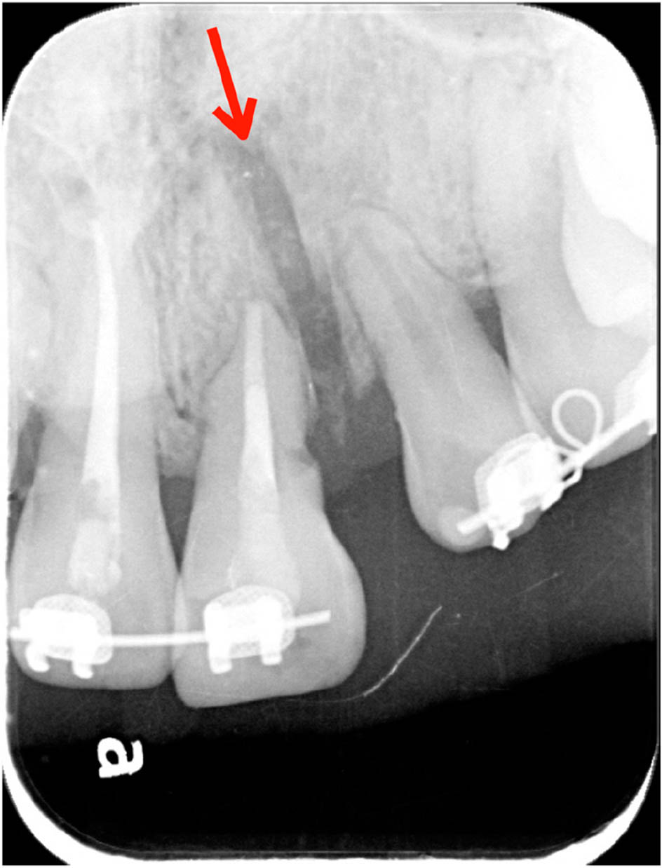
Figure 12.
Periapical radiograph after the surgery showing the bony gap and spacing between teeth 21 and 22 (red arrow).
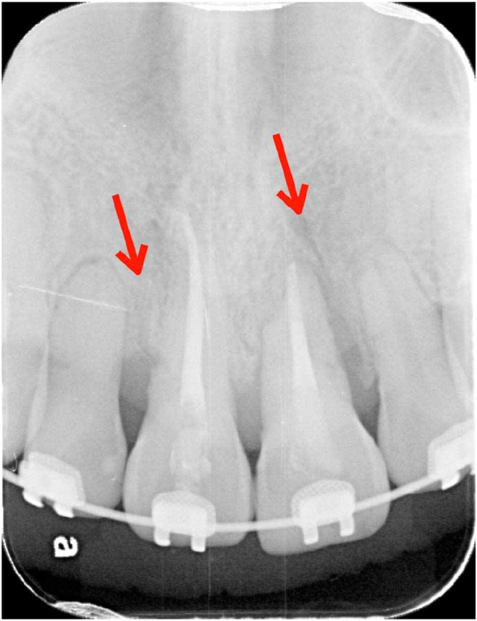
Figure 13.
Periapical radiograph four months postoperatively showing good alveolar bone healing and the closing of the space between central and lateral incisors (red arrow).
Treatment results
Treatment yielded substantial dental, skeletal, and esthetic improvement. The ankylosed maxillary central incisors were successfully retracted and impacted, reducing the overjet from 8.0 mm to 2.0 mm and the overbite from 6.5 mm to 2.0 mm. Cephalometric analysis demonstrated a corresponding improvement in skeletal relationships, with the ANB angle decreasing from 10.0° to 5.5° and the maxillary incisor inclination (U1–SN) reducing from 107.0° to 102.5° (Figures 14–20, Table I). Soft-tissue analysis also showed notable improvement: the nasolabial angle increased from 98.5° to 106.1°, and facial convexity improved from 154.8° to 162.8°. In addition, the upper and lower lips relative to the E-line were reduced to within normal limits (Figures 14–20, Table I). These changes contributed to a more favorable sagittal profile and improved lip posture.
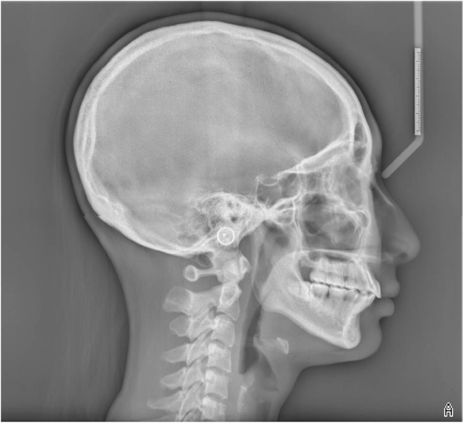
Figure 14.
Posttreatment cephalometric radiograph.
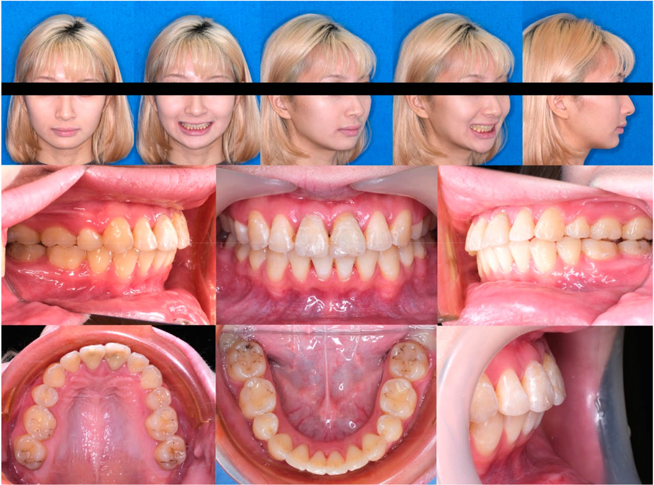
Figure 15.
Posttreatment extraoral and intraoral photographs.
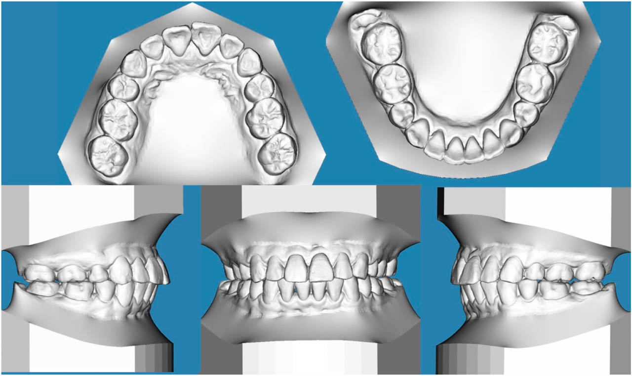
Figure 16.
Posttreatment digital study model.
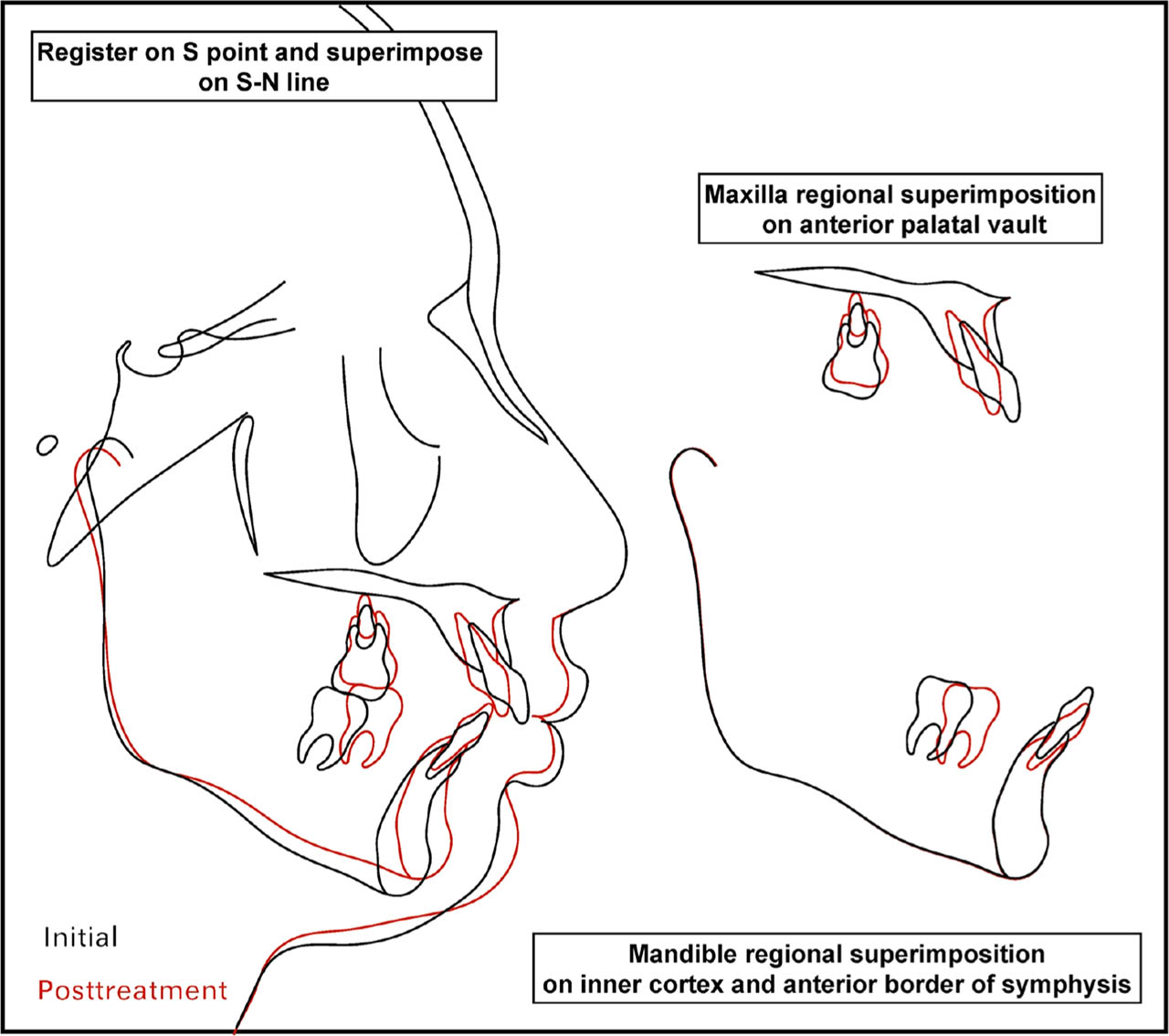
Figure 17.
Cephalometric superimposition.
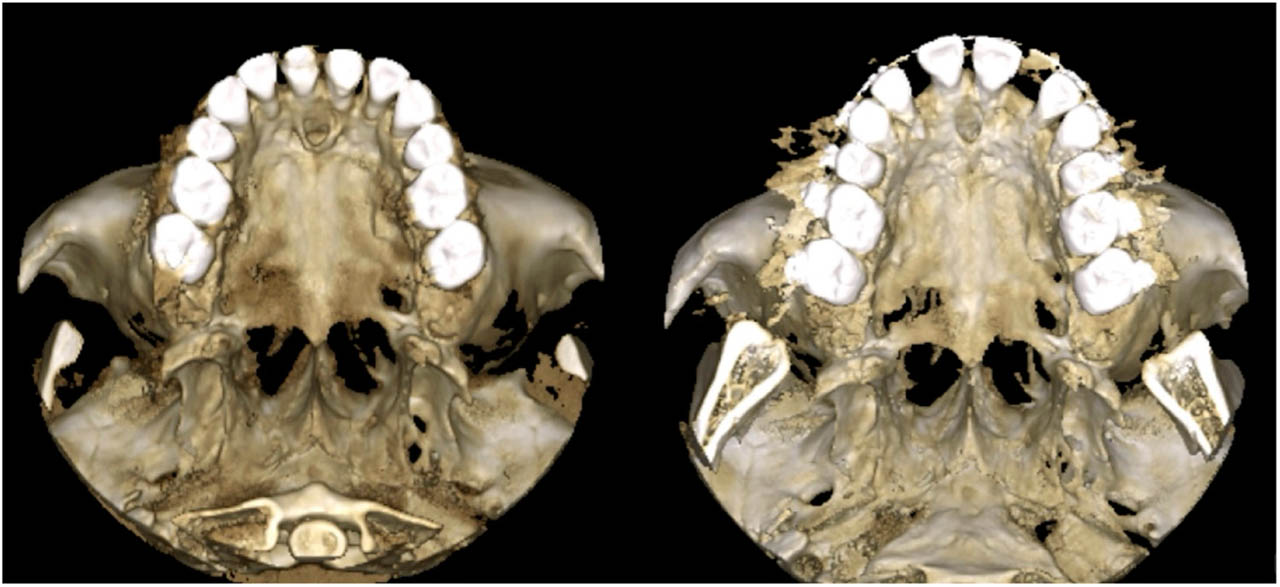
Figure 18.
Presurgical and posttreatment CBCT 3D model in axial view.
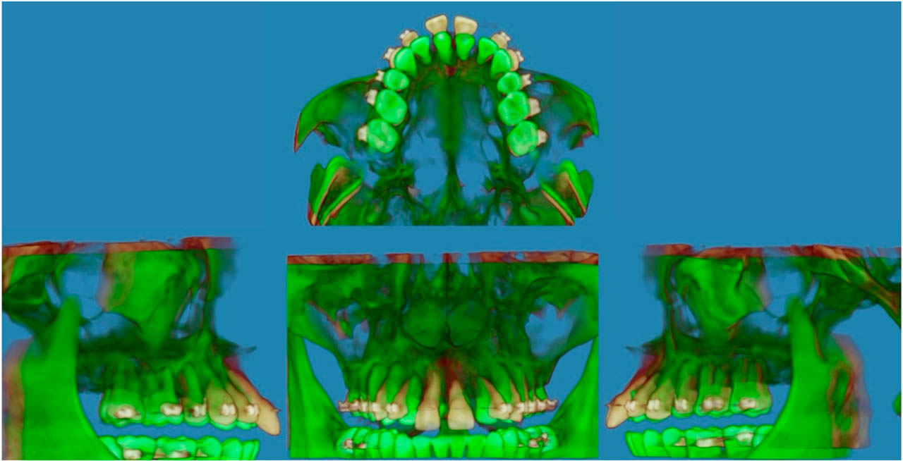
Figure 19.
CBCT superimposition.
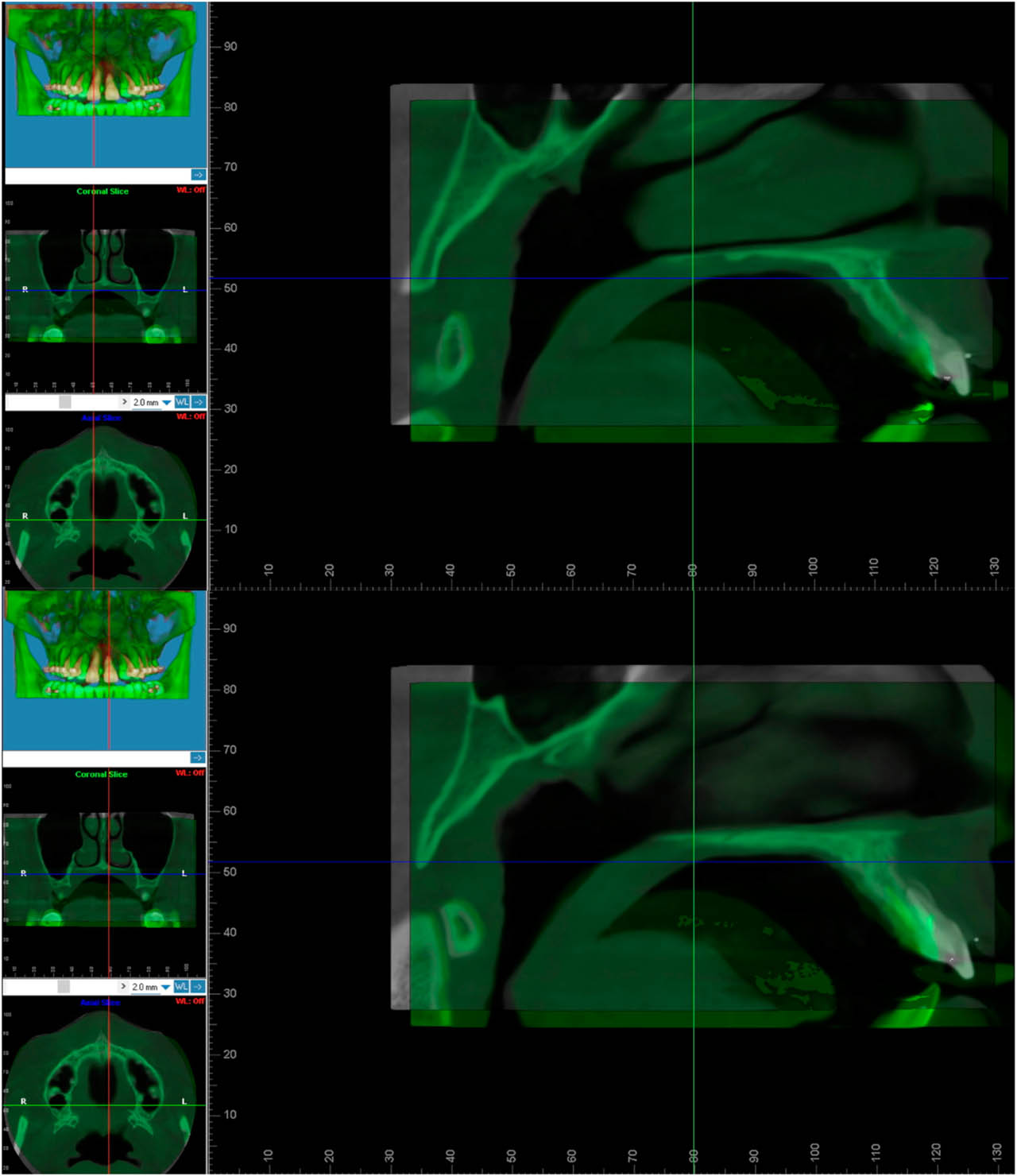
Figure 20.
Superimposed sagittal CBCT sections at the levels of teeth 11 and 21 demonstrating retraction and impaction of the dento-osseous segment.
Radiographic follow-up confirmed healing of the osteotomy site, with closure of the surgical gap and re-establishment of continuous alveolar bone architecture around the repositioned segment (Figure 13). The maxillary dentition achieved stable transverse alignment, resolving the initial crowding and improving the upper–lower midline relationship. Although the mandibular midline remained 1.0 mm to the left, the inter-arch discrepancy was considerably reduced. The final occlusion demonstrated a stable Angle Class I relationship 12 months posttreatment (compare Figures 15, 16 to Figure 21).
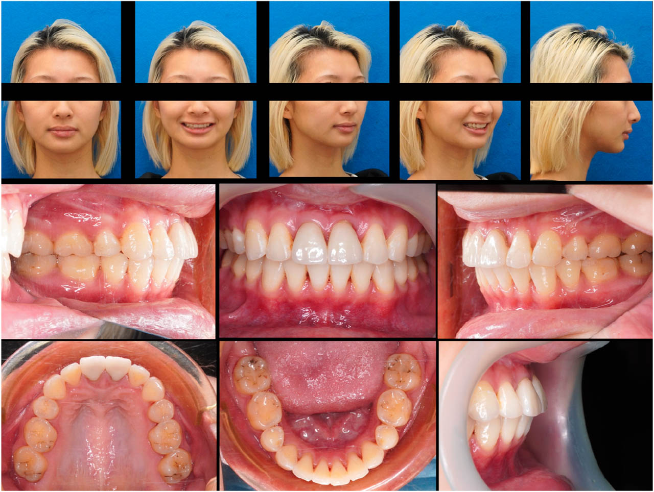
Figure 21.
One-year follow-up extraoral and intraoral photographs.
Facial esthetics improved markedly. Counterclockwise mandibular rotation and controlled incisor repositioning transformed the profile from convex to nearly straight, aligning with the patient's esthetic goals (Figure 15). The total active treatment time was 48 months. No signs of necrosis or soft-tissue compromise were observed, confirming adequate vascular preservation throughout healing.
At the one-year follow-up after debonding, teeth 11 and 21 showed normal clinical findings, including the absence of mobility, symptoms, or periodontal deterioration. Periodontal tissues remained stable, and prosthetic rehabilitation of teeth 11 and 21 was subsequently completed for optimal esthetics and structural support (Figures 21, 22).
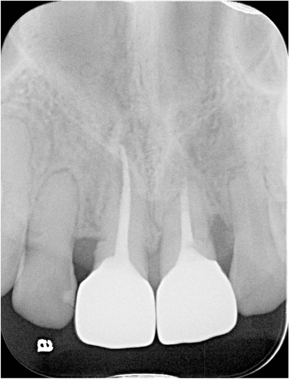
Figure 22.
One-year follow-up periapical radiograph.
Discussion
Ankylosed maxillary incisors present substantial therapeutic challenges, and management typically falls into three categories: extraction with orthodontic space closure, extraction followed by prosthetic or implant rehabilitation, and segmental osteotomy to reposition the involved tooth–bone unit.11 Each modality has inherent advantages and limitations. Space closure preserves natural dentition but risks alveolar bone loss and unfavorable anterior esthetics, particularly in the maxillary region.12 Extraction with prosthetic replacement resolves ankylosis quickly but sacrifices alveolar and basal bone, may complicate future implant placement, and imposes a greater financial and restorative burden.13,14 Segmental osteotomy maintains the natural tooth and alveolar bone height but introduces surgical morbidity and the need for precise intraoperative control to preserve vascularity and ensure predictable healing. Selecting an appropriate approach therefore requires balancing esthetic demands, periodontal considerations, and long-term functional stability (Table II).
TABLE II.
Comparison of treatment approaches for ankylosed maxillary incisors: extraction, prosthetic replacement, and segmental osteotomy
| Extracting the ankylosed tooth and orthodontically closing the space | Extracting the ankylosed tooth, maintaining the space, then utilizing a prosthesis for rehabilitation | Segmental osteotomy to move the ankylosed tooth | |
|---|---|---|---|
| Advantages |
|
|
|
| Disadvantages |
|
|
|
In the present case, piezoelectric-assisted segmental osteotomy was chosen because the ankylosed segment required combined retraction and impaction—movements that cannot be achieved through conventional orthodontic mechanics. This method enabled three-dimensional repositioning of the dento-osseous block while preserving alveolar architecture and soft-tissue contours. The technique's minimally invasive nature, including a single vestibular incision and preservation of both facial and palatal pedicles, provided a favorable compromise between surgical precision and biological safety.
This movement also contributed to the improvement in the sagittal skeletal relationship observed in this case. Superimposition of sagittal CBCT sections demonstrated that the segmental osteotomy enabled actual retraction and impaction of the dento-osseous segment (Figure 20). This repositioning of the anterior maxillary dento-osseous block contributed to the reduction of the ANB angle from 10.0° to 5.5°. In addition to the cephalometric improvement, this skeletal and dentoalveolar repositioning also contributed to favorable soft-tissue changes, including improvements in facial convexity and the nasolabial angle (Figures 14–20, Table I).
The single vestibular incision provided a wide horizontal surgical field, which was crucial for controlled removal of interdental and apical bone during the osteotomy. This flap design also preserved the principal blood supply to the dento-osseous segment through the palatal pedicle while minimizing soft-tissue disruption on the facial aspect (Figures 7, 8). The enhanced surgical visibility allowed precise execution of the osteotomy and facilitated careful inspection of the bone interface following apical bone reduction. This ensured that the segment remained continuous and free of unintended gaps or step deformities—factors essential for predictable postoperative bone healing (Figures 8, 9). The combination of preserved vascularity and improved visualization contributed to the favorable healing response observed in this case.
Postoperative evaluation further supported the maintenance of vascular integrity. Flap and gingival coloration remained within normal limits throughout healing, and serial radiographs demonstrated progressive bone fill and consolidation without signs of ischemia or necrosis. These findings underscore the importance of preserving both facial and palatal pedicles during anterior maxillary osteotomy.
Piezoelectric instrumentation contributed to predictable outcomes by enabling selective cutting of mineralized tissue while sparing adjacent soft tissues. This precision reduced the risk of palatal flap trauma, minimized thermal injury near the roots, and allowed controlled removal of apical and interdental bone in a confined anatomical space 15,16. Such control was particularly advantageous for tooth 21, which was compromised by external root resorption and required atraumatic handling during segmental repositioning.
Post-treatment radiographs demonstrated external root resorption not only in tooth 21 but also in teeth 12 and 22 (Figure 4), suggesting an inherent susceptibility of this patient to orthodontically induced root resorption. In this context, alternative treatment approaches that rely solely on orthodontic tooth movement might have required greater displacement of tooth 21, potentially exacerbating root resorption and compromising its prognosis. This observation further supports the rationale for selecting a segmental osteotomy approach, which allowed controlled repositioning of the ankylosed dento-osseous segment while minimizing excessive orthodontic forces on the already compromised incisor.
Regarding long-term prognosis, previous studies have suggested that teeth with pronounced external root resorption can remain functional for many years, provided that adequate periodontal support is preserved and occlusal loading is carefully controlled.16,17 Although continued monitoring is warranted, the absence of clinical symptoms and the stability observed at the one-year follow-up indicate a favorable short- to medium-term prognosis for tooth 21 in the present case (Figure 22).
However, piezoelectric-assisted osteotomy also presents practical limitations that must be considered.18 The equipment cost is higher than that of conventional rotary instruments, and operators may experience a steeper learning curve, particularly during early adoption. In addition, operative time can be longer in inexperienced hands. These drawbacks should be weighed against the potential benefits when selecting this technique for anterior maxillary ankylosis.
Despite the minimally invasive surgical approach, the overall treatment duration was prolonged due to several contributing factors. The pre-surgical alignment phase required extended anchorage control in the skeletal Class II pattern, and repeated attempts to orthodontically mobilize tooth 11 confirmed complete ankylosis. A cautious force application protocol was adopted after surgery to preserve vascularity of the repositioned segment. In addition, tooth 21 exhibited progressive external root resorption, necessitating slower and more controlled mechanics to avoid further compromise. Intermittent limitations in patient availability also contributed to the extended timeline. These factors collectively explain the prolonged 48-month duration and are not inherent limitations of the osteotomy technique itself.
Segmental osteotomy carries potential risks, including pulp vitality loss, periodontal changes, irregular bone healing, and relapse. In this case, vitality testing, periodontal assessment, and radiographic monitoring were performed throughout treatment and follow-up, with no evidence of adverse outcomes. The relatively rapid movement of the dento-osseous segment after surgery is consistent with the regional acceleratory phenomenon (RAP), which enhances transient bone remodeling following corticotomy or osteotomy. Controlled retraction with a power chain allowed the segment to gradually settle into functional occlusion despite initial palatal soft-tissue tension, which limited immediate postoperative positioning but diminished as healing progressed.
Aesthetically, the preservation of proximal bone supported stable interdental papillae and harmonious gingival contours. Improved torque control of teeth 11 and 21 contributed to refinement of the smile arc and enhanced lip posture, and the patient reported satisfaction with overall facial balance.
Notably, the treatment also resulted in a marked improvement in the facial profile, with a reduction in lip protrusion and a transition from a convex to a nearly straight profile. This improvement was supported by favorable soft-tissue cephalometric changes, including increases in the nasolabial angle and facial convexity. This degree of profile enhancement was more favorable than initially anticipated, given that no orthognathic surgery was performed and the correction relied primarily on dentoalveolar compensation combined with segmental osteotomy.
A principal limitation of this report is the absence of long-term follow-up beyond one-year after debonding. Although early healing and periodontal health were stable, longer-term observations are necessary to assess relapse potential and periodontal stability. Future studies with extended follow-up would help clarify the durability of this technique.
Conclusions
This case demonstrates that piezoelectric-assisted segmental osteotomy can be a feasible approach for repositioning ankylosed maxillary incisors when conventional orthodontic mechanics are ineffective. The technique allowed controlled three-dimensional movement of the dento-osseous segment while preserving facial and palatal pedicles, maintaining soft-tissue contours, and supporting favorable bone healing. These advantages contributed to improvements in occlusion, dental alignment and facial aesthetics in a patient with a complex skeletal Class II presentation.
However, conclusions regarding treatment predictability or long-term stability cannot be drawn from a single case. Although early periodontal and radiographic findings were favorable, extended follow-up is necessary to assess long-term periodontal health and relapse risk. Further studies with larger samples and longer observation periods are needed to refine treatment guidelines and validate the outcomes of this minimally invasive approach.
Acknowledgements
The authors would like to express their gratitude to the patient who cooperated with us in preparing this article.
Notes
[1] Conflicts of interest Conflict of interest
Disclosure of interests: No potential conflict of interest was reported by the authors.
[2] Contributed by Author's contribution
Sheng-Yang Lee contributed to conceptualisation, supervision, and editing. Chih-Yuan Fang contributed to project administration, surgery, supervision, and editing. Chang-Yu Chen contributed to orthodontic treatment. Zhong-Lin Tsai contributed to visualisation, and writing-original draft & manuscript. All authors have read and approved this article for submission.