
Introduction
Today, nasal and respiratory function, as well as their relationship to craniofacial growth, are of great interest not only as a practical concern for pediatricians, otorhinolaryngologists, allergists, speech physiologists, orthodontists, and other members of the healthcare community, but also as an example of the fundamental biological relationship between form and function.1 One essential component of the correct development of the craniofacial features is a functional airway. There are notable connections between the dentofacial and craniofacial structures and the pharyngeal structures.2 The human face and airway morphology are mostly determined by heredity, but the aetiology of nasal blockage also seems to be largely influenced by the environment.2
Because they are both a part of the unit that performs breathing and deglutition, the nasopharynx and the oropharynx have significant roles and locations.3 It has recently been demonstrated that patients with Obstructive Sleep Apnoea have abnormal skeletal and soft tissue patterns that reduce airway space.4 Examining the soft tissues, such as tonsils, adenoids, nasal polyps, neuromuscular functional jaw patterns, and facial contours, should be a crucial part of treatment planning to help with the stability and aesthetics of the orthodontic or orthopaedic results. Skeletal features such as retrusion of the maxilla, mandible, and vertical maxillary excess in hyperdivergent patients may result in narrower anteroposterior dimensions of the airway.5
By altering the size, shape, and position of the facial bones, orthognathic surgery seeks to restore harmonious facial features and appropriate dental occlusion. Large alterations in the pharyngeal airway space (PAS) dimensions and face appearance are brought about by these new soft tissue interactions, particularly when a large anteroposterior component is present.6
Numerous methods, such as magnetic resonance imaging, cine-magnetic resonance imaging, computed tomography (CT), endoscopy, acoustic rhinometry, fluoroscopy, nasopharyngoscopy, MRI, cephalometry and tomography, and optical coherence tomography, can be used in the medical field to evaluate the airway. Cone-beam CT (CBCT), on the other hand, has made it possible to assess the airway with a quick, low-radiation, non-invasive scan.6
The literature on various techniques for evaluating the airway in orthodontics has very little information. Therefore, we have attempted to provide a thorough explanation of the various techniques for evaluating the airway and its clinical implications in orthodontics through this review article.
Methods To measure airway
General assessment7
A global assessment should include the following:
A comprehensive evaluation of the airway is essential for effective orthodontic diagnosis and treatment planning.
Assessing nasal patency is the first step to finding any obstructions that could interfere with proper breathing, such as nasal polyps or a deviated nasal septum. To guarantee effective oral space, an adult's mouth opening should be checked to make sure it is at least two finger breadths between the upper and lower incisors.
Both airway volume and orthodontic considerations may be impacted by anatomical abnormalities, such as a high-arched palate or a long or small mouth cavity. Assessing mandibular prognathism, or the lower incisors' capacity to protrude past the upper, offers information about skeletal relationships that could affect the size of the airways.
The temporomandibular joint should also be examined for limitations in movement caused by conditions such as ankylosis, fibrosis, or tumorous growths.
Measurements of submental space, ideally with a hyomental or thyromental distance greater than 6 cm, can show sufficient mobility and airway space. While examining the patient's neck, a short, thick neck is often associated with difficulty intubation. Neck extension, the presence of masses, and the capacity to adopt the “sniffing” position should all be carefully noted. Additionally, a short, thick neck or limited cervical mobility is frequently associated with airway issues, making a neck examination essential. Airway stenosis may be the cause of a history of stridor, hoarseness, or tracheostomy.
Last but not least, active airway infections like pneumonia, bronchitis, abscesses, croup, or epiglottitis, as well as systemic or congenital conditions like respiratory disorders, cardiovascular disease, or acromegaly, need special attention because they can all have a major impact on airway assessment and management.
Specific tests for assessment
Mallampatti test7
Pharyngeal and tongue sizes are correlated by the Mallampati classification (Figure 1). The patient sits with their head in a neutral position, their mouth open, and their tongue sticking out as far as it can during this exam. Three groups are distinguished based on how well the base of the tongue obscures the appearance of pharyngeal structures:
Class I: The anterior and posterior pillars, uvula, fauces, and soft palate are visualised.
Class II: Uvula, fauces, and soft palate visualisation.
Class III: Uvula base and soft palate visualization.
In Samsoon and Young's modification (1987)3 of the Mallampati classification, class IV was added.
Class IV: Only the hard palate is visible. The soft palate is not visible at all.
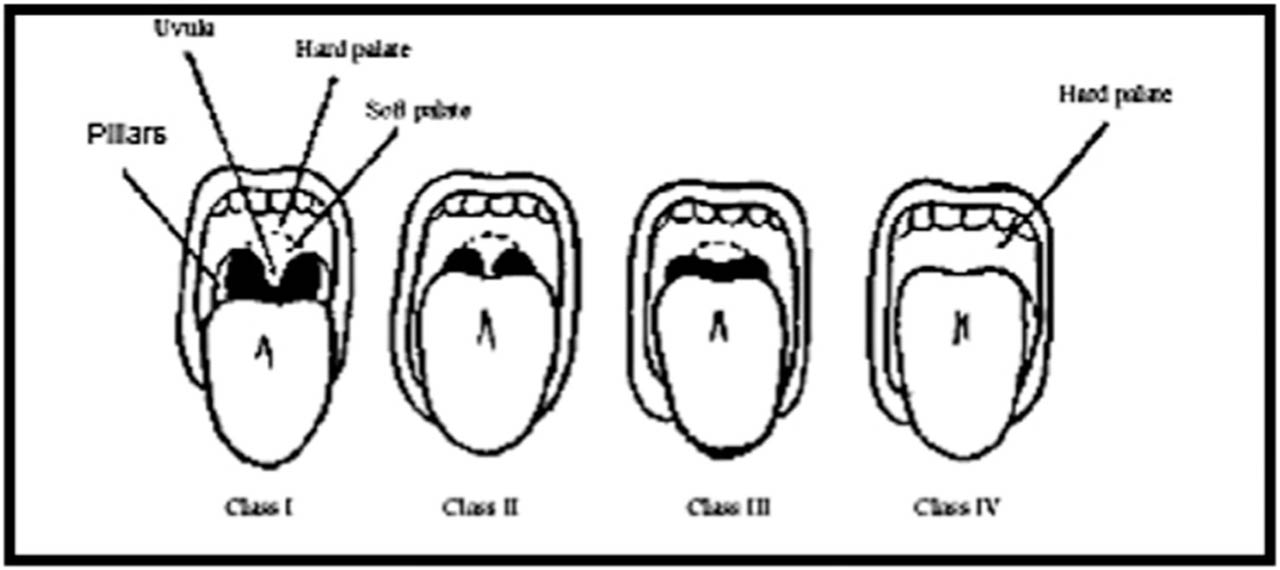
Figure 1.
Mallampatti test.
LEMON airway assessment method8
Each of the following LEMON criteria is worth one point, for a total score of up to ten points (Figure 2):
L: Examine the outside (larger incisors, beard or moustache, large tongue, facial damage).
E: Assess the 3-3-2 rule (thyroid-to-mouth distance-two finger breadths; incisor distance, three finger breadths, and hyoid-mental distance-three finger breadths).
M: Mallampati (score greater than 3).
O: Obstruction (any illness, such as trauma, peritonsillar abscess, or epiglottitis).
N: Stands for neck mobility (limited).
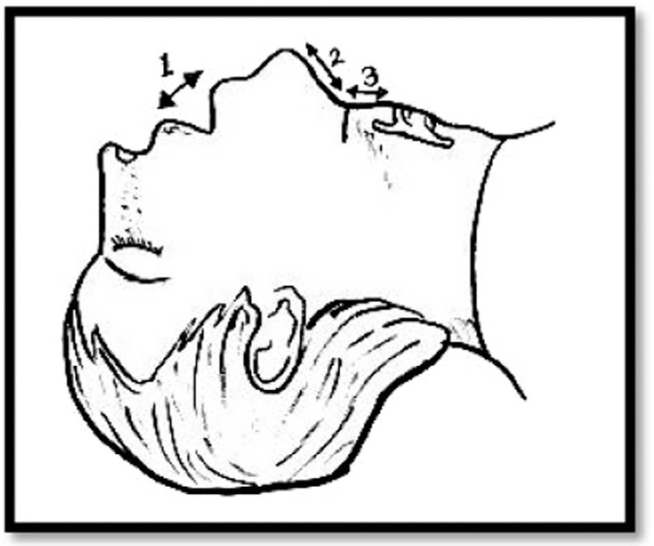
Figure 2.
LEMON Airway Assessment Method.
Patients in the difficult intubation group have higher LEMON scores.
Predicting the difficulty of intubating patients during the preoperative phase is one of the main issues in the practice of general anaesthesia. Patients who are pregnant, have intra-oral pathology, such as infections or tumours, or have suffered facial or maxillary trauma are more prone to have problems during intubation. Additionally, it is observed in people with maxillofacial abnormalities such as TMJ ankylosis, which limits mouth opening, retrognathia, micrognathia, and facial asymmetry. One of the crucial elements to take into account during orthognathic procedures is the size of the tongue's base, which also affects how difficult direct laryngoscopy is.
Other methods to measure airway
Nasometer9
The Nasometer is a recently released device (Kay Elemetrics, Pine Brook, N.J.) (Figures 3 and 4). A sound-separator plate that sits on the upper lip is surrounded by microphones on either side of this microcomputer-based device. Custom electronic modules filter and digitise the signal from each microphone. A personal computer then processes the data. A ratio of nasal acoustic energy to nasal-plus-oral acoustic energy is the resulting signal. This ratio is converted to a “nasalance” score by multiplying it by 100. If nasal airway blockage genuinely reduces nasal resonance during nasal consonant production, then people with nasal obstruction should have low “nasalance” scores when asked to generate a speech sequence that includes the speech sounds “m,” “n,” and “ng.”
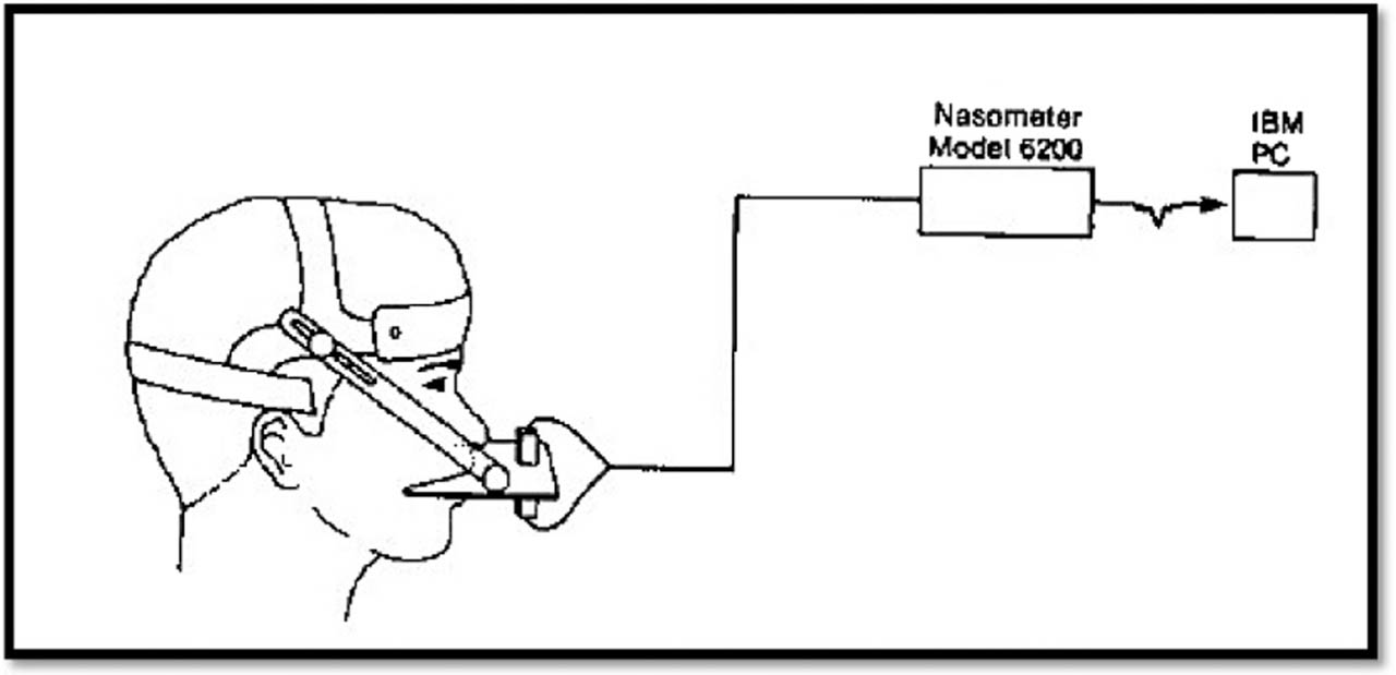
Figure 3.
Nasometer.
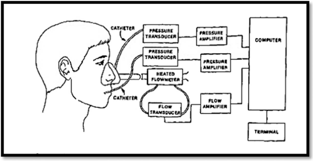
Figure 4.
Nasometer parameters.
Cleft lip and palate is characterised by nasal air emission, articulation difficulties, and resonance abnormalities. The Nasometer is used to do an instrumental assessment of velopharyngeal closure and resonance for speech.
Polysomnograph10
It is a diagnostic technique and a gold test. Sleep, breathing, and oxygenation are recorded during the course of the test. Apnoea, oxygen saturation, body posture, heart rate, snoring, desaturation relations, and sleep staging are all analysed in the study. Electroencephalography (EEG), electrooculography (EOG), electromyography (EMG), and electrocardiography (ECG) are all used in sleep staging. According to the International 10–20 system, electrodes are placed in C3-A2 and C4-A1 to gather EEG data, while the submental muscles are used to capture EMG activity.
To find cardiac arrhythmias, a single electrocardiogram (ECG) is tracked. An ear oximeter is used to continuously assess arterial oxygen saturation (SaO2). Respiratory Inductive Plethysmography is used to track the movement of the chest wall. A 15-channel polygraph is used to record the data at a paper speed of 10 mm/sec. Respiratory and sleep stage characteristics are examined separately. Total apnoea time/total sleep time (TST) and number of apnea/TST (apnoea index) are used to measure the severity of sleep apnoea. Following mandibular advancement or setback, the inferior pharyngeal air way space increases or decreases respectively. Polysomnographs can be used to evaluate the airway changes in orthognathic surgery in patients with obstructive sleep apnoea.
Cephalometric radiography
Grandy11,12,13 provided the first radiographic demonstration of the adenoids and the nasopharyngeal airway in 1925.
Various cephalographic airway analyses are as follows:
Airway analysis by McNamara14
Airway analysis by Athanasios E.11
Airway analysis by Preston CB, Lampasso JD, Tobias PV12
Airway analysis by de Vasconcellos Vilella et al17
Airway analysis by Shigetoshi Hiyama et al18
The cephalometric landmarks used in all these analyses are explained in Table I
Table I.
Cephalometric landmarks
| Cephalometric Landmark | Description |
|---|---|
| AA | Posterior wall of nasopharynx |
| Ba | Basion |
| C2i | The most anteroroinferior point on the body of the second cervical vertebra |
| FH | Frankfurt horizontal plane |
| LPW | Lower pharyngeal wall : Lower pharyngeal wall, intersection of the perpendicular line from V to the posterior pharyngeal wall |
| MPW | Middle pharyngeal wall, intersection of the perpendicular line from Uvula to the posterior pharyngeal wall |
| P | Porion |
| PNS | Posterior nasal spine |
| N | Nasion |
| UPW | Upper pharyngeal wall, point of intersection of the line perpendicular to the posterior pharyngeal wall from the posterior nasal spine. |
Airway analysis by McNamara
In 1984, James A. McNamara employed a cephalometric analysis technique to arrange orthodontic and orthognathic surgery patients' treatments (Figure 5).12
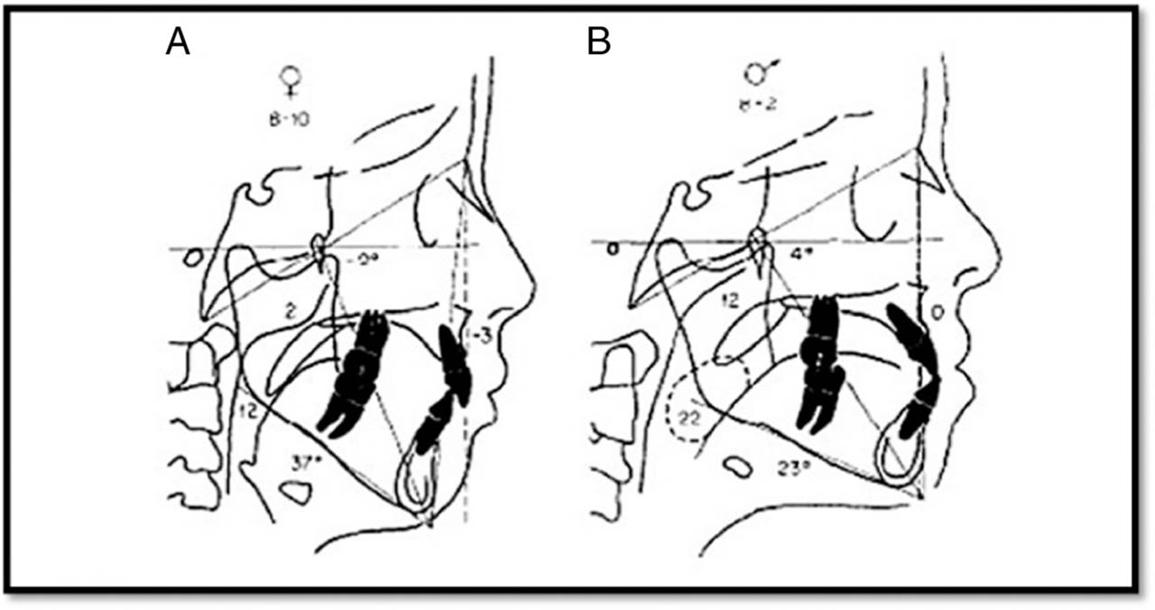
Figure 5.
Airway analysis by McNamara.
The likelihood of an airway impairment is assessed using two measurements. Upper pharynx: The distance between a point on the rear soft palate outline and the nearest point on the posterior pharyngeal wall is the upper pharyngeal width. Because the region directly next to the nose's posterior entrance is crucial for assessing upper respiratory patency, this measurement is made on the anterior half of the soft palate outline. This measurement's typical value is between 16 and 18 mm.
Lower pharynx: The distance between the closest point on the posterior pharyngeal wall and the junction of the inferior border of the mandible and the posterior border of the tongue is the lower pharyngeal width. The average value for this measurement, based on the measurements taken from the Ann Arbor sample, is between 10 and 12 mm, and it does not significantly alter with age.
Airway analysis by Preston CB, Lampasso JD, Tobias PV
Often described as a trapezoid, the region of the bony nasopharynx is bordered by the lines below: A line drawn through AA that is parallel to the pterygoid vertical and extends to intersect the ba-N line; the portion of the ba-N line that runs between the pterygoid vertical and the vertical erected through point AA; the pterygoid vertical between PNS and the intersection of this vertical line and the ba-N line.
The part of the adenoid tissue that represents the nasopharynx and is enclosed by the trapezoid (Figure 6).
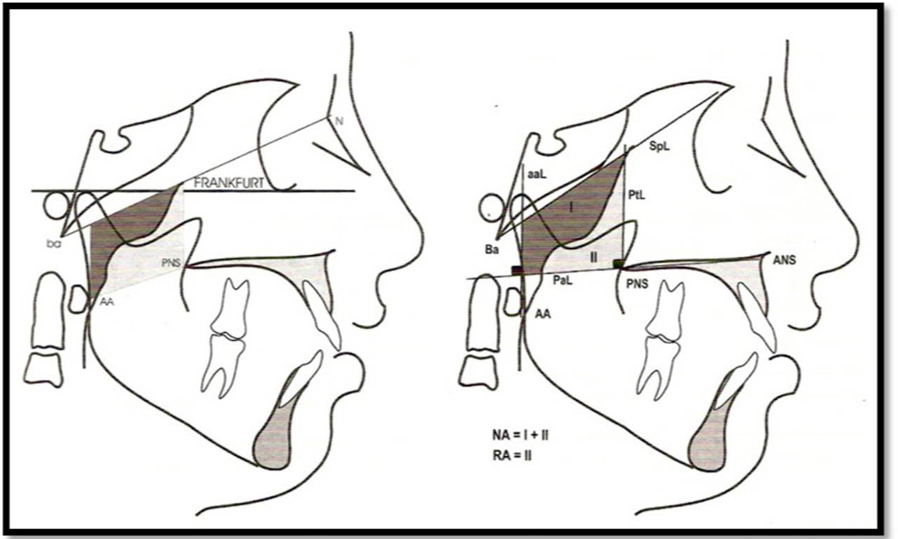
Figure 6.
Airway analysis by Preston CB, Lampasso JD, Tobias PV.
Airway analysis by Shigetoshi Hiyama et al. (Figure 7)
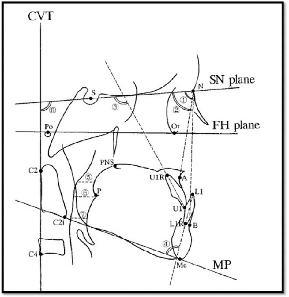
Figure 7.
Airway analysis by Shigetoshi Hiyama et al.
Superior pharyngeal space: Anteroposterior breadth of the pharynx measured between the dorsum of the soft palate and the posterior pharyngeal wall on a line parallel to the FH plane that passes through the centre of the line from PNS to P is known as the superior pharyngeal space.
Middle pharyngeal space: On a line parallel to the FH plane that passes through P, the middle pharyngeal space is the anteroposterior width of the pharynx, measured between the dorsum of the tongue and the posterior pharyngeal wall.
Inferior pharyngeal space: The anteroposterior breadth of the pharynx, measured on a line parallel to the FH plane that passes through C2i, between the dorsum of the tongue and the posterior pharyngeal wall, is known as the inferior pharyngeal space.
Airway analysis by Lyberg et al
From the pterygo-maxillare to the upper pharyngeal wall (UPW) is, the nasopharyngeal airway depth.
The distance between the middle pharyngeal wall (MPW) and the uvula is the oropharyngeal airway space depth. From the Vallecula to the lower pharyngeal wall (LPW), is the hypopharyngeal airway space depth.
Acoustic Reflection19,20
The usage of an acoustic reflection switch, which is based on the idea that sound is reflected by changes in impedance brought on by variations in the pharyngeal cross-sectional areas, was initially described by Jackson et al. and Fredberg et al.
Orthodontists and otorhinolaryngologists have recently begun using acoustic pharyngometry in clinical settings. Like ship sonar, it is a noninvasive process that uses acoustic reflection technology. The Pharyngometer software analyses and measures changes in the airway cross-sectional area by projecting sound waves down the airway and reflecting them out. From the oropharyngeal junction to the glottis, it enables users to rapidly and simply quantify the size and patency of the patient's pharyngeal airway.
A higher propensity for airway collapsibility is known to result in sleep disordered breathing, including snoring and obstructive sleep apnoea (OSA). Craniofacial features, such as relative mandibular retrognathism, increased hyoid distance, decreased posterior airway space, increased tongue volume, enlargement of the palatine or adenoidal tissues, etc. are linked to greater airway collapsibility. Accurately identifying these situations is crucial to ensure orthodontic therapy does not further reduce airway dimensions.
Cone-Beam CT devices21,22,23,24
CBCT can be used to study the sinuses and airway, frontal and lateral cephalometric views, three-dimensional skeletal images, etc.
The upper airway can be seen and measured as a solid structure, which makes volumetric studies achievable with a small margin of error. CBCT technology is now the recommended option to evaluate the airways due to its lower prices, quicker scanning times, and resulting lower radiation doses.
Three-Dimensional Computed Tomography (3DCT)21,22,23,24 is widely used in Orthodontics for many reasons:
Real measurements can be taken;
A spatial image of the craniofacial structures can be created;
Rotating the 3D image is simple by adjusting the rotational axis; and
By eliminating the outside surfaces, the interior structures may be seen.
Finite Element Analysis25
Researchers studying upper airway mechanics and pathology commonly measure the Upper Airway Cross-Sectional Area (CSA). Research investigations would benefit from a noninvasive assessment of the pharyngeal cross-sectional area (CSA) during sleep. A fiber-optic scope is used to see the retropalatal airway to determine the measured CSA (mCSA). A pressure catheter near the palatal rim was used to monitor pharyngeal pressure, and a pneumotachometer was used to assess flow.
A mesh of finite elements is produced using a three-dimensional picture of the upper airway. Using the measured pressure and flow, specialised software enables the simultaneous computation of area and velocity for each element.
Magnetic resonance imaging28-28
An MRI (Magnetic Resonance Imaging) scanner takes images of your internal organs using radio waves, magnetic fields, and a computer. Similar to a CT scanner, with the exception that it uses no radiation. It obtains several images of a cross-section of the body. An orthodontist can benefit from MRI's use for both airway and temporomandibular joint imaging.
Functional MRI29,30,31,32
To our knowledge, only functional MRI (fMRI) has been used in functional neuroimaging studies of obstructive sleep apnoea, despite the fact that there are multiple distinct functional neuroimaging techniques. Examining brain activation in response to an external probe is made possible by fMRI. Typically, the blood-oxygen level-dependent (BOLD) approach is used to obtain fMRI images. The acquisition of images that rely on the sensitivity of the Magnetic Resonance signal to an excess of cerebral blood flow linked to an increase in synaptic activity in the brain is made possible by this noninvasive, high spatial and temporal resolution technology. Investigating acute alterations in the brain linked to completing different tasks is best done with functional neuroimaging techniques.
Additionally, people with vascular pathology may have impaired fMRI, an indirect indicator of vascular function. In summary, cognitive challenges trigger the neural mechanisms involved in performance on complex mental tasks, while respiratory challenges force changes in breathing effort to elicit changes in the autonomic nervous system similar to those experienced by people with breathing disorders.
Clinical implications of airway assessment in orthodontics
It is important to enquire about and evaluate any airway-related difficulties in every patient who visits the orthodontic clinic. An important consideration in the decision-making process should be the evaluation of how orthodontic treatment will affect the dimensions of the upper airway. There is substantial evidence to support the beneficial association and effects of many therapy approaches, such as RME, functional jaw orthopaedics, and particularly maxillomandibular advancement procedures, on issues related to the airways. Additionally, some procedures, particularly those involving mandibular setback, should be performed as sparingly as possible after careful examination.33
A study by Ciavarella D et al. found a statistically significant inverse relationship between the oxygen desaturation index (ODI) and posterior maxillary width. Posterior maxillary breadth in patients with severe OSA was inversely correlated with the ODI and significantly correlated with the minimal airway area, transverse diameter of the limited airway area, and total airway volume. Additionally, there was a statistically significant inverse relationship between the ODI and the minimal airway area, total airway volume, and the cross-sectional diameter of the minimal airway region.34
Class I, II, and III skeletal malocclusion types did not significantly differ in upper and lower pharyngeal airway dimensions, according to the study by Sheraffin RH et al. Within the malocclusion groups, no statistically significant correlations between age or sex and airway morphometrics were found. Although the trend of upper pharyngeal dimensions growing with age was more pronounced than that of lower pharyngeal dimensions, the difference was not statistically significant.35
Regarding the approach of the airway assessment following various orthodontic therapies, there is a great deal of variation in the literature that is currently available. Alswairki HJ et al. concluded that while maxillary protraction, extraction therapy, orthognathic surgery, and the use of a functional appliance have no discernible effects on Class III patients, rapid maxillary expansion and surgical advancement for Class II patients are linked to a notable improvement in the upper airway.36
According to a study by Ertugrul BY, individuals with Class I and Class II malocclusion may exhibit an increase in the air way; thus, orthodontic treatment should be properly planned. Similar to this, orthodontic therapy can be planned for people with Class III malocclusion based on the fact that their upper airways will narrow. Knowledge of how fixed orthodontic treatment affects the airways, early-life airway issues can be avoided.37
Airway volume and skeletal patterns were found to be substantially associated in a study by Le LN et al. Patients with skeletal Class II malocclusion who had a vertical development pattern, a retrognathic jaw, and a high ANB angle had significantly narrower airways. When performing mandibular retrusion on skeletal Class II patients, doctors should exercise caution to avoid aggravating airway obstruction.38 These findings were consistent with research by Mahmoud YM et al. that found that, out of all the study groups, patients with skeletal class II malocclusion had the lowest cross-sectional airway variables and volume values. An accurate indicator of airway volume was the airway's maximal cross-sectional area.39 Among the various skeletal malocclusions, the Nasopharynx showed the largest variation in the linear dimensions of the upper pharyngeal airway space. According to Rodrigues J et al., Skeletal Class III had the most prominent dimensions, followed by Class I, and Class II had the least.40
Upper airway volume increased statistically significantly in growing patients treated with the Twin-block device. Condylar volume, width, and length also significantly increased, and the condyle was moved more anteriorly within the glenoid fossa. Nevertheless, no statistically significant association between upper airway measures and TMJ was discovered as shown by Anagnostopoulos I et al.41
Kurnaz M.'s study revealed that in orthodontic diagnosis and treatment planning, it is essential to take gender-specific differences in Class I and III patients into consideration and assess pharyngeal airway characteristics. Compared to Class III males, Class III girls had bigger OA and RG regions as well as superior and inferior pharyngeal spaces.42
It is important to keep in mind that although orthodontists can identify craniofacial characteristics related to airway morphology, formal diagnosis and treatment of airway disorders are outside the purview of orthodontic practice.
When necessary, ENT specialists, sleep doctors, and anesthesiologists should do a thorough airway assessment that includes nasoendoscopy, polysomnography, nasal patency assessment, and evaluation of upper airway pathology. When airway dysfunction is detected, orthodontists should refer patients to the appropriate specialists and restrict their evaluation to standard dental and craniofacial evaluations.
Although some orthodontic procedures may have secondary effects on airway dimensions, orthodontic treatment is not meant to address severe airway issues. Therefore, ENT and sleep medicine professionals should be the main decision-makers when it comes to the treatment of airway disorders. When necessary, these experts may include orthodontists in a multidisciplinary treatment plan, offering guidance on how orthodontic procedures (like mandibular advancement or maxillary expansion) could promote general airway health.
Conclusion
Clarifying the factors influencing facial morphology has been a goal of researchers and clinicians treating dentofacial abnormalities. There has been much discussion in the literature regarding the connection between respiratory pattern disorders and alterations in facial morphology. Moderate distortions or imbalances of the natural muscle forces cause most malocclusions and dentofacial abnormalities. An understanding of the physiology and pathophysiology of blockage requires comprehension of the shape and function of the soft tissue and skeletal structures that comprise the upper airway space. Orthodontists should have access to reliable diagnostic instruments and analyses that yield information about airway problems that are helpful for their treatment planning as well as for other medical professionals.