
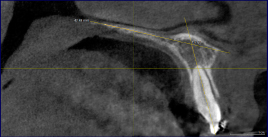
Figure 1.
Shows the measurement of the axial inclination of the maxillary central incisor using the palatal plane as reference on the CBCT scan.
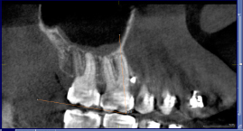
Figure 2.
Shows the measurement of the inclination of the maxillary first permanent molar using the molar occlusal plane as reference on the CBCT scan.
Table I.
Shows the demographics of the patients included in the study
| Male | Female | Total | |
|---|---|---|---|
| N | 6 | 9 | 15 |
| Mean age (SD) in years | 22.17 | 17.89 | 19.6 |
| (5.27) | (2.76) | (4.36) |
Table II.
Showing the comparison of pre-treatment and post-treatment measurements between the CBCT image and cephalogram in the maxillary arch
| Pre-distalisation measurements | Post-distalisation measurements | |||||||||||
|---|---|---|---|---|---|---|---|---|---|---|---|---|
| N | Side | Parameters | CBCT Mean (SD) | Lateral Cephalogram Mean (SD) | Difference Mean (SE) | 95% Cl | Sig. (2- tailed) | CBCT Mean (SD) | Lateral Cephalogram Mean (SD) | Difference Mean (SE) | 95% Cl | Sig. (2- tailed) |
| 15 | Right side | Axial inclination of U1 (°) | 118.04 (6.18) | 120.80 (5.48) | –2.76 (2.13) | –7.13 –1.61 | 0.206 | 1 16.22 (7.77) | 1 18.27 (4.98) | –2.047 (2.38) | –6.93 –2.83 | 0.398 |
| Axial inclination of M1 (°) | 80.32 (4.86) | 90.60 (5.91) | –0.28 (1.98) | –14.33 –6.23 | 0.000* | 82.33 (5.26) | 93.80 (6.61) | –11.47 (2.18) | –15.94 –6.99 | 0.000* | ||
| Left Side | Axial inclination of Ul (°) | 1 18.96 (6.56) | 120.80 (5.48) | –1.84 (2.21) | –6.36 –2.68 | 0.41 1 | 1 17.23 (6.23) | 1 18.27 (4.98) | –1.03 (2.06) | –5.25 –3.18 | 0.620 | |
| Axial inclination of M1 (°) | 78.65 (4.73) | 90.60 (5.91) | –11.95 (1.96) | –15.96 –7.95 | 0.000* | 80.33 (6.58) | 93.80 (6.61) | –13.47 (2.41) | –18.41 –8.54 | 0.000* | ||
1 Axial inclination of U1 – Long axis of the maxillary central incisor to the palatal plane.
Axial inclination of M1 – Long axis of the maxillary first permanent molar to the molar occlusal plane on CBCT and long axis of the maxillary first permanent molar to the palatal plane on lateral cephalogram.
Table III.
Showing the comparison treatment change achieved and level of significance when measured with lateral cephalogram and CBCT
| Right side | Left side | ||||||||||
|---|---|---|---|---|---|---|---|---|---|---|---|
| N | Treatment change (T1-T2) | CBCT Mean (SD) | Lateral Cephalogram Mean (SD) | Difference Mean (SE) | 95% Cl | Sig. (2- tailed) | CBCT Mean (SD) | Lateral Cephalogram Mean (SD) | Difference Mean (SE) | 95% Cl | Sig. (2- tailed) |
| 15 | Change in axial inclination of M1 (°) | –2.01 (3.49) | –3.20 (4.48) | 1.19 (1.47) | –1.82 –4.19 | 0.425 | –1.68 (4.29) | –3.20 (4.48) | 1.52 (1.60) | –1.76 –4.79 | 0.350 |
| Change in axial inclination of U1 (°) | 1.82 (5.49) | 2.53 (4.52) | –.71 (1.84) | –4.48 –3.05 | 0.701 | 1.72 (5.09) | 2.53 (4.52) | –0.81 (1.76) | –4.41 –2.79 | 0.650 | |
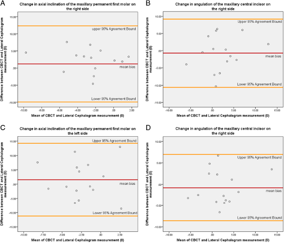
Figure 3.
Shows the Bland–Altman plot to evaluate method of agreement of the various parameters evaluated in the study.
Table IV.
Shows the linear regression analysis done after the Bland Altman test depicted in fig 3 and the presence or absence of proportional bias for each of the parameters between CBCT and lateral cephalogram
| Unstandardized Coefficients | Standardized Coefficients | ||||||
|---|---|---|---|---|---|---|---|
| Parameters | B | Std. Error | Beta | t | Sig. | R2 | Standard error of estimate |
| Change in axial inclination of the maxillary permanent first molar on the right side | –0.487 | 0.538 | –0.243 | –0.905 | 0.382 | 0.059 | 5.71 |
| Change in angulation of the maxillary central incisor on the right side | 0.258 | 0.313 | 0.223 | 0.824 | 0.425 | 0.050 | 5.11 |
| Change in axial inclination of the maxillary permanent first molar on the left side | –0.096 | 0.613 | –0.043 | –0.156 | 0.878 | 0.002 | 6.74 |
| Change in angulation of the maxillary central incisor on the left side | 0.143 | 0.247 | 0.159 | 0.580 | 0.572 | 0.025 | 4.06 |